RNASEH2B Antibody (673) - Azide and BSA Free
Novus Biologicals | Catalog # NBP2-43776
![Western Blot: RNASEH2B Antibody (673) [NBP2-43776]](https://resources.rndsystems.com/images/products/RNASEH2B-Antibody-673-Western-Blot-NBP2-43776-img0002.jpg "Western Blot: RNASEH2B Antibody (673) [NBP2-43776]")
Loading...
Key Product Details
Species Reactivity
Human
Applications
Western Blot, Immunocytochemistry/ Immunofluorescence
Label
Unconjugated
Antibody Source
Monoclonal Mouse IgG1 Clone # 673
Format
Azide and BSA Free
Loading...
Product Specifications
Immunogen
Recombinant protein encompassing a sequence within the center region of human RNASEH2B. The exact sequence is proprietary.
Reactivity Notes
Immunogen displays the following percentage of sequence identity for non-tested species: Porcine (85%).
Clonality
Monoclonal
Host
Mouse
Isotype
IgG1
Theoretical MW
35 kDa.
Disclaimer note: The observed molecular weight of the protein may vary from the listed predicted molecular weight due to post translational modifications, post translation cleavages, relative charges, and other experimental factors.
Disclaimer note: The observed molecular weight of the protein may vary from the listed predicted molecular weight due to post translational modifications, post translation cleavages, relative charges, and other experimental factors.
Scientific Data Images for RNASEH2B Antibody (673) - Azide and BSA Free
Western Blot: RNASEH2B Antibody (673) [NBP2-43776]
Western Blot: RNASEH2B Antibody (673) [NBP2-43776] - Analysis of Ferroportin in human fibroblasts using Ferroportin antibody. Image from verified customer review.![Immunocytochemistry/ Immunofluorescence: RNASEH2B Antibody (673) [NBP2-43776]](https://resources.rndsystems.com/images/products/RNASEH2B-Antibody-673-Immunocytochemistry-Immunofluorescence-NBP2-43776-img0003.jpg "Immunocytochemistry/ Immunofluorescence: RNASEH2B Antibody (673) [NBP2-43776]")
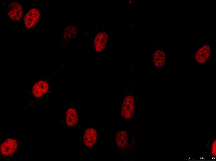
Immunocytochemistry/ Immunofluorescence: RNASEH2B Antibody (673) [NBP2-43776]
Immunocytochemistry/Immunofluorescence: RNASEH2B Antibody (673) [NBP2-43776] - Staining in fibroblasts. Image submitted by a verified customer review.![Western Blot: RNASEH2B Antibody (673) [NBP2-43776]](https://resources.rndsystems.com/images/products/RNASEH2B-Antibody-673-Western-Blot-NBP2-43776-img0001.jpg "Western Blot: RNASEH2B Antibody (673) [NBP2-43776]")
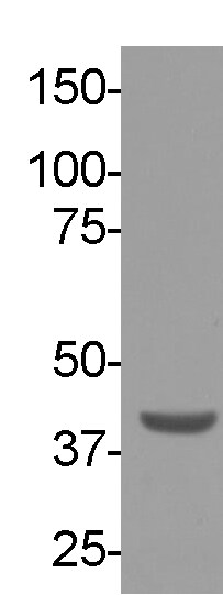
Western Blot: RNASEH2B Antibody (673) [NBP2-43776]
Western Blot: RNASEH2B Antibody (673) [NBP2-43776] - Analysis of HeLa whole cell extracts and nuclear extracts (30 ug) were separated by 10% SDS-PAGE, and the membrane was blotted with RNaseH2B antibody at a dilution of 1:1000.Applications for RNASEH2B Antibody (673) - Azide and BSA Free
Application
Recommended Usage
Immunocytochemistry/ Immunofluorescence
Validated from a verified customer review
Western Blot
1:500-1:3000
Reviewed Applications
Read 2 reviews rated 4.5 using NBP2-43776 in the following applications:
Formulation, Preparation, and Storage
Purification
Protein G purified
Formulation
PBS
Format
Azide and BSA Free
Preservative
No Preservative
Concentration
Concentrations vary lot to lot. See vial label for concentration. If unlisted please contact technical services.
Shipping
The product is shipped with polar packs. Upon receipt, store it immediately at the temperature recommended below.
Stability & Storage
Aliquot and store at -20C or -80C. Avoid freeze-thaw cycles.
Background: RNASEH2B
Alternate Names
ribonuclease H2, subunit B
Gene Symbol
RNASEH2B
Additional RNASEH2B Products
Product Documents for RNASEH2B Antibody (673) - Azide and BSA Free
Certificate of Analysis
To download a Certificate of Analysis, please enter a lot or batch number in the search box below.
Product Specific Notices for RNASEH2B Antibody (673) - Azide and BSA Free
This product is for research use only and is not approved for use in humans or in clinical diagnosis. Primary Antibodies are guaranteed for 1 year from date of receipt.
Customer Reviews for RNASEH2B Antibody (673) - Azide and BSA Free (2)
4.5 out of 5
2 Customer Ratings
Have you used RNASEH2B Antibody (673) - Azide and BSA Free?
Submit a review and receive an Amazon gift card!
$25/€18/£15/$25CAN/¥2500 Yen for a review with an image
$10/€7/£6/$10CAN/¥1110 Yen for a review without an image
Submit a review
Customer Images
Showing
1
-
2 of
2 reviews
Showing All
Filter By:
-
Application: ImmunocytochemistrySample Tested: fibroblastsSpecies: HumanVerified Customer | Posted 11/18/2017RNASEH2B in fibroblastsBlocked 1H 25C in 1X PBS/5% normal serum/0.3% Triton™ X-100 Diluted 1:250 in 1X PBS/1% BSA/0.3% Triton™ X-100, 16 hours at 4C incubation. 2 hours (25C) secondary (1.5:1000 dilution)

-
Application: Western BlotSample Tested: fibroblastsSpecies: HumanVerified Customer | Posted 08/12/2016RNASEH2B
There are no reviews that match your criteria.
Protocols
Find general support by application which include: protocols, troubleshooting, illustrated assays, videos and webinars.
- Appropriate Fixation of IHC/ICC Samples
- Cellular Response to Hypoxia Protocols
- ClariTSA™ Fluorophore Kits
- Detection & Visualization of Antibody Binding
- ICC Cell Smear Protocol for Suspension Cells
- ICC Immunocytochemistry Protocol Videos
- ICC for Adherent Cells
- Immunocytochemistry (ICC) Protocol
- Immunocytochemistry Troubleshooting
- Immunofluorescence of Organoids Embedded in Cultrex Basement Membrane Extract
- Immunohistochemistry (IHC) and Immunocytochemistry (ICC) Protocols
- Preparing Samples for IHC/ICC Experiments
- Preventing Non-Specific Staining (Non-Specific Binding)
- Primary Antibody Selection & Optimization
- Protocol for VisUCyte™ HRP Polymer Detection Reagent
- Protocol for the Fluorescent ICC Staining of Cell Smears - Graphic
- Protocol for the Fluorescent ICC Staining of Cultured Cells on Coverslips - Graphic
- Protocol for the Preparation and Fluorescent ICC Staining of Cells on Coverslips
- Protocol for the Preparation and Fluorescent ICC Staining of Non-adherent Cells
- Protocol for the Preparation and Fluorescent ICC Staining of Stem Cells on Coverslips
- Protocol for the Preparation of a Cell Smear for Non-adherent Cell ICC - Graphic
- R&D Systems Quality Control Western Blot Protocol
- TUNEL and Active Caspase-3 Detection by IHC/ICC Protocol
- The Importance of IHC/ICC Controls
- Troubleshooting Guide: Western Blot Figures
- Western Blot Conditions
- Western Blot Protocol
- Western Blot Protocol for Cell Lysates
- Western Blot Troubleshooting
- Western Blot Troubleshooting Guide
- View all Protocols, Troubleshooting, Illustrated assays and Webinars
Loading...