
The Coronavirus Spike protein (S Protein) is one of four major structural proteins (S, M, E, N) present in each viral particle. Spike proteins are abundantly expressed on the surface forming its characteristic crown-like halo structure. The Spike protein is a highly glycosylated, type I transmembrane protein that plays a critical role in receptor recognition and host cell entry. There are two subunits, S1 and S2, that divide the protein roughly in half. Within the N-terminal S1 subunit is the N-terminal domain and receptor binding domain (Spike RBD protein). The Spike RBD binds to the host cell and undergoes several conformational changes and proteolysis leading to fusion of the virus particle to the host cell membrane allowing transfer of the nucleocapsid into the host cell cytoplasm. For SARS-CoV-2, the virus responsible for the COVID-19 pandemic, the Spike RBD tightly associates with human ACE-2. Recombinant SARS-CoV-2 proteins and antibodies that recognize these proteins are essential for studying the molecular mechanisms of the coronavirus life cycle.

Scientific images may be resized, cropped, or adjusted for brightness or contrast for presentation purposes. These updates are limited to presentation and do not affect the underlying scientific interpretation of the data. Where available, additional source data may be provided upon request.
Key Product Details
Species Reactivity
Applications
Label
Antibody Source
Product Specifications
Immunogen
Sequence:
ADYSVLYNSTFFSTFKCYGVSATKLNDLCFSNVYADSFVVKGDDVRQIAPGQTGVIADYNYKLPDDFMGCVLAWNTRNIDATSTGNYNYKYRYLRHGKLRP
Clonality
Host
Isotype
Theoretical MW
Disclaimer note: The observed molecular weight of the protein may vary from the listed predicted molecular weight due to post translational modifications, post translation cleavages, relative charges, and other experimental factors.
Scientific Data Images for Spike Antibody (8H6J10)
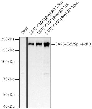
Western Blot: Spike Antibody (8H6J10) [NBP3-33299] -
Western Blot: Spike Antibody (8H6J10) [NBP3-33299] - Western blot analysis of various lysates, using Spike Rabbit mAb at 1:2000 dilution.Secondary antibody: HRP-conjugated Goat anti-Rabbit IgG (H+L) at 1:10000 dilution.
Lysates/proteins: 25ug per lane.
Blocking buffer: 3% nonfat dry milk in TBST.
Detection: ECL Basic Kit.
Exposure time: 10s.
Applications for Spike Antibody (8H6J10)
ELISA
Western Blot
Formulation, Preparation, and Storage
Purification
Formulation
Preservative
Concentration
Shipping
Stability & Storage
Background: Spike
Long Name
Alternate Names
Additional Spike Products
Product Documents for Spike Antibody (8H6J10)
Certificate of Analysis
To download a Certificate of Analysis, please enter a lot or batch number in the search box below.
Product Specific Notices for Spike Antibody (8H6J10)
This product is for research use only and is not approved for use in humans or in clinical diagnosis. Primary Antibodies are guaranteed for 1 year from date of receipt.
Related Research Areas
Customer Reviews for Spike Antibody (8H6J10)
There are currently no reviews for this product. Be the first to review Spike Antibody (8H6J10) and earn rewards!
Have you used Spike Antibody (8H6J10)?
Submit a review and receive an Amazon gift card!
$25/€18/£15/$25CAN/¥2500 for a review with an image
$10/€7/£6/$10CAN/¥1110 for a review without an image
Submit a review
Protocols
Find general support by application which include: protocols, troubleshooting, illustrated assays, videos and webinars.
- Cellular Response to Hypoxia Protocols
- ELISA Sample Preparation & Collection Guide
- ELISA Troubleshooting Guide
- How to Run an R&D Systems DuoSet ELISA
- How to Run an R&D Systems Quantikine ELISA
- How to Run an R&D Systems Quantikine™ QuicKit™ ELISA
- Quantikine HS ELISA Kit Assay Principle, Alkaline Phosphatase
- Quantikine HS ELISA Kit Principle, Streptavidin-HRP Polymer
- R&D Systems Quality Control Western Blot Protocol
- Sandwich ELISA (Colorimetric) – Biotin/Streptavidin Detection Protocol
- Sandwich ELISA (Colorimetric) – Direct Detection Protocol
- Troubleshooting Guide: ELISA
- Troubleshooting Guide: Western Blot Figures
- Western Blot Conditions
- Western Blot Protocol
- Western Blot Protocol for Cell Lysates
- Western Blot Troubleshooting
- Western Blot Troubleshooting Guide
- View all Protocols, Troubleshooting, Illustrated assays and Webinars