SUR1 Antibody - BSA Free
Novus Biologicals | Catalog # NBP2-34077
![Immunocytochemistry/ Immunofluorescence: SUR1 Antibody [NBP2-34077]](https://resources.rndsystems.com/images/products/SUR1-Antibody-Immunocytochemistry-Immunofluorescence-NBP2-34077-img0008.jpg "Immunocytochemistry/ Immunofluorescence: SUR1 Antibody [NBP2-34077]")
Loading...
Key Product Details
Species Reactivity
Validated:
Human
Cited:
Mouse
Predicted:
Rat (96%). Backed by our 100% Guarantee.
Applications
Validated:
Immunohistochemistry, Immunohistochemistry-Paraffin, Immunocytochemistry/ Immunofluorescence, Knockdown Validated
Cited:
Immunocytochemistry/ Immunofluorescence, Knockdown Validated
Label
Unconjugated
Antibody Source
Polyclonal Rabbit IgG
Format
BSA Free
Loading...
Product Specifications
Immunogen
This antibody was developed against a recombinant protein corresponding to amino acids: DHLGKENDVFQPKTQFLGVYFVSSQ
Reactivity Notes
Mouse reactivity reported in scientific literature (PMID: 31067750).
Clonality
Polyclonal
Host
Rabbit
Isotype
IgG
Scientific Data Images for SUR1 Antibody - BSA Free
Immunocytochemistry/ Immunofluorescence: SUR1 Antibody [NBP2-34077]
Immunocytochemistry/Immunofluorescence: SUR1 Antibody [NBP2-34077] - Staining of human cell line U-2 OS shows localization to nucleoli, cytosol & the Golgi apparatus. Antibody staining is shown in green.![Immunohistochemistry-Paraffin: SUR1 Antibody [NBP2-34077]](https://resources.rndsystems.com/images/products/SUR1-Antibody-Immunohistochemistry-Paraffin-NBP2-34077-img0013.jpg "Immunohistochemistry-Paraffin: SUR1 Antibody [NBP2-34077]")
Immunohistochemistry-Paraffin: SUR1 Antibody [NBP2-34077]
Immunohistochemistry-Paraffin: SUR1 Antibody [NBP2-34077] - Staining of human cerebellum shows strong cytoplasmic positivity in purkinje cells.![Immunohistochemistry: SUR1 Antibody [NBP2-34077]](https://resources.rndsystems.com/images/products/SUR1-Antibody-Immunohistochemistry-NBP2-34077-img0007.jpg "Immunohistochemistry: SUR1 Antibody [NBP2-34077]")
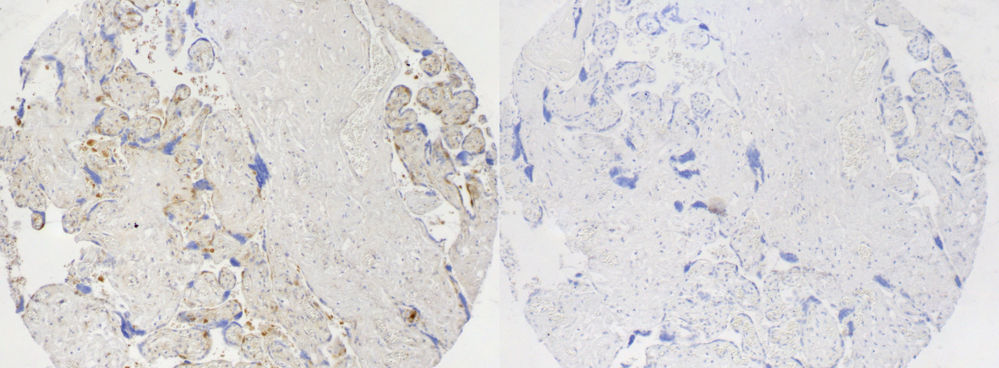
Immunohistochemistry: SUR1 Antibody [NBP2-34077]
Immunohistochemistry: SUR1 Antibody [NBP2-34077] - IHC using NBP2-34077 at a dilution of 1:50 on FFPE human placenta. Primary antibody was incubated overnight at 4 C, then secondary HRP conjugate applied for 1 hour RT and visualized with DAB. HIER performed with citrate buffer pH 6.0 at 95 C for 15 minutes. Antibody incubation with 10x immunogenic peptide shows no staining. Image submitted by a verified customer review.![Immunohistochemistry-Paraffin: SUR1 Antibody [NBP2-34077]](https://resources.rndsystems.com/images/products/SUR1-Antibody-Immunohistochemistry-Paraffin-NBP2-34077-img0011.jpg "Immunohistochemistry-Paraffin: SUR1 Antibody [NBP2-34077]")
Immunohistochemistry-Paraffin: SUR1 Antibody [NBP2-34077]
Immunohistochemistry-Paraffin: SUR1 Antibody [NBP2-34077] - Staining of human pancreas shows weak cytoplasmic positivity in islets of Langerhans.![Immunohistochemistry-Paraffin: SUR1 Antibody [NBP2-34077]](https://resources.rndsystems.com/images/products/SUR1-Antibody-Immunohistochemistry-Paraffin-NBP2-34077-img0012.jpg "Immunohistochemistry-Paraffin: SUR1 Antibody [NBP2-34077]")
Immunohistochemistry-Paraffin: SUR1 Antibody [NBP2-34077]
Immunohistochemistry-Paraffin: SUR1 Antibody [NBP2-34077] - Staining of human cerebral cortex shows moderate cytoplasmic positivity in neurons.![Immunohistochemistry-Paraffin: SUR1 Antibody [NBP2-34077]](https://resources.rndsystems.com/images/products/SUR1-Antibody-Immunohistochemistry-Paraffin-NBP2-34077-img0014.jpg "Immunohistochemistry-Paraffin: SUR1 Antibody [NBP2-34077]")
Immunohistochemistry-Paraffin: SUR1 Antibody [NBP2-34077]
Immunohistochemistry-Paraffin: SUR1 Antibody [NBP2-34077] - Staining of human liver shows no positivity in hepatocytes as expected.Applications for SUR1 Antibody - BSA Free
Application
Recommended Usage
Immunocytochemistry/ Immunofluorescence
0.25-2 ug/ml
Immunohistochemistry
1:50 - 1:200
Immunohistochemistry-Paraffin
1:50 - 1:200
Application Notes
For IHC-Paraffin, HIER pH 6 retrieval is recommended. ICC/IF Fixation Permeabilization: Use PFA/Triton X-100. Use in Knockdown Validated reported in scientific literature (PMID: 31067750). SUR1 antibody validated for IHC from a verified customer review.
Reviewed Applications
Read 1 review rated 4 using NBP2-34077 in the following applications:
Formulation, Preparation, and Storage
Purification
Affinity purified
Formulation
PBS (pH 7.2) and 40% Glycerol
Format
BSA Free
Preservative
0.02% Sodium Azide
Concentration
Concentrations vary lot to lot. See vial label for concentration. If unlisted please contact technical services.
Shipping
The product is shipped with polar packs. Upon receipt, store it immediately at the temperature recommended below.
Stability & Storage
Store at 4C short term. Aliquot and store at -20C long term. Avoid freeze-thaw cycles.
Background: SUR1
Long Name
ATP-binding cassette sub-family C member 8
Alternate Names
ABCC8, HRINS, SUR
Gene Symbol
ABCC8
UniProt
Additional SUR1 Products
Product Documents for SUR1 Antibody - BSA Free
Certificate of Analysis
To download a Certificate of Analysis, please enter a lot or batch number in the search box below.
Product Specific Notices for SUR1 Antibody - BSA Free
This product is for research use only and is not approved for use in humans or in clinical diagnosis. Primary Antibodies are guaranteed for 1 year from date of receipt.
Citations for SUR1 Antibody - BSA Free
 Powered by Bioz
Customer Reviews for SUR1 Antibody - BSA Free (1)
4 out of 5
1 Customer Rating
Have you used SUR1 Antibody - BSA Free?
Submit a review and receive an Amazon gift card!
$25/€18/£15/$25CAN/¥2500 Yen for a review with an image
$10/€7/£6/$10CAN/¥1110 Yen for a review without an image
Submit a review
Customer Images
Showing
1
-
1 of
1 review
Showing All
Filter By:
-
Application: ImmunohistochemistrySample Tested: Placental tissueSpecies: HumanVerified Customer | Posted 04/03/2017IHC using NBP2-34077 at a dilution of 1:50 on FFPE human placenta. Primary antibody was incubated overnight at 4 C, then secondary HRP conjugate applied for 1 hour RT and visualized with DAB. HIER performed with citrate buffer pH 6.0 at 95 C for 15 minutes. Antibody incubation with 10x immunogenic peptide shows no staining.

There are no reviews that match your criteria.
Protocols
Find general support by application which include: protocols, troubleshooting, illustrated assays, videos and webinars.
- Antigen Retrieval Protocol (PIER)
- Antigen Retrieval for Frozen Sections Protocol
- Appropriate Fixation of IHC/ICC Samples
- Cellular Response to Hypoxia Protocols
- Chromogenic IHC Staining of Formalin-Fixed Paraffin-Embedded (FFPE) Tissue Protocol
- Chromogenic Immunohistochemistry Staining of Frozen Tissue
- ClariTSA™ Fluorophore Kits
- Detection & Visualization of Antibody Binding
- Fluorescent IHC Staining of Frozen Tissue Protocol
- Graphic Protocol for Heat-induced Epitope Retrieval
- Graphic Protocol for the Preparation and Fluorescent IHC Staining of Frozen Tissue Sections
- Graphic Protocol for the Preparation and Fluorescent IHC Staining of Paraffin-embedded Tissue Sections
- Graphic Protocol for the Preparation of Gelatin-coated Slides for Histological Tissue Sections
- ICC Cell Smear Protocol for Suspension Cells
- ICC Immunocytochemistry Protocol Videos
- ICC for Adherent Cells
- IHC Sample Preparation (Frozen sections vs Paraffin)
- Immunocytochemistry (ICC) Protocol
- Immunocytochemistry Troubleshooting
- Immunofluorescence of Organoids Embedded in Cultrex Basement Membrane Extract
- Immunofluorescent IHC Staining of Formalin-Fixed Paraffin-Embedded (FFPE) Tissue Protocol
- Immunohistochemistry (IHC) and Immunocytochemistry (ICC) Protocols
- Immunohistochemistry Frozen Troubleshooting
- Immunohistochemistry Paraffin Troubleshooting
- Preparing Samples for IHC/ICC Experiments
- Preventing Non-Specific Staining (Non-Specific Binding)
- Primary Antibody Selection & Optimization
- Protocol for Heat-Induced Epitope Retrieval (HIER)
- Protocol for Making a 4% Formaldehyde Solution in PBS
- Protocol for VisUCyte™ HRP Polymer Detection Reagent
- Protocol for the Fluorescent ICC Staining of Cell Smears - Graphic
- Protocol for the Fluorescent ICC Staining of Cultured Cells on Coverslips - Graphic
- Protocol for the Preparation & Fixation of Cells on Coverslips
- Protocol for the Preparation and Chromogenic IHC Staining of Frozen Tissue Sections
- Protocol for the Preparation and Chromogenic IHC Staining of Frozen Tissue Sections - Graphic
- Protocol for the Preparation and Chromogenic IHC Staining of Paraffin-embedded Tissue Sections
- Protocol for the Preparation and Chromogenic IHC Staining of Paraffin-embedded Tissue Sections - Graphic
- Protocol for the Preparation and Fluorescent ICC Staining of Cells on Coverslips
- Protocol for the Preparation and Fluorescent ICC Staining of Non-adherent Cells
- Protocol for the Preparation and Fluorescent ICC Staining of Stem Cells on Coverslips
- Protocol for the Preparation and Fluorescent IHC Staining of Frozen Tissue Sections
- Protocol for the Preparation and Fluorescent IHC Staining of Paraffin-embedded Tissue Sections
- Protocol for the Preparation of Gelatin-coated Slides for Histological Tissue Sections
- Protocol for the Preparation of a Cell Smear for Non-adherent Cell ICC - Graphic
- TUNEL and Active Caspase-3 Detection by IHC/ICC Protocol
- The Importance of IHC/ICC Controls
- Troubleshooting Guide: Immunohistochemistry
- View all Protocols, Troubleshooting, Illustrated assays and Webinars
Loading...