Tat-Beclin 1 D11 Autophagy Inducing Peptide - Retroinverso form
Novus Biologicals | Catalog # NBP2-49888
![Immunocytochemistry/ Immunofluorescence: Tat-Beclin 1 D11 Autophagy Inducing Peptide - Retroinverso form [NBP2-49888]](https://resources.rndsystems.com/images/products/Tat-Beclin-1-D11-Autophagy-Inducing-Peptide-Immunocytochemistry-Immunofluorescence-NBP2-49888-img0002.jpg "Immunocytochemistry/ Immunofluorescence: Tat-Beclin 1 D11 Autophagy Inducing Peptide - Retroinverso form [NBP2-49888]")
Key Product Details
Applications
Product Specifications
Description
Background
Predicted Molecular Mass
Disclaimer note: The observed molecular weight of the protein may vary from the listed predicted molecular weight due to post translational modifications, post translation cleavages, relative charges, and other experimental factors.
Protein / Peptide Type
Reviewed Applications
Read 1 review rated 5 using NBP2-49888 in the following applications:
Scientific Data Images for Tat-Beclin 1 D11 Autophagy Inducing Peptide - Retroinverso form
Immunocytochemistry/ Immunofluorescence: Tat-Beclin 1 D11 Autophagy Inducing Peptide - Retroinverso form [NBP2-49888]
Immunocytochemistry/Immunofluorescence: Tat-Beclin 1 D11 Autophagy Inducing Peptide [NBP2-49888] - HeLa GFP-LC3B cells were treated with Tat-D11, Tat-L11, Tat-Beclin 1 or Tat-L11S for 1.5 hours. Thereafter, the cells were stained using NeuroTrace Red or DAPI and analyzed employing fluorescent microscopy. Note the higher number of autophagosomes/GFP-LC3B+ puncta in the images of Tat-D11 and Tat-L11 treated cells when compared to Tat-Beclin 1 and Tat-L11S treated cells.![In vitro assay: Tat-Beclin 1 D11 Autophagy Inducing Peptide - Retroinverso form [NBP2-49888]](https://resources.rndsystems.com/images/products/Tat-Beclin-1-D11-Autophagy-Inducing-Peptide---Retroinverso-form-Western-Blot-NBP2-49888-img0001.jpg "In vitro assay: Tat-Beclin 1 D11 Autophagy Inducing Peptide - Retroinverso form [NBP2-49888]")
In vitro assay: Tat-Beclin 1 D11 Autophagy Inducing Peptide - Retroinverso form [NBP2-49888]
In vitro assay: Tat-Beclin 1 D11 Autophagy Inducing Peptide - Retroinverso form [NBP2-49888] - Analysis of lysates from HeLa cells that were left untreated (blank) or were treated with 10-20 uM each of Tat-D11, Tat-L11, Tat-Beclin 1 or Tat-L11S. The lysates were analyzed for the expression of LC3-1/LC3-II and SQSTM1/p62 using 2 ug/mL each of anti-LC3B (NB100-2220) and anti-SQSTM1/p62 (MAB8028) respectively. Anti-Actin (AF4000) was used as a loading control. TatD11 exhibited superior induction of LC3-II and down-regulation of p62 protein when compared to other treatment and control groups.![In vivo assay: Tat-Beclin 1 D11 Autophagy Inducing Peptide - Retroinverso form [NBP2-49888]](https://resources.rndsystems.com/images/products/Tat-Beclin-1-D11-Autophagy-Inducing-Peptide---Retroinverso-form-In-vivo-assay-NBP2-49888-img0004.jpg "In vivo assay: Tat-Beclin 1 D11 Autophagy Inducing Peptide - Retroinverso form [NBP2-49888]")
In vivo assay: Tat-Beclin 1 D11 Autophagy Inducing Peptide - Retroinverso form [NBP2-49888]
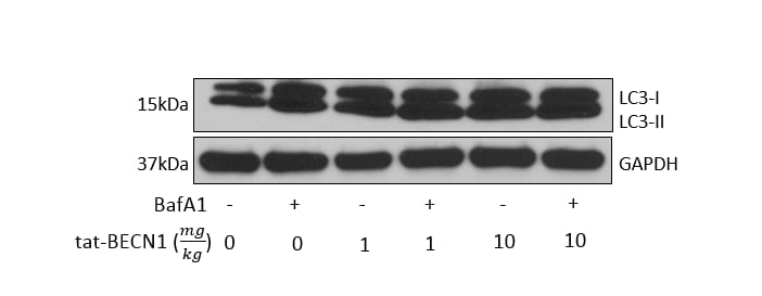
In vivo assay: Tat-Beclin 1 D11 Autophagy Inducing Peptide - Retroinverso form [NBP2-49888] - In vivo dose study in 10wk old C57BL/6J mice. Either 1mg/kg or 10mg/kg IP once daily was administered for 2 days, mice were sacrificed, kidneys prepared for Western blot analysis. Lysosomal inhibitor bafilomycin A1 was used to provide a measurement of autophagic flux. *vehicle is scrambled tat-beclin (NBP2-49887-5mg). Image from verified customer review.Formulation, Preparation, and Storage
NBP2-49888
| Formulation | This product is supplied lyophilized. Purity is >= to 97% (HPLC) |
| Concentration | Lyoph |
| Reconstitution | Reconstitute with DMSO or water to desired concetration. Note:- D11 should NOT be reconstituted at a concentration greater than 5 mM. |
| Shipping | The product is shipped with polar packs. Upon receipt, store it immediately at the temperature recommended below. |
| Stability & Storage | Store at -20C in powder form. Store at -80C once reconstituted. |
Calculators
Background: Tat-Beclin 1
Alternate Names
Additional Tat-Beclin 1 Products
Product Documents for Tat-Beclin 1 D11 Autophagy Inducing Peptide - Retroinverso form
Certificate of Analysis
To download a Certificate of Analysis, please enter a lot or batch number in the search box below.
Product Specific Notices for Tat-Beclin 1 D11 Autophagy Inducing Peptide - Retroinverso form
Note: Tat-L11 and Tat-D11, are exclusively available from Novus Biologicals (Bio-Techne's exclusive patent license: US Patent 8,802,633).
This product is for research use only and is not approved for use in humans or in clinical diagnosis. This product is guaranteed for 1 year from date of receipt.
Citations for Tat-Beclin 1 D11 Autophagy Inducing Peptide - Retroinverso form
 Powered by Bioz
Customer Reviews for Tat-Beclin 1 D11 Autophagy Inducing Peptide - Retroinverso form (1)
Have you used Tat-Beclin 1 D11 Autophagy Inducing Peptide - Retroinverso form?
Submit a review and receive an Amazon gift card!
$25/€18/£15/$25CAN/¥2500 Yen for a review with an image
$10/€7/£6/$10CAN/¥1110 Yen for a review without an image
Submit a review
Customer Images
-
Application: In vivo treatment + immunoblot analysisVerified Customer | Posted 01/07/2019In vivo treatment, then immunoblot analysis on kidneysI used this peptide for an in vivo dose study in 10wk old C57BL/6J mice. I administered either 1mg/kg or 10mg/kg IP once daily for 2 days, sacrificed the mice, and prepared the kidneys for western blot analysis (image shown). Additionally, the lysosomal inhibitor bafilomycin A1 was used to provide a measurement of autophagic flux. *vehicle is scrambled tat-beclin (NBP2-49887-5mg)

There are no reviews that match your criteria.
Protocols
View specific protocols for Tat-Beclin 1 D11 Autophagy Inducing Peptide - Retroinverso form (NBP2-49888):
ICC/IF protocol to induce autophagy in HeLa cells using Tat-Beclin 1 L11 or Tat-Beclin 1 D11 peptides.
Important Notes
1. Peptides Tat-Beclin 1 L11 [NBP2-49886] and Tat-Beclin 1 D11 [NBP2-49888] are useful for induction of autophagy.
2. Tat-Beclin L11S [NBP2-49887], an inactive/scrambled control peptide derived from Tat-Beclin L11, is useful as a negative control when analyzing NBP2-49886 and/or NBP2-49888 for autophagy induction experiments.
3. Molecular weights for L11, L11S and D11 are 3.08 kDa each.
4. Tat-Beclin D11 should NOT be reconstituted at a concentration greater than 5 mM.
5. Antibodies against LC3B or p62/SQSTM1 may be used for detecting the induction of autophagy. An increased number of LC3 stained vacuoles or decreased levels of p62 indicate autophagy induction.
Day 1 - Culturing HeLa Cells
1. Plate cells at a density of 1-1.5 x 10^5 cells per mL and 100 uL per well in DMEM with 10% FBS and 1X Pen/Strep into a black-welled Perkin Elmer Cell Carrier 96-well plate (6005550).
2. Incubate the cultured plate overnight at 37C with 5 % CO2.
Day 2 - Treatments
1. Check cells for confluency. Cells should be 60-80% confluent before treatments.
2. Wash cells 3 times with 1X PBS.
3. Resuspend 1 mM of each peptide in OptiMEM (Life Technologies: 11058021) acidified with 0.15 % 6 N HCl.
Optimization of peptide concentration and incubation time
i. To determine the most effective concentration for your cell line of interest, perform a 1:2 serial dilution with the 1 mM peptides. Start with at least 20 uM and dilute to 0 uM final testing concentration in each well.
ii. Duration of induction can be determined by collecting images at every 15 min up to 2 hrs. Fix the cells from one well at each time point according to the instructions starting from Step 6 below.
4. Add 50 uL to each well (in triplicate)
5. Incubate for 1.5 hrs at 37C with 5 % CO2.
6. Remove remaining liquid from wells and fix cells with 4% paraformaldehyde for 20 minutes at room temperature.
7. Wash cells 3 times with 1X PBS.
8. Block cells with blocking buffer (1X PBS, 0.1 % Triton-X, 5 % Normal Donkey Serum) for 1 hr at room temperature (alternately, cells can be blocked overnight at 4 C).
Day 2/3 - Primary Antibody Staining
1. Dilute the primary antibody in blocking buffer to the specifications listed in the antibody datasheet.
2. Incubate the primary antibodies overnight at 4 C or 2 hrs at room temperature.
Day 2/3 - Secondary Antibody Staining
1. Wash cells 3 times with 1X PBS.
2. Dilute the secondary antibodies to specifications in the blocking buffer.
3. Incubate for 1 hr at room temperature in dark.
4. Wash cells 3 times with 1X PBS.
5. Stain with DAPI, cytosolic stain [NeuroTrace (R)], and Northern Lights Guard (R&D Systems, Inc. NL996).
a. NeuroTrace (R)- 1:200 dilution in 1X PBS.
b. DAPI- 1:10000 dilution into NeuroTrace (R)/1X PBS solution.
c. Northern Lights Guard - add 1:1 to NeuroTrace (R)/DAPI/PBS solution
6. Leave solution in wells and seal with a foil plate sealer.
7. Image plate. Store plate at 4 C in dark.
Useful Resources:
1. Troubleshooting for Autophagy and LC3:
2. Autophagy Research Sub-topics:
3. Support by Application, Protocols:
WB Protocol for Tat-Beclin 1 L11 or Tat-Beclin 1 D11 Induced Autophagy in HeLa Cells
Important Notes
1. Peptides Tat-Beclin 1 L11 [NBP2-49886] and Tat-Beclin 1 D11 [NBP2-49888] are useful for induction of autophagy.
2. Tat-Beclin 1 L11S [NBP2-49887], an inactive/scrambled control peptide derived from Tat-Beclin 1 L11, is useful as a negative control when analyzing NBP2-49886 and/or NBP2-49888 for autophagy induction experiments.
3. Molecular weights for L11, L11S and D11 are 3.08 kDa each.
4. Tat-Beclin 1 D11 should NOT be reconstituted at a concentration greater than 5 mM.
5. An increased levels of LC3B-II band or a decreased levels of p62 indicate autophagy induction.
Cell Culture and Treatments
1. Plate HeLa cells overnight in 12 well plates and check for confluency. Cells should be 60-80 % confluent before treatments.
2. Wash cells 3 times with 1X PBS.
3. Re-suspend 1 mM of each peptide in OptiMEM (Life Technologies: 11058021) acidified with 0.15 % 6 N HCl.
Optimization of peptide concentration and incubation time
i. To determine the most effective concentration for your cell line perform a 1:2 serial dilution with the 1 mM peptides starting with 20 uM and diluting to 0 uM final concentration in each well.
ii. Duration of induction would be concentration and cell line dependent. HeLa cells may be incubated with the peptides up to 20 uM /up to 2 hrs.
Cell Lysate Preparation
4. Remove the medium from one well at each time point and add cold lysis buffer immediately. Cell lysis may be performed using 150 uL of M-PER (TM) Mammalian Protein Extraction Reagent (Thermo 78501) with 1:100 Halt (TM) Protease and Phosphatase Inhibitor Single-Use Cocktail (Thermo 78442) per well.
5. Incubate the plates at room temperature for 10 min with gentle agitation.
6. Scrape the cells from the plate and spin down at 13500 rpm for 10 min at 4C. Save the supernatant (lysate) and discard the pellet (cell debris).
Western Blot
7. Add 30 uL of 6X Lamelli Reducing SDS loading buffer to 150 uL of the lysate. Boil the solution for 5 min at 95C and then cool to room temperature before loading.
8. Use a 5-20% gradient gel and load the wells with 10 uL of reduced sample. Run the gel at 130V for 1 hour.
9. Transfer the proteins from gel to a nitrocellulose membrane at 100V for 1 hour.
10. Block the membranes for 1-2 hours at room temperature in Pierce (TM) Protein-Free (PBS) Blocking Buffer (Thermo 37584).
11. Incubate the membranes for overnight in blocking buffer with the respective antibodies: rabbit anti-LC3B (Novus NB100-2220) at 2 ug/mL, mouse anti-SQSTM1/p62 (Novus MAB8028) at 2 ug/mL, and sheep anti-actin (Novus AF4000) at 1 ug/mL.
12. Next day, rinse the membranes with DI water and wash with 1X TBST for 1 hour at room temperature. Probe the membranes with a secondary antibodies for 1 hour at room temperature; goat anti-rabbit IgG HRP (Novus HAF008) at 1:1000, donkey anti-mouse IgG HRP (Novus HAF018) at 1:1000, and donkey anti-sheep IgG HRP (Novus HAF016) at 1:1000.
13. Wash the membranes for 2 hours with 1X TBST and then develop using a 1:1 solution of WesternGlo A and B for 1 min with a 1 min exposure time on a Kodak Chemiluminescent imager.
Useful Resources:
1. Troubleshooting for Autophagy and LC3:-
2. Autophagy Research Sub-topics:-
3. Support by Application, Protocols:-