TLR4 Inhibitor Peptide Set
Novus Biologicals | Catalog # NBP2-26244
Loading...
Key Product Details
Species
Human, Mouse, Rat
Applications
Block/Neutralize, Flow Cytometry, Functional Assay
Product Summary for TLR4 Inhibitor Peptide Set
TLR4 inhibitor and control peptides are quality controlled in vitro using the TLR4/MD-2/CD14/NF-kB/SEAP cell line with SEAP as a readout assay (Image 3).
Loading...
Product Specifications
Applications
Binding Inhibition (reported in scientific literature (PMID 27897392))
Block/Neutralize (reported in multiple pieces of scientific literature)
Functional (reported in scientific literature (PMID 24630524))
Block/Neutralize (reported in multiple pieces of scientific literature)
Functional (reported in scientific literature (PMID 24630524))
Application Notes
The inhibitor is used in assays to inhibit TLR4 activation; see Image 3 and also refer to Lysakova-Devine et al (2010) for examples. Optimal inhibitor concentrations should be established through titration and may vary between model systems. We recommend an initial titration from 0-30 uM for in vitro assays (Image 3). Control concentrations should mirror inhibitor concentrations. Inhibitor and control should be preincubated with cells prior to ligand activation to allow sufficient time for the peptides to enter from the media into the cell. We typically preincubate with inhibitor and control peptides for 2 h prior to TLR4 activation with LPS (Image 3); however, optimal preincubation times may vary between model systems.
The TLR4/MD-2/CD14 stably transfected cell line is a useful positive control model system for studying inhibition of TLR4 activation by VIPER (Image 3). SEAP is used as a readout assay in Image 3 to measure TLR4 inhibition.
A novel model system is shown in Image 1 where TLR4 inhibitor peptide, but not CP7, inhibited TLR4 activation in Mal-deficient immortalized mouse bone marrow-derived macrophages (iBMDMs). In these iBMDMs, the inhibitor targets TLR4-TRAM, but not TLR-Mal, interactions as Mal is not expressed. TNF-alpha is used as a readout assay in Image 1 to measure inhibition.
Reactivity Notes
Rat reactivity reported in scientific literature (PMID: 25811788)
Inhibitor Content
VIPER: A TLR4 Inhibitory Peptide: 1 mg (lyophilized) KYSFKLILAEYRRRRRRRRR (VIPER sequence: KYSFKLILAEY). Molecular weight: 2780.3
CP7, Control Peptide: 1 mg (lyophilized) RNTISGNIYSARRRRRRRRR (Control sequence: RNTISGNIYSA). Molecular weight: 2601
CP7, Control Peptide: 1 mg (lyophilized) RNTISGNIYSARRRRRRRRR (Control sequence: RNTISGNIYSA). Molecular weight: 2601
Scientific Data Images for TLR4 Inhibitor Peptide Set
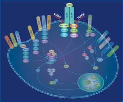
TLR4 Inhibitor Peptide Set [NBP2-26244] - The inhibitor peptide inhibits TLR4 signaling by blocking interactions between TLR4 and its adaptors Mal/TIRAP and TRAM.
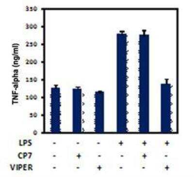
Blocking/Neutralizing: TLR4 Inhibitor Peptide Set [NBP2-26244] - iBMDM cultures were pretreated with 5uM of inhibitor or control prior to stimulation with 20 ng/ml LPS. Negative (LPS-, LPS-/CP7+, LPS-/CP7+) and positive (LPS+) controls were also included. Supernatants were harvested after 6 h and murine TNF-alpha measured by ELISA. TLR4 inhibitor, but not the control, inhibited LPS-induced TNF-alpha production. Image courtesy of Dr. Julianne Stack and Dr. Andrew Bowie, Trinity College, Dublin, Ireland.
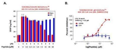
TLR4 Inhibitor Peptide Set [NBP2-26244] - TLR4/MD-2/CD14/NF-kB/SEAPorter stably transfected cells were plated in 96-well plates at 1x10^5 cells/well. After 16 h, cells were preincubated with various concentrations of inhibitor or control peptides (CP) for 1 h. Cells were then stimulated with 10 ng/ml LPS for 24 h. The secreted alkaline phosphatase (SEAP) was analyzed using the SEAPorter Assay Kit. Inhibitory effect on the LPS-mediated TLR4 activation by the VIPER peptide is shown in panel A, and IC50 of the VIPER peptide is described in panel B.
Formulation, Preparation, and Storage
Preparation Method
Preparation of 5 mM VIPER and CP7 Stock Solutions
Note: Bring the peptides to room temperature and quick spin the tubes before opening the caps.
VIPER: A final volume of 72 ul will make a 5 mM stock solution. Add 72 ul sterile H20 to the tube of peptide. Carefully pipet to ensure all of the peptide is dissolved.
CP7: A final volume of 76 ul will make a 5 mM stock solution. Add 76 ul sterile H20 to the tube of peptide. Carefully pipet to ensure all of the peptide is dissolved.
The stock solutions may be diluted further to make working solutions. Dilute according to the needs for your assay. For example dilute 5 mM stock solutions 1:10 in sterile 1X PBS or cell culture media to make 500 uM working solutions. Working solutions should be made fresh daily and not stored.
Please contact technical support for detailed reconstitution instructions.
Purification
>95%, by HPLC.
Concentration
Lyoph
Shipping
The product is shipped with polar packs. Upon receipt, store it immediately at the temperature recommended below.
Storage
Store at 4C short term. Aliquot and store at -20C long term. Avoid freeze-thaw cycles.
Background: TLR4
TLR4 signaling occurs through two distinct pathways: The MyD88 (myeloid differentiation primary response gene 88)-dependent pathway and the MyD88-independent (TRIF-dependent, TIR domain-containing adaptor inducing IFN-beta) pathway (3, 5-7). The MyD88-dependent pathway occurs mainly at the plasma membrane and involves the binding of MyD88-adaptor-like (MAL) protein followed by a signaling cascade that results in the activation of transcription factors including nuclear factor-kappaB (NF-kappaB) that promote the secretion of inflammatory molecules and increased phagocytosis (5-7). Conversely, the MyD88-independent pathway occurs after TLR4-MD2 complex internalization in the endosomal compartment. This pathway involves the binding of adapter proteins TRIF and TRIF-related adaptor molecule (TRAM), a signaling activation cascade resulting in IFN regulatory factor 3 (IRF3) translocation into the nucleus, and secretion of interferon-beta (INF-beta) genes and increased phagocytosis (5-7).
Given its expression on immune-related cells and its role in inflammation, TLR4 activation can contribute to various diseases (6-8). For instance, several studies have found that TLR4 activation is associated with neurodegeneration and several central nervous system (CNS) pathologies, including Alzheimer's disease, Parkinson's disease, and Huntington's disease (6, 7). Furthermore, TLR4 mutations have been shown to lead to higher rates of infections and increased susceptibility to sepsis (7-8). One potential therapeutic approach aimed at targeting TLR4 and neuroinflammation is polyphenolic compounds which include flavonoids and phenolic acids and alcohols (8).
Alternative names for TLR4 includes 76B357.1, ARMD10, CD284 antigen, CD284, EC 3.2.2.6, homolog of Drosophila toll, hToll, toll like receptor 4 protein, TOLL, toll-like receptor 4.
References
1. Vaure, C., & Liu, Y. (2014). A comparative review of toll-like receptor 4 expression and functionality in different animal species. Frontiers in immunology. https://doi.org/10.3389/fimmu.2014.00316
2. Park, B. S., & Lee, J. O. (2013). Recognition of lipopolysaccharide pattern by TLR4 complexes. Experimental & molecular medicine. https://doi.org/10.1038/emm.2013.97
3. Krishnan, J., Anwar, M.A., & Choi, S. (2016) TLR4 (Toll-Like Receptor 4). In: Choi S. (eds) Encyclopedia of Signaling Molecules. Springer, New York, NY. https://doi.org/10.1007/978-1-4614-6438-9_592-1
4. Botos, I., Segal, D. M., & Davies, D. R. (2011). The structural biology of Toll-like receptors. Structure. https://doi.org/10.1016/j.str.2011.02.004
5. Lu, Y. C., Yeh, W. C., & Ohashi, P. S. (2008). LPS/TLR4 signal transduction pathway. Cytokine. https://doi.org/10.1016/j.cyto.2008.01.006
6. Leitner, G. R., Wenzel, T. J., Marshall, N., Gates, E. J., & Klegeris, A. (2019). Targeting toll-like receptor 4 to modulate neuroinflammation in central nervous system disorders. Expert opinion on therapeutic targets. https://doi.org/10.1080/14728222.2019.1676416
7. Molteni, M., Gemma, S., & Rossetti, C. (2016). The Role of Toll-Like Receptor 4 in Infectious and Noninfectious Inflammation. Mediators of inflammation. https://doi.org/10.1155/2016/6978936
8. Rahimifard, M., Maqbool, F., Moeini-Nodeh, S., Niaz, K., Abdollahi, M., Braidy, N., Nabavi, S. M., & Nabavi, S. F. (2017). Targeting the TLR4 signaling pathway by polyphenols: A novel therapeutic strategy for neuroinflammation. Ageing research reviews. https://doi.org/10.1016/j.arr.2017.02.004
Long Name
Toll-like Receptor 4
Alternate Names
CD284
Gene Symbol
TLR4
Additional TLR4 Products
Product Documents for TLR4 Inhibitor Peptide Set
Certificate of Analysis
To download a Certificate of Analysis, please enter a lot or batch number in the search box below.
Product Specific Notices for TLR4 Inhibitor Peptide Set
This product is for research use only and is not approved for use in humans or in clinical diagnosis. Inhibitors are guaranteed for 1 year from date of receipt.
Citations for TLR4 Inhibitor Peptide Set
 Powered by Bioz
Customer Reviews for TLR4 Inhibitor Peptide Set (1)
5 out of 5
1 Customer Rating
Have you used TLR4 Inhibitor Peptide Set?
Submit a review and receive an Amazon gift card!
$25/€18/£15/$25CAN/¥2500 Yen for a review with an image
$10/€7/£6/$10CAN/¥1110 Yen for a review without an image
Submit a review
Customer Images
-(2-Vials)_NBP2-26244_10101.jpg)
Showing
1
-
1 of
1 review
Showing All
Filter By:
-
Application: ELISASample Tested: THP-1 cell culture supernatantSpecies: HumanVerified Customer | Posted 09/15/2014

There are no reviews that match your criteria.
Protocols
View specific protocols for TLR4 Inhibitor Peptide Set (NBP2-26244):
Hazard Information
Chemical Name: Non hazardous products
Chemical Formula: N/A
CAS Number: N/A
EEC-No: N/A
Hazard Identification
None
First Aid Measures
Eye Contact: None
Skin Contact: None
Inhalation: None
Ingestion: None
Accidental Release Measures
This product either does not contain hazardous constituents or the concentration of all chemical constituents are below the regulatory threshold limits described by Occupational Safety Health Administration Hazard Communication Standard 29 CFR 1910.1200 and the European Directive 91/155/EEC. 88/379/EEC, and 67/546/EEC.
Handling and Storage
Exposure Controls / Personal Protection
Other Precautions: None
Physical and Chemical Properties
Form: N/A
Color: N/A
Odor: N/A
Melting Point: N/A
Boiling Temperature: N/A
Density: N/A
Vapor Pressure: N/A
Solubility in Water: N/A
Flash Point: N/A
Explosion limits: N/A
Ignition Temperature: N/A
Copyright Novus Biologicals, LLC
Find general support by application which include: protocols, troubleshooting, illustrated assays, videos and webinars.
- 7-Amino Actinomycin D (7-AAD) Cell Viability Flow Cytometry Protocol
- Extracellular Membrane Flow Cytometry Protocol
- Flow Cytometry Protocol for Cell Surface Markers
- Flow Cytometry Protocol for Staining Membrane Associated Proteins
- Flow Cytometry Staining Protocols
- Flow Cytometry Troubleshooting Guide
- Intracellular Flow Cytometry Protocol Using Alcohol (Methanol)
- Intracellular Flow Cytometry Protocol Using Detergents
- Intracellular Nuclear Staining Flow Cytometry Protocol Using Detergents
- Intracellular Staining Flow Cytometry Protocol Using Alcohol Permeabilization
- Intracellular Staining Flow Cytometry Protocol Using Detergents to Permeabilize Cells
- Propidium Iodide Cell Viability Flow Cytometry Protocol
- Protocol for Liperfluo
- Protocol for the Characterization of Human Th22 Cells
- Protocol for the Characterization of Human Th9 Cells
- Protocol: Annexin V and PI Staining by Flow Cytometry
- Protocol: Annexin V and PI Staining for Apoptosis by Flow Cytometry
- Troubleshooting Guide: Fluorokine Flow Cytometry Kits
- View all Protocols, Troubleshooting, Illustrated assays and Webinars


