
ZFP41 Antibody - BSA Free
Novus Biologicals | Catalog # NBP2-83801

![Western Blot: ZFP41 Antibody [NBP2-83801]](https://resources.rndsystems.com/images/products/ZFP41-Antibody-Western-Blot-NBP2-83801-img0001.jpg "Western Blot: ZFP41 Antibody [NBP2-83801]")
Loading...
Key Product Details
Species Reactivity
Human
Applications
Western Blot
Label
Unconjugated
Antibody Source
Polyclonal Rabbit
Format
BSA Free
Loading...
Product Specifications
Immunogen
The immunogen is a synthetic peptide directed towards the N-terminal region of Human ZFP41. Peptide sequence: PRTEPCLSPEDEEHVFDAFDASFKDDFEGVPVFIPFQRKKPYECSECGRI The peptide sequence for this immunogen was taken from within the described region.
Clonality
Polyclonal
Host
Rabbit
Scientific Data Images for ZFP41 Antibody - BSA Free
Western Blot: ZFP41 Antibody [NBP2-83801]
Western Blot: ZFP41 Antibody [NBP2-83801] - Host: Rabbit. Target Name: ZFP41. Sample Type: Liver Tumor lysates. Antibody Dilution: 1.0ug/ml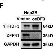
Western Blot: ZFP41 Antibody - BSA Free [NBP2-83801] -
YTHDF3‐mediated m6A modification of ZFP41 mRNA and decays its mRNA stability. (A) qPCR results showed that efficiencies of siRNA‐mediated knockdown of common m6A regulators in MHCC‐97H cells. (B) qPCR results showed that expression of ZFP41 after silencing these m6A regulators in MHCC‐97H cells. (C) Overexpression of YTHDF3 notably suppress ZFP41 expression on transcription level in MHCC‐97H and Hep3B cells. (D) Knockdown of YTHDF3 obviously increase ZFP41 expression on mRNA level in MHCC‐97H and Hep3B cells. (E) Western blots results demonstrated that the ZFP41 protein level in MHCC‐97H and HLF cells after silencing YTHDF3 with siRNA of YTHDF3. (F) Western blots results demonstrated that the ZFP41 protein level in Hep3B cells after overexpression of YTHDF3. (G) The diagram of the potential site of m6A modification on the CDS (Coding Sequence) area. (H) The luciferase activity in both MHCC‐97H and Hep3B cells cotransfected with relative plasmids. (I) MeRIP results showed that ZFP41‐wt or ZFP41‐mut in both MHCC‐97H and Hep3B cells. (J) The rate of ZFP41 mRNA degradation in MHCC‐97H and Hep3B cells with YTHDF3 overexpression or knockdown. (K) The binding of ZFP41 mRNA and YTHDF3 was tested in MHCC‐97H and Hep3B cells by RIP‐qPCR analyses. (L) The relationship between ZFP41 and YTHDF3 was confirmed by RNA pull‐down assay. Image collected and cropped by CiteAb from the following open publication (https://pubmed.ncbi.nlm.nih.gov/39473907), licensed under a CC-BY license. Not internally tested by Novus Biologicals.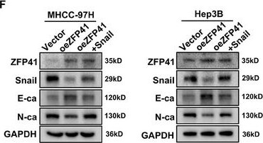
Western Blot: ZFP41 Antibody - BSA Free [NBP2-83801] -
ZFP41 suppresses the proliferation and invasion of HCC cells by inhibiting Snail expression and EMT pathway. (A) Representative images of subcutaneous xenograft tumors formation of the Vector group, oeZFP41 group and oeZFP41+Snail group in MHCC‐97H cells. The dissected tumors from two groups were photographed. (B) Volumes and Weights of subcutaneous xenograft tumors in the Vector group, oeZFP41 group and oeZFP41+Snail group. (C and D) The lung fluorescence and the number of lung metastatic nodules was calculated in each group. (E) HE staining of lung tissue in each group were presented. (F) Western blots results showed the protein level of Snail and EMT‐related targets with Vector, oeZFP41, and oeZFP41+Snail groups in MHCC‐97H and Hep3B cells. Image collected and cropped by CiteAb from the following open publication (https://pubmed.ncbi.nlm.nih.gov/39473907), licensed under a CC-BY license. Not internally tested by Novus Biologicals.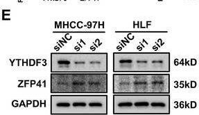
Western Blot: ZFP41 Antibody - BSA Free [NBP2-83801] -
YTHDF3‐mediated m6A modification of ZFP41 mRNA and decays its mRNA stability. (A) qPCR results showed that efficiencies of siRNA‐mediated knockdown of common m6A regulators in MHCC‐97H cells. (B) qPCR results showed that expression of ZFP41 after silencing these m6A regulators in MHCC‐97H cells. (C) Overexpression of YTHDF3 notably suppress ZFP41 expression on transcription level in MHCC‐97H and Hep3B cells. (D) Knockdown of YTHDF3 obviously increase ZFP41 expression on mRNA level in MHCC‐97H and Hep3B cells. (E) Western blots results demonstrated that the ZFP41 protein level in MHCC‐97H and HLF cells after silencing YTHDF3 with siRNA of YTHDF3. (F) Western blots results demonstrated that the ZFP41 protein level in Hep3B cells after overexpression of YTHDF3. (G) The diagram of the potential site of m6A modification on the CDS (Coding Sequence) area. (H) The luciferase activity in both MHCC‐97H and Hep3B cells cotransfected with relative plasmids. (I) MeRIP results showed that ZFP41‐wt or ZFP41‐mut in both MHCC‐97H and Hep3B cells. (J) The rate of ZFP41 mRNA degradation in MHCC‐97H and Hep3B cells with YTHDF3 overexpression or knockdown. (K) The binding of ZFP41 mRNA and YTHDF3 was tested in MHCC‐97H and Hep3B cells by RIP‐qPCR analyses. (L) The relationship between ZFP41 and YTHDF3 was confirmed by RNA pull‐down assay. Image collected and cropped by CiteAb from the following open publication (https://pubmed.ncbi.nlm.nih.gov/39473907), licensed under a CC-BY license. Not internally tested by Novus Biologicals.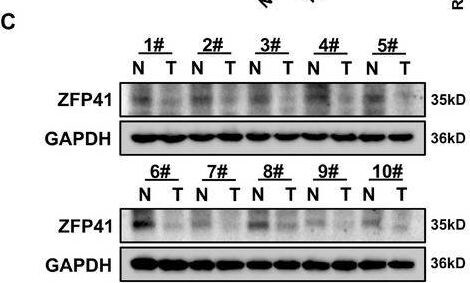
Western Blot: ZFP41 Antibody - BSA Free [NBP2-83801] -
Downregulated ZFP41 is correlated with poor survival in HCC patients. (A) The IHC staining results of tissue microarray from Tongji hospital HCC patients. (B) qPCR results showed that ZFP41 was highly expressed in normal tissues rather than tumor tissues in 63 pairs samples from HCC patients. The relevance of 63 pairs of HCC patient samples was calculated. (C) Western blots results showed the ZFP41 protein level in both tumor and normal tissues. The relevance of 40 pairs of HCC patient samples was demonstrated. (D) qPCR results showed that the mRNA level of ZFP41 in normal hepatocyte cell and other HCC cells. (E) Western blots results showed that the protein level of ZFP41 in normal hepatocyte cell and other HCC cells. (F) The overall survival prognosis of patients determined on the basis of ZFP41 expression in Tongji cohort and TCGA database. Image collected and cropped by CiteAb from the following open publication (https://pubmed.ncbi.nlm.nih.gov/39473907), licensed under a CC-BY license. Not internally tested by Novus Biologicals.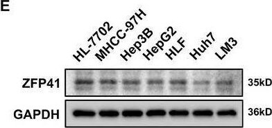
Western Blot: ZFP41 Antibody - BSA Free [NBP2-83801] -
Downregulated ZFP41 is correlated with poor survival in HCC patients. (A) The IHC staining results of tissue microarray from Tongji hospital HCC patients. (B) qPCR results showed that ZFP41 was highly expressed in normal tissues rather than tumor tissues in 63 pairs samples from HCC patients. The relevance of 63 pairs of HCC patient samples was calculated. (C) Western blots results showed the ZFP41 protein level in both tumor and normal tissues. The relevance of 40 pairs of HCC patient samples was demonstrated. (D) qPCR results showed that the mRNA level of ZFP41 in normal hepatocyte cell and other HCC cells. (E) Western blots results showed that the protein level of ZFP41 in normal hepatocyte cell and other HCC cells. (F) The overall survival prognosis of patients determined on the basis of ZFP41 expression in Tongji cohort and TCGA database. Image collected and cropped by CiteAb from the following open publication (https://pubmed.ncbi.nlm.nih.gov/39473907), licensed under a CC-BY license. Not internally tested by Novus Biologicals.Applications for ZFP41 Antibody - BSA Free
Application
Recommended Usage
Western Blot
1.0 ug/ml
Formulation, Preparation, and Storage
Purification
Affinity purified
Formulation
PBS, 2% Sucrose
Format
BSA Free
Preservative
0.09% Sodium Azide
Concentration
0.5 mg/ml
Shipping
The product is shipped with polar packs. Upon receipt, store it immediately at the temperature recommended below.
Stability & Storage
Store at 4C short term. Aliquot and store at -20C long term. Avoid freeze-thaw cycles.
Background: ZFP41
Alternate Names
ZFP41 zinc finger protein, zinc finger protein 41 homolog, ZNF753
Gene Symbol
ZFP41
Additional ZFP41 Products
Product Documents for ZFP41 Antibody - BSA Free
Certificate of Analysis
To download a Certificate of Analysis, please enter a lot or batch number in the search box below.
Product Specific Notices for ZFP41 Antibody - BSA Free
This product is for research use only and is not approved for use in humans or in clinical diagnosis. Primary Antibodies are guaranteed for 1 year from date of receipt.
Citations for ZFP41 Antibody - BSA Free
 Powered by Bioz
Customer Reviews for ZFP41 Antibody - BSA Free
There are currently no reviews for this product. Be the first to review ZFP41 Antibody - BSA Free and earn rewards!
Have you used ZFP41 Antibody - BSA Free?
Submit a review and receive an Amazon gift card!
$25/€18/£15/$25CAN/¥2500 Yen for a review with an image
$10/€7/£6/$10CAN/¥1110 Yen for a review without an image
Submit a review
Protocols
Find general support by application which include: protocols, troubleshooting, illustrated assays, videos and webinars.
- Cellular Response to Hypoxia Protocols
- R&D Systems Quality Control Western Blot Protocol
- Troubleshooting Guide: Western Blot Figures
- Western Blot Conditions
- Western Blot Protocol
- Western Blot Protocol for Cell Lysates
- Western Blot Troubleshooting
- Western Blot Troubleshooting Guide
- View all Protocols, Troubleshooting, Illustrated assays and Webinars
Loading...