
Beta Dystroglycan Antibody (P38G1) - BSA Free
Novus Biologicals | Catalog # NBP3-43569

Key Product Details
Species Reactivity
Applications
Label
Antibody Source
Format
Product Specifications
Immunogen
Specificity
Clonality
Host
Isotype
Scientific Data Images for Beta Dystroglycan Antibody (P38G1) - BSA Free
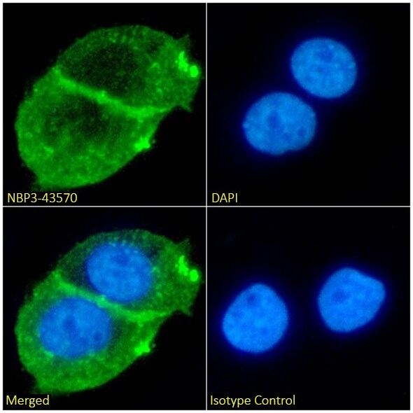
Immunocytochemistry/ Immunofluorescence: Beta Dystroglycan Antibody (P38G1) - BSA Free [NBP3-43569] -
Immunocytochemistry/ Immunofluorescence: Beta Dystroglycan Antibody (P38G1) - BSA Free [NBP3-43569] - Immunofluorescence analysis of paraformaldehyde fixed A431 cells on coverslips stained with the chimeric verison of this antibody, NBP3-43570 (1:100 dilution) for 1h followed by Alexa Fluor® 488 secondary antibody (1:1000 dilution), showing cell membrane/junction and cytoplasmic staining. The nuclear stain is DAPI (blue). Panels show, from left-right, top-bottom, NBP3-43570, DAPI, merged channels, and an isotype control. The isotype control was an unknown specificity antibody followed by staining with Alexa Fluor® 488 secondary antibody.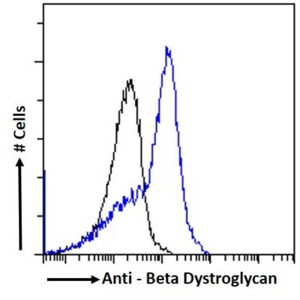
Flow Cytometry: Beta Dystroglycan Antibody (P38G1) - BSA Free [NBP3-43569] -
Flow Cytometry: Beta Dystroglycan Antibody (P38G1) - BSA Free [NBP3-43569] - MCF7 cells were stained with the anti-unknown specificity antibody (isotype control, black line) or the chimeric version of this antibody, NBP3-43570 (blue line) at a dilution of 1:100 for 1h at RT. After washing, the bound antibody was detected using a goat anti-rabbit IgG AlexaFluor® 488 antibody at a dilution of 1:1000, and the cells were analyzed using a FACSCanto flow-cytometer.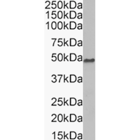
Western Blot: Beta Dystroglycan Antibody (P38G1) - BSA Free [NBP3-43569] -
Western Blot: Beta Dystroglycan Antibody (P38G1) - BSA Free [NBP3-43569] - Human skeletal muscle tissue lysate (35µg protein in RIPA buffer) was resolved on an SDS-PAGE gel, and blots were probed with the chimeric verison of this antibody, NBP3-43570 at 0.1µg/ml before detection using an anti-rabbit secondary antibody. A primary incubation of 1 hour was used, and protein was detected by chemiluminescence.Applications for Beta Dystroglycan Antibody (P38G1) - BSA Free
ELISA
Flow Cytometry
Flow Cytometry Panel Builder
Bio-Techne Knows Flow Cytometry
Save time and reduce costly mistakes by quickly finding compatible reagents using the Panel Builder Tool.
Advanced Features
- Spectra Viewer - Custom analysis of spectra from multiple fluorochromes
- Spillover Popups - Visualize the spectra of individual fluorochromes
- Antigen Density Selector - Match fluorochrome brightness with antigen density

Formulation, Preparation, and Storage
Purification
Formulation
Format
Preservative
Concentration
Shipping
Stability & Storage
Background: Beta Dystroglycan
Alternate Names
Gene Symbol
Additional Beta Dystroglycan Products
Product Documents for Beta Dystroglycan Antibody (P38G1) - BSA Free
Certificate of Analysis
To download a Certificate of Analysis, please enter a lot or batch number in the search box below.
Product Specific Notices for Beta Dystroglycan Antibody (P38G1) - BSA Free
This product is for research use only and is not approved for use in humans or in clinical diagnosis. Primary Antibodies are guaranteed for 1 year from date of receipt.
Customer Reviews for Beta Dystroglycan Antibody (P38G1) - BSA Free
There are currently no reviews for this product. Be the first to review Beta Dystroglycan Antibody (P38G1) - BSA Free and earn rewards!
Have you used Beta Dystroglycan Antibody (P38G1) - BSA Free?
Submit a review and receive an Amazon gift card!
$25/€18/£15/$25CAN/¥2500 for a review with an image
$10/€7/£6/$10CAN/¥1110 for a review without an image
Submit a review
Protocols
Find general support by application which include: protocols, troubleshooting, illustrated assays, videos and webinars.
- 7-Amino Actinomycin D (7-AAD) Cell Viability Flow Cytometry Protocol
- ELISA Sample Preparation & Collection Guide
- ELISA Troubleshooting Guide
- Extracellular Membrane Flow Cytometry Protocol
- Flow Cytometry Protocol for Cell Surface Markers
- Flow Cytometry Protocol for Staining Membrane Associated Proteins
- Flow Cytometry Staining Protocols
- Flow Cytometry Troubleshooting Guide
- How to Run an R&D Systems DuoSet ELISA
- How to Run an R&D Systems Quantikine ELISA
- How to Run an R&D Systems Quantikine™ QuicKit™ ELISA
- Intracellular Flow Cytometry Protocol Using Alcohol (Methanol)
- Intracellular Flow Cytometry Protocol Using Detergents
- Intracellular Nuclear Staining Flow Cytometry Protocol Using Detergents
- Intracellular Staining Flow Cytometry Protocol Using Alcohol Permeabilization
- Intracellular Staining Flow Cytometry Protocol Using Detergents to Permeabilize Cells
- Propidium Iodide Cell Viability Flow Cytometry Protocol
- Protocol for Liperfluo
- Protocol for the Characterization of Human Th22 Cells
- Protocol for the Characterization of Human Th9 Cells
- Protocol: Annexin V and PI Staining by Flow Cytometry
- Protocol: Annexin V and PI Staining for Apoptosis by Flow Cytometry
- Quantikine HS ELISA Kit Assay Principle, Alkaline Phosphatase
- Quantikine HS ELISA Kit Principle, Streptavidin-HRP Polymer
- Sandwich ELISA (Colorimetric) – Biotin/Streptavidin Detection Protocol
- Sandwich ELISA (Colorimetric) – Direct Detection Protocol
- Troubleshooting Guide: ELISA
- Troubleshooting Guide: Fluorokine Flow Cytometry Kits
- View all Protocols, Troubleshooting, Illustrated assays and Webinars