CBARA1 Antibody - BSA Free
Novus Biologicals | Catalog # NBP1-86663
![Simple Western: CBARA1 Antibody [NBP1-86663]](https://resources.rndsystems.com/images/products/CBARA1-Antibody-Simple-Western-NBP1-86663-img0008.jpg "Simple Western: CBARA1 Antibody [NBP1-86663]")
Loading...
Key Product Details
Species Reactivity
Validated:
Human
Cited:
Human, Mouse
Predicted:
Rat (98%). Backed by our 100% Guarantee.
Applications
Validated:
Immunohistochemistry-Paraffin, Simple Western
Cited:
Immunohistochemistry-Paraffin, Western Blot
Label
Unconjugated
Antibody Source
Polyclonal Rabbit IgG
Format
BSA Free
Loading...
Product Specifications
Immunogen
This antibody was developed against Recombinant Protein corresponding to amino acids: LKGKLTIKNFLEFQRKLQHDVLKLEFERHDPVDGRITERQFGGMLLAYSGVQSKKLTAMQRQLKKHFKEGKGLTFQEVENFFTFL
Reactivity Notes
Use in Mouse reported in scientific literature (PMID:32075766).
Clonality
Polyclonal
Host
Rabbit
Isotype
IgG
Theoretical MW
54 kDa.
Disclaimer note: The observed molecular weight of the protein may vary from the listed predicted molecular weight due to post translational modifications, post translation cleavages, relative charges, and other experimental factors.
Disclaimer note: The observed molecular weight of the protein may vary from the listed predicted molecular weight due to post translational modifications, post translation cleavages, relative charges, and other experimental factors.
Scientific Data Images for CBARA1 Antibody - BSA Free
Simple Western: CBARA1 Antibody [NBP1-86663]
Simple Western: CBARA1 Antibody [NBP1-86663] - Electropherogram image(s) of corresponding Simple Western lane view. CBARA1 antibody was used at 1:20 dilution on RT-4 and U-251 lysate(s).![CBARA1 Antibody - BSA Free Immunohistochemistry-Paraffin: CBARA1 Antibody [NBP1-86663]](https://resources.rndsystems.com/images/products/nbp1-86663_-immunohistochemistry-paraffin-639173124783803933.jpg "Immunohistochemistry-Paraffin: CBARA1 Antibody [NBP1-86663]")
Immunohistochemistry-Paraffin: CBARA1 Antibody [NBP1-86663]
Staining of human kidney shows strong granular positivity in cytoplasm in cells in tubules.![CBARA1 Antibody - BSA Free Immunohistochemistry-Paraffin: CBARA1 Antibody [NBP1-86663]](https://resources.rndsystems.com/images/products/nbp1-86663_-immunohistochemistry-paraffin-639173124845418922.jpg "Immunohistochemistry-Paraffin: CBARA1 Antibody [NBP1-86663]")
Immunohistochemistry-Paraffin: CBARA1 Antibody [NBP1-86663]
Staining of human liver shows moderate granular positivity in cytoplasm in hepatocytes.![CBARA1 Antibody - BSA Free Immunohistochemistry-Paraffin: CBARA1 Antibody [NBP1-86663]](https://resources.rndsystems.com/images/products/nbp1-86663_-immunohistochemistry-paraffin-639173125279194705.jpg "Immunohistochemistry-Paraffin: CBARA1 Antibody [NBP1-86663]")
Immunohistochemistry-Paraffin: CBARA1 Antibody [NBP1-86663]
Staining of human rectum shows strong granular positivity in cytoplasm in glandular cells.![CBARA1 Antibody - BSA Free Immunohistochemistry-Paraffin: CBARA1 Antibody [NBP1-86663]](https://resources.rndsystems.com/images/products/nbp1-86663_-immunohistochemistry-paraffin-639173132690259298.jpg "Immunohistochemistry-Paraffin: CBARA1 Antibody [NBP1-86663]")
Immunohistochemistry-Paraffin: CBARA1 Antibody [NBP1-86663]
Staining of human endometrium shows strong granular positivity in cytoplasm in glandular cells.Applications for CBARA1 Antibody - BSA Free
Application
Recommended Usage
Immunohistochemistry-Paraffin
1:50 - 1:200
Simple Western
1:20
Application Notes
For IHC-Paraffin, HIER pH 6 retrieval is recommended.
In Simple Western only 10 - 15 uL of the recommended dilution is used per data point.
See Simple Western Antibody Database for Simple Western validation: Tested in RT-4, U-251, separated by Size, antibody dilution of 1:20, apparent MW was 52 kDa. Separated by Size-Wes, Sally Sue/Peggy Sue.
In Simple Western only 10 - 15 uL of the recommended dilution is used per data point.
See Simple Western Antibody Database for Simple Western validation: Tested in RT-4, U-251, separated by Size, antibody dilution of 1:20, apparent MW was 52 kDa. Separated by Size-Wes, Sally Sue/Peggy Sue.
Reviewed Applications
Read 1 review rated 5 using NBP1-86663 in the following applications:
Formulation, Preparation, and Storage
Purification
Affinity purified
Formulation
PBS (pH 7.2) and 40% Glycerol
Format
BSA Free
Preservative
0.02% Sodium Azide
Concentration
Concentrations vary lot to lot. See vial label for concentration. If unlisted please contact technical services.
Shipping
The product is shipped with polar packs. Upon receipt, store it immediately at the temperature recommended below.
Stability & Storage
Store at 4C short term. Aliquot and store at -20C long term. Avoid freeze-thaw cycles.
Background: CBARA1
Long Name
Calcium uptake protein 1, mitochondrial
Alternate Names
allergen Hom s 4, ara CALC, CALC, MICU1
Entrez Gene IDs
10367 (Human)
Gene Symbol
MICU1
UniProt
Additional CBARA1 Products
Product Documents for CBARA1 Antibody - BSA Free
Certificate of Analysis
To download a Certificate of Analysis, please enter a lot or batch number in the search box below.
Product Specific Notices for CBARA1 Antibody - BSA Free
This product is for research use only and is not approved for use in humans or in clinical diagnosis. Primary Antibodies are guaranteed for 1 year from date of receipt.
Citations for CBARA1 Antibody - BSA Free
 Powered by Bioz
Customer Reviews for CBARA1 Antibody - BSA Free (1)
5 out of 5
1 Customer Rating
Have you used CBARA1 Antibody - BSA Free?
Submit a review and receive an Amazon gift card!
$25/€18/£15/$25CAN/¥2500 Yen for a review with an image
$10/€7/£6/$10CAN/¥1110 Yen for a review without an image
Submit a review
Customer Images
Showing
1
-
1 的
1 review
Showing All
Filter By:
-
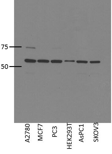
Application: Western BlotSample Tested: HUMAN CANCER CELL LINES, WHOLE CELL LYSATESSpecies: HumanVerified Customer | Posted 06/11/2014

There are no reviews that match your criteria.
Protocols
Find general support by application which include: protocols, troubleshooting, illustrated assays, videos and webinars.
- Antigen Retrieval Protocol (PIER)
- Antigen Retrieval for Frozen Sections Protocol
- Appropriate Fixation of IHC/ICC Samples
- Cellular Response to Hypoxia Protocols
- Chromogenic IHC Staining of Formalin-Fixed Paraffin-Embedded (FFPE) Tissue Protocol
- Chromogenic Immunohistochemistry Staining of Frozen Tissue
- ClariTSA™ Fluorophore Kits
- Detection & Visualization of Antibody Binding
- Fluorescent IHC Staining of Frozen Tissue Protocol
- Graphic Protocol for Heat-induced Epitope Retrieval
- Graphic Protocol for the Preparation and Fluorescent IHC Staining of Frozen Tissue Sections
- Graphic Protocol for the Preparation and Fluorescent IHC Staining of Paraffin-embedded Tissue Sections
- Graphic Protocol for the Preparation of Gelatin-coated Slides for Histological Tissue Sections
- IHC Sample Preparation (Frozen sections vs Paraffin)
- Immunofluorescent IHC Staining of Formalin-Fixed Paraffin-Embedded (FFPE) Tissue Protocol
- Immunohistochemistry (IHC) and Immunocytochemistry (ICC) Protocols
- Immunohistochemistry Frozen Troubleshooting
- Immunohistochemistry Paraffin Troubleshooting
- Preparing Samples for IHC/ICC Experiments
- Preventing Non-Specific Staining (Non-Specific Binding)
- Primary Antibody Selection & Optimization
- Protocol for Heat-Induced Epitope Retrieval (HIER)
- Protocol for Making a 4% Formaldehyde Solution in PBS
- Protocol for VisUCyte™ HRP Polymer Detection Reagent
- Protocol for the Preparation & Fixation of Cells on Coverslips
- Protocol for the Preparation and Chromogenic IHC Staining of Frozen Tissue Sections
- Protocol for the Preparation and Chromogenic IHC Staining of Frozen Tissue Sections - Graphic
- Protocol for the Preparation and Chromogenic IHC Staining of Paraffin-embedded Tissue Sections
- Protocol for the Preparation and Chromogenic IHC Staining of Paraffin-embedded Tissue Sections - Graphic
- Protocol for the Preparation and Fluorescent IHC Staining of Frozen Tissue Sections
- Protocol for the Preparation and Fluorescent IHC Staining of Paraffin-embedded Tissue Sections
- Protocol for the Preparation of Gelatin-coated Slides for Histological Tissue Sections
- TUNEL and Active Caspase-3 Detection by IHC/ICC Protocol
- The Importance of IHC/ICC Controls
- Troubleshooting Guide: Immunohistochemistry
- View all Protocols, Troubleshooting, Illustrated assays and Webinars
Loading...