Key Product Details
Species Reactivity
Validated:
Human
Predicted:
Mouse (99%), Rat (100%). Backed by our 100% Guarantee.
Applications
Immunohistochemistry, Immunohistochemistry-Paraffin, Western Blot, Immunocytochemistry/ Immunofluorescence, Chromatin Immunoprecipitation
Label
Unconjugated
Antibody Source
Monoclonal Mouse IgG2B Clone # CL0307
Format
BSA Free
Loading...
Product Specifications
Immunogen
This antibody was developed using a recombinant protein derived from P49711, with the exact immunogen sequence remaining proprietary.
Reactivity Notes
Please note that this antibody is reactive to Mouse and derived from the same host, Mouse. Mouse-On-Mouse blocking reagent may be needed for IHC and ICC experiments to reduce high background signal. You can find these reagents under catalog numbers PK-2200-NB and MP-2400-NB. Please contact Technical Support if you have any questions
Clonality
Monoclonal
Host
Mouse
Isotype
IgG2B
Scientific Data Images for CTCF Antibody (CL0307) - BSA Free
![Western Blot: CTCF Antibody (CL0307) [NBP2-52911]](https://resources.rndsystems.com/images/products/CTCF-Antibody-CL0307-Western-Blot-NBP2-52911-img0007.jpg "Western Blot: CTCF Antibody (CL0307) [NBP2-52911]")
Western Blot: CTCF Antibody (CL0307) [NBP2-52911]
Western Blot: CTCF Antibody (CL0307) [NBP2-52911] - Lane 1: Marker [kDa], Lane 2: Human cell line U-251 MG![Immunohistochemistry-Paraffin: CTCF Antibody (CL0307) [NBP2-52911]](https://resources.rndsystems.com/images/products/CTCF-Antibody-CL0307-Immunohistochemistry-Paraffin-NBP2-52911-img0013.jpg "Immunohistochemistry-Paraffin: CTCF Antibody (CL0307) [NBP2-52911]")
Immunohistochemistry-Paraffin: CTCF Antibody (CL0307) [NBP2-52911]
Immunohistochemistry-Paraffin: CTCF Antibody (CL0307) [NBP2-52911] - Analysis of CTCF antibody on Human fallopian tube tissue. Antigen retrieval was done enzymatically. Image from verified customer review.![Immunohistochemistry-Paraffin: CTCF Antibody (CL0307) [NBP2-52911]](https://resources.rndsystems.com/images/products/CTCF-Antibody-CL0307-Immunohistochemistry-Paraffin-NBP2-52911-img0008.jpg "Immunohistochemistry-Paraffin: CTCF Antibody (CL0307) [NBP2-52911]")
Immunohistochemistry-Paraffin: CTCF Antibody (CL0307) [NBP2-52911]
Immunohistochemistry-Paraffin: CTCF Antibody (CL0307) [NBP2-52911] - Staining of human endometrium shows strong nuclear positivity in glandular and stromal cells![Immunohistochemistry-Paraffin: CTCF Antibody (CL0307) [NBP2-52911]](https://resources.rndsystems.com/images/products/CTCF-Antibody-CL0307-Immunohistochemistry-Paraffin-NBP2-52911-img0009.jpg "Immunohistochemistry-Paraffin: CTCF Antibody (CL0307) [NBP2-52911]")
Immunohistochemistry-Paraffin: CTCF Antibody (CL0307) [NBP2-52911]
Immunohistochemistry-Paraffin: CTCF Antibody (CL0307) [NBP2-52911] - Staining of human kidney shows strong nuclear positivity in cells in glomeruli and tubuli.![Immunohistochemistry-Paraffin: CTCF Antibody (CL0307) [NBP2-52911]](https://resources.rndsystems.com/images/products/CTCF-Antibody-CL0307-Immunohistochemistry-Paraffin-NBP2-52911-img0010.jpg "Immunohistochemistry-Paraffin: CTCF Antibody (CL0307) [NBP2-52911]")
Immunohistochemistry-Paraffin: CTCF Antibody (CL0307) [NBP2-52911]
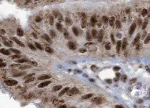
Immunohistochemistry-Paraffin: CTCF Antibody (CL0307) [NBP2-52911] - Staining of human small intestine shows strong nuclear positivity in glandular cells.![Immunohistochemistry-Paraffin: CTCF Antibody (CL0307) [NBP2-52911]](https://resources.rndsystems.com/images/products/CTCF-Antibody-CL0307-Immunohistochemistry-Paraffin-NBP2-52911-img0011.jpg "Immunohistochemistry-Paraffin: CTCF Antibody (CL0307) [NBP2-52911]")
Immunohistochemistry-Paraffin: CTCF Antibody (CL0307) [NBP2-52911]
Immunohistochemistry-Paraffin: CTCF Antibody (CL0307) [NBP2-52911] - Staining of human testis shows moderate nuclear positivity in a subset of cells in seminiferous ducts.![CTCF Antibody (CL0307) - BSA Free Chromatin Immunoprecipitation-exo-Seq: CTCF Antibody (CL0307) - BSA Free [NBP2-52911]](https://resources.rndsystems.com/images/products/nbp2-52911_mouse-monoclonal-ctcf-antibody-cl0307-2562025911406.jpg "Chromatin Immunoprecipitation-exo-Seq: CTCF Antibody (CL0307) - BSA Free [NBP2-52911]")
Chromatin Immunoprecipitation-exo-Seq: CTCF Antibody (CL0307) - BSA Free [NBP2-52911]
ChIP-Exo-Seq composite graph for Anti-CTCF (NBP2-52911) tested in K562 cells. Strand-specific reads (blue: forward, red: reverse) and IgG controls (black: forward, grey: reverse) are plotted against the distance from a composite set of reference binding sites. The antibody exhibits robust target enrichment compared to a non-specific IgG control and precisely reveals its structural organization around the binding site. Data generated by Prof. B. F. Pugh´s Lab at Cornell University.![CTCF Antibody (CL0307) - BSA Free Immunocytochemistry/ Immunofluorescence: CTCF Antibody (CL0307) [NBP2-52911]](https://resources.rndsystems.com/images/products/nbp2-52911_-immunocytochemistry-immunofluorescence-639174076992195058.jpg "Immunocytochemistry/ Immunofluorescence: CTCF Antibody (CL0307) [NBP2-52911]")
Immunocytochemistry/ Immunofluorescence: CTCF Antibody (CL0307) [NBP2-52911]
Staining of MCF7 cells, showing specific staining in the nucleoplasm in green. Microtubule- and nuclear probes are visualized in red and blue, respectively (where available).Applications for CTCF Antibody (CL0307) - BSA Free
Application
Recommended Usage
Chromatin Immunoprecipitation
1-10µg per reaction
Immunocytochemistry/ Immunofluorescence
2-10 ug/ml
Immunohistochemistry
1:200 - 1:500
Immunohistochemistry-Paraffin
1:200 - 1:500
Western Blot
1 ug/ml
Application Notes
IHC-Paraffin, HIER pH 6 retrieval is recommended. ICC/IF, PFA/Triton X-100 is recommended for fixation/permeabilization.
Reviewed Applications
Read 1 review rated 5 using NBP2-52911 in the following applications:
Formulation, Preparation, and Storage
Purification
Protein A purified
Formulation
PBS (pH 7.2) and 40% Glycerol
Format
BSA Free
Preservative
0.02% Sodium Azide
Concentration
1 mg/ml
Shipping
The product is shipped with polar packs. Upon receipt, store it immediately at the temperature recommended below.
Stability & Storage
Store at 4C short term. Aliquot and store at -20C long term. Avoid freeze-thaw cycles.
Background: CTCF
Long Name
CCCTC-binding Factor
Alternate Names
11-Zinc Finger Protein2, CCCTC-binding Factor
Gene Symbol
CTCF
Additional CTCF Products
Product Documents for CTCF Antibody (CL0307) - BSA Free
Certificate of Analysis
To download a Certificate of Analysis, please enter a lot or batch number in the search box below.
Product Specific Notices for CTCF Antibody (CL0307) - BSA Free
This product is for research use only and is not approved for use in humans or in clinical diagnosis. Primary Antibodies are guaranteed for 1 year from date of receipt.
Related Research Areas
Customer Reviews for CTCF Antibody (CL0307) - BSA Free (1)
5 out of 5
1 Customer Rating
Have you used CTCF Antibody (CL0307) - BSA Free?
Submit a review and receive an Amazon gift card!
$25/€18/£15/$25CAN/¥2500 Yen for a review with an image
$10/€7/£6/$10CAN/¥1110 Yen for a review without an image
Submit a review
Customer Images
Showing
1
-
1 的
1 review
Showing All
Filter By:
-
Application: Immunohistochemistry-ParaffinSample Tested: fallopian tube tissueSpecies: HumanVerified Customer | Posted 02/21/2022Human fallopian tube tissueantigen retrieval was done enzymatically. Incubated O/N in 4 C with the 1ry antibody.

There are no reviews that match your criteria.
Protocols
Find general support by application which include: protocols, troubleshooting, illustrated assays, videos and webinars.
- Antigen Retrieval Protocol (PIER)
- Antigen Retrieval for Frozen Sections Protocol
- Appropriate Fixation of IHC/ICC Samples
- Cellular Response to Hypoxia Protocols
- Chromogenic IHC Staining of Formalin-Fixed Paraffin-Embedded (FFPE) Tissue Protocol
- Chromogenic Immunohistochemistry Staining of Frozen Tissue
- ClariTSA™ Fluorophore Kits
- Detection & Visualization of Antibody Binding
- Fluorescent IHC Staining of Frozen Tissue Protocol
- Graphic Protocol for Heat-induced Epitope Retrieval
- Graphic Protocol for the Preparation and Fluorescent IHC Staining of Frozen Tissue Sections
- Graphic Protocol for the Preparation and Fluorescent IHC Staining of Paraffin-embedded Tissue Sections
- Graphic Protocol for the Preparation of Gelatin-coated Slides for Histological Tissue Sections
- ICC Cell Smear Protocol for Suspension Cells
- ICC Immunocytochemistry Protocol Videos
- ICC for Adherent Cells
- IHC Sample Preparation (Frozen sections vs Paraffin)
- Immunocytochemistry (ICC) Protocol
- Immunocytochemistry Troubleshooting
- Immunofluorescence of Organoids Embedded in Cultrex Basement Membrane Extract
- Immunofluorescent IHC Staining of Formalin-Fixed Paraffin-Embedded (FFPE) Tissue Protocol
- Immunohistochemistry (IHC) and Immunocytochemistry (ICC) Protocols
- Immunohistochemistry Frozen Troubleshooting
- Immunohistochemistry Paraffin Troubleshooting
- Preparing Samples for IHC/ICC Experiments
- Preventing Non-Specific Staining (Non-Specific Binding)
- Primary Antibody Selection & Optimization
- Protocol for Heat-Induced Epitope Retrieval (HIER)
- Protocol for Making a 4% Formaldehyde Solution in PBS
- Protocol for VisUCyte™ HRP Polymer Detection Reagent
- Protocol for the Fluorescent ICC Staining of Cell Smears - Graphic
- Protocol for the Fluorescent ICC Staining of Cultured Cells on Coverslips - Graphic
- Protocol for the Preparation & Fixation of Cells on Coverslips
- Protocol for the Preparation and Chromogenic IHC Staining of Frozen Tissue Sections
- Protocol for the Preparation and Chromogenic IHC Staining of Frozen Tissue Sections - Graphic
- Protocol for the Preparation and Chromogenic IHC Staining of Paraffin-embedded Tissue Sections
- Protocol for the Preparation and Chromogenic IHC Staining of Paraffin-embedded Tissue Sections - Graphic
- Protocol for the Preparation and Fluorescent ICC Staining of Cells on Coverslips
- Protocol for the Preparation and Fluorescent ICC Staining of Non-adherent Cells
- Protocol for the Preparation and Fluorescent ICC Staining of Stem Cells on Coverslips
- Protocol for the Preparation and Fluorescent IHC Staining of Frozen Tissue Sections
- Protocol for the Preparation and Fluorescent IHC Staining of Paraffin-embedded Tissue Sections
- Protocol for the Preparation of Gelatin-coated Slides for Histological Tissue Sections
- Protocol for the Preparation of a Cell Smear for Non-adherent Cell ICC - Graphic
- R&D Systems Quality Control Western Blot Protocol
- TUNEL and Active Caspase-3 Detection by IHC/ICC Protocol
- The Importance of IHC/ICC Controls
- Troubleshooting Guide: Immunohistochemistry
- Troubleshooting Guide: Western Blot Figures
- Western Blot Conditions
- Western Blot Protocol
- Western Blot Protocol for Cell Lysates
- Western Blot Troubleshooting
- Western Blot Troubleshooting Guide
- View all Protocols, Troubleshooting, Illustrated assays and Webinars
Loading...