CTCF Antibody
Novus Biologicals | Catalog # NB500-177
![Western Blot: CTCF Antibody [NB500-177]](https://resources.rndsystems.com/images/products/CTCF-Antibody-Western-Blot-NB500-177-img0011.jpg "Western Blot: CTCF Antibody [NB500-177]")
Loading...
Key Product Details
Species Reactivity
Validated:
Human, Mouse
Cited:
Human
Predicted:
Rat (100%). Backed by our 100% Guarantee.
Applications
Validated:
Immunohistochemistry, Immunohistochemistry-Paraffin, Western Blot, Immunoprecipitation, Chromatin Immunoprecipitation (ChIP)
Cited:
Western Blot, Immunoprecipitation, Chemotaxis
Label
Unconjugated
Antibody Source
Polyclonal Rabbit IgG
Loading...
Product Specifications
Immunogen
The immunogen recognized by this antibody maps to a region between residues 650 and 700 of human CCCTC-binding factor using the numbering given in entry NP_006556.1 (GeneID 10664).
Clonality
Polyclonal
Host
Rabbit
Isotype
IgG
Scientific Data Images for CTCF Antibody
Western Blot: CTCF Antibody [NB500-177]
Western Blot: CTCF Antibody [NB500-177] - Detection of human CTCF by western blot. Samples: Whole cell lysate (50 ug) from Jurkat, HEK293T, and HeLa cells prepared using NETN lysis buffer. Antibody: Affinity purified rabbit anti-CTCF antibody NB500-177 used for WB at 0.02 ug/ml. Detection: Chemiluminescence with an exposure time of 10 seconds.![Immunohistochemistry-Paraffin: CTCF Antibody [NB500-177]](https://resources.rndsystems.com/images/products/CTCF-Antibody-Immunohistochemistry-NB500-177-img0013.jpg "Immunohistochemistry-Paraffin: CTCF Antibody [NB500-177]")
Immunohistochemistry-Paraffin: CTCF Antibody [NB500-177]
Immunohistochemistry-Paraffin: CTCF Antibody [NB500-177] - Section of human small cell lung cancer. Antibody: Affinity purified rabbit anti-CTCF antibody (NB500-177) used at 1:1000. Secondary: HRP-conjugated goat anti-rabbit IgG.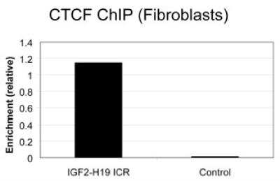
Chromatin Immunoprecipitation: CTCF Antibody [NB500-177] - Analysis of Human CTCF by Chromatin Immunoprecipitation (ChIP). Primary human fibroblasts from lung (5 x 10⁷ cells) were cross-linked with formaldehyde, sonicated, and immunoprecipitated with 20 micrograms of rabbit anti-CTCF antibody NB500-177. The resulting ChIP DNA was quantified using real-time PCR with primers against the IGF2-H19 ICR or a control region in the HoxA cluster.
![Western Blot: CTCF Antibody [NB500-177]](https://resources.rndsystems.com/images/products/CTCF-Antibody-Western-Blot-NB500-177-img0010.jpg "Western Blot: CTCF Antibody [NB500-177]")
Western Blot: CTCF Antibody [NB500-177]
Western Blot: CTCF Antibody [NB500-177] - Detection of mouse CTCF by western blot. Samples: Whole cell lysate (50 ug) from NIH 3T3 cells prepared using NETN lysis buffer. Antibody: Affinity purified rabbit anti-CTCF antibody NB500-177 used for WB at 0.02 ug/ml. Detection: Chemiluminescence with an exposure time of 75 seconds.![Immunohistochemistry-Paraffin: CTCF Antibody [NB500-177]](https://resources.rndsystems.com/images/products/CTCF-Antibody-Immunohistochemistry-NB500-177-img0012.jpg "Immunohistochemistry-Paraffin: CTCF Antibody [NB500-177]")
Immunohistochemistry-Paraffin: CTCF Antibody [NB500-177]
Immunohistochemistry-Paraffin: CTCF Antibody [NB500-177] - Section of mouse renal cell carcinoma. Antibody: Affinity purified rabbit anti-CTCF antibody (NB500-177) used at 1:1000. Secondary: HRP-conjugated goat anti-rabbit IgG.![Immunoprecipitation: CTCF Antibody [NB500-177]](https://resources.rndsystems.com/images/products/CTCF-Antibody-Immunoprecipitation-NB500-177-img0009.jpg "Immunoprecipitation: CTCF Antibody [NB500-177]")
Immunoprecipitation: CTCF Antibody [NB500-177]
Immunoprecipitation: CTCF Antibody [NB500-177] - Detection of human CTCF by western blot of immunoprecipitates. Samples: Whole cell lysate (1.0 mg per IP reaction; 20% of IP loaded) from Jurkat cells prepared using NETN lysis buffer. Antibodies: Affinity purified rabbit anti-CTCF antibody NB500-177 (lot NB500-177-6) used for IP at 6 ug per reaction. CTCF was also immunoprecipitated by a previous lot of this antibody (lot NB500-177-5). For blotting immunoprecipitated CTCF, NB500-177 was used at 0.02 ug/ml. Detection: Chemiluminescence with an exposure time of 10 seconds.Applications for CTCF Antibody
Application
Recommended Usage
Chromatin Immunoprecipitation (ChIP)
1:10-1:500
Immunohistochemistry
1:500 - 1:2000
Immunohistochemistry-Paraffin
1:500 - 1:2000
Immunoprecipitation
2-10 ug/mg lysate
Western Blot
1:2500-1:10000
Application Notes
Epitope retrieval with Tris-EDTA pH9.0 is recommended for FFPE tissue sections. Chip, WB, IP reacivity reported in (PMID: 24741094).
Formulation, Preparation, and Storage
Purification
Immunogen affinity purified
Formulation
TBS and 0.1% BSA
Preservative
0.09% Sodium Azide
Concentration
0.2 mg/ml
Shipping
The product is shipped with polar packs. Upon receipt, store it immediately at the temperature recommended below.
Stability & Storage
Store at 4C. Do not freeze.
Background: CTCF
Long Name
CCCTC-binding Factor
Alternate Names
11-Zinc Finger Protein2, CCCTC-binding Factor
Entrez Gene IDs
10664 (Human)
Gene Symbol
CTCF
UniProt
Additional CTCF Products
Product Documents for CTCF Antibody
Certificate of Analysis
To download a Certificate of Analysis, please enter a lot or batch number in the search box below.
Product Specific Notices for CTCF Antibody
This product is for research use only and is not approved for use in humans or in clinical diagnosis. Primary Antibodies are guaranteed for 1 year from date of receipt.
Related Research Areas
Citations for CTCF Antibody
 Powered by Bioz
Customer Reviews for CTCF Antibody
There are currently no reviews for this product. Be the first to review CTCF Antibody and earn rewards!
Have you used CTCF Antibody?
Submit a review and receive an Amazon gift card!
$25/€18/£15/$25CAN/¥2500 Yen for a review with an image
$10/€7/£6/$10CAN/¥1110 Yen for a review without an image
Submit a review
Protocols
Find general support by application which include: protocols, troubleshooting, illustrated assays, videos and webinars.
- Antigen Retrieval Protocol (PIER)
- Antigen Retrieval for Frozen Sections Protocol
- Appropriate Fixation of IHC/ICC Samples
- Cellular Response to Hypoxia Protocols
- ChIP Protocol Video
- Chromatin Immunoprecipitation (ChIP) Protocol
- Chromatin Immunoprecipitation Protocol
- Chromogenic IHC Staining of Formalin-Fixed Paraffin-Embedded (FFPE) Tissue Protocol
- Chromogenic Immunohistochemistry Staining of Frozen Tissue
- ClariTSA™ Fluorophore Kits
- Detection & Visualization of Antibody Binding
- Fluorescent IHC Staining of Frozen Tissue Protocol
- Graphic Protocol for Heat-induced Epitope Retrieval
- Graphic Protocol for the Preparation and Fluorescent IHC Staining of Frozen Tissue Sections
- Graphic Protocol for the Preparation and Fluorescent IHC Staining of Paraffin-embedded Tissue Sections
- Graphic Protocol for the Preparation of Gelatin-coated Slides for Histological Tissue Sections
- IHC Sample Preparation (Frozen sections vs Paraffin)
- Immunofluorescent IHC Staining of Formalin-Fixed Paraffin-Embedded (FFPE) Tissue Protocol
- Immunohistochemistry (IHC) and Immunocytochemistry (ICC) Protocols
- Immunohistochemistry Frozen Troubleshooting
- Immunohistochemistry Paraffin Troubleshooting
- Immunoprecipitation Protocol
- Preparing Samples for IHC/ICC Experiments
- Preventing Non-Specific Staining (Non-Specific Binding)
- Primary Antibody Selection & Optimization
- Protocol for Heat-Induced Epitope Retrieval (HIER)
- Protocol for Making a 4% Formaldehyde Solution in PBS
- Protocol for VisUCyte™ HRP Polymer Detection Reagent
- Protocol for the Preparation & Fixation of Cells on Coverslips
- Protocol for the Preparation and Chromogenic IHC Staining of Frozen Tissue Sections
- Protocol for the Preparation and Chromogenic IHC Staining of Frozen Tissue Sections - Graphic
- Protocol for the Preparation and Chromogenic IHC Staining of Paraffin-embedded Tissue Sections
- Protocol for the Preparation and Chromogenic IHC Staining of Paraffin-embedded Tissue Sections - Graphic
- Protocol for the Preparation and Fluorescent IHC Staining of Frozen Tissue Sections
- Protocol for the Preparation and Fluorescent IHC Staining of Paraffin-embedded Tissue Sections
- Protocol for the Preparation of Gelatin-coated Slides for Histological Tissue Sections
- R&D Systems Quality Control Western Blot Protocol
- TUNEL and Active Caspase-3 Detection by IHC/ICC Protocol
- The Importance of IHC/ICC Controls
- Troubleshooting Guide: Immunohistochemistry
- Troubleshooting Guide: Western Blot Figures
- Western Blot Conditions
- Western Blot Protocol
- Western Blot Protocol for Cell Lysates
- Western Blot Troubleshooting
- Western Blot Troubleshooting Guide
- View all Protocols, Troubleshooting, Illustrated assays and Webinars
Loading...