DEC2/SHARP1 Antibody - BSA Free
Novus Biologicals | Catalog # NBP1-19613
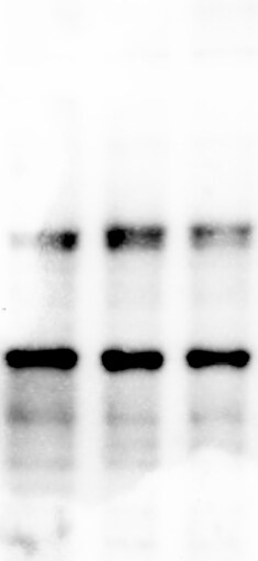
![Western Blot: DEC2/SHARP1 AntibodyBSA Free [NBP1-19613]](https://resources.rndsystems.com/images/products/DEC2-SHARP1-Antibody-Western-Blot-NBP1-19613-img0006.jpg "Western Blot: DEC2/SHARP1 AntibodyBSA Free [NBP1-19613]")
Key Product Details
Species Reactivity
Validated:
Cited:
Predicted:
Applications
Validated:
Cited:
Label
Antibody Source
Format
Product Specifications
Immunogen
Reactivity Notes
Localization
Clonality
Host
Isotype
Theoretical MW
Disclaimer note: The observed molecular weight of the protein may vary from the listed predicted molecular weight due to post translational modifications, post translation cleavages, relative charges, and other experimental factors.
Scientific Data Images for DEC2/SHARP1 Antibody - BSA Free
Western Blot: DEC2/SHARP1 AntibodyBSA Free [NBP1-19613]
Western Blot: DEC2/SHARP1 Antibody [NBP1-19613] - Huh7 cell lysates loaded from left to right at concentrations of 20, 30, and 40 ug of total protein. Image from verified customer review.![Immunohistochemistry-Paraffin: DEC2/SHARP1 Antibody - BSA Free [NBP1-19613]](https://resources.rndsystems.com/images/products/DEC2-SHARP1-Antibody-Immunohistochemistry-Paraffin-NBP1-19613-img0002.jpg "Immunohistochemistry-Paraffin: DEC2/SHARP1 Antibody - BSA Free [NBP1-19613]")
Immunohistochemistry-Paraffin: DEC2/SHARP1 Antibody - BSA Free [NBP1-19613]
Immunohistochemistry-Paraffin: DEC2/SHARP1 Antibody [NBP1-19613] - Staining of SHARP1 in mouse pancreas.![Western Blot: DEC2/SHARP1 AntibodyBSA Free [NBP1-19613]](https://resources.rndsystems.com/images/products/DEC2-SHARP1-Antibody-Western-Blot-NBP1-19613-img0003.jpg "Western Blot: DEC2/SHARP1 AntibodyBSA Free [NBP1-19613]")
Western Blot: DEC2/SHARP1 AntibodyBSA Free [NBP1-19613]
Western Blot: DEC2/SHARP1 Antibody [NBP1-19613] - Analysis of SHARP1 in HeLa whole cell extract.![Western Blot: DEC2/SHARP1 AntibodyBSA Free [NBP1-19613]](https://resources.rndsystems.com/images/products/DEC2-SHARP1-Antibody-Western-Blot-NBP1-19613-img0004.jpg "Western Blot: DEC2/SHARP1 AntibodyBSA Free [NBP1-19613]")
Western Blot: DEC2/SHARP1 AntibodyBSA Free [NBP1-19613]
Western Blot: DEC2/SHARP1 Antibody [NBP1-19613] - Analysis of HeLa (1), Raw 264.7 (2), A431 (3), MCF7 (4), and PC12 (5) using SHARP1 antibody at 2 ug/mL.![Simple Western: DEC2/SHARP1 AntibodyBSA Free [NBP1-19613]](https://resources.rndsystems.com/images/products/DEC2-SHARP1-Antibody-Simple-Western-NBP1-19613-img0005.jpg "Simple Western: DEC2/SHARP1 AntibodyBSA Free [NBP1-19613]")
Simple Western: DEC2/SHARP1 AntibodyBSA Free [NBP1-19613]
Simple Western: DEC2/SHARP1 Antibody [NBP1-19613] - Image shows a specific band for SHARP1 in 0.5 mg/mL of HeLa lysate. This experiment was performed under reducing conditions using the 12-230 kDa separation system.
Western Blot: DEC2/SHARP1 Antibody - BSA Free [NBP1-19613] -
Characterization of SHARP1 degradation.A, B Parental MDA-MB231 cells A or MDA-MB231 cells stably expressing HA-tagged SHARP1 B were treated with the proteasome inhibitor MG132 for 6 hours. Cells were lysed and whole-cell extracts were subjected to immunoblotting with antibodies for the indicated proteins. C MDA-MB231 cells were treated with cycloheximide (CHX) with or without the proteasome inhibitor MG132, collected at the indicated times, and lysed. Whole-cell extracts were subjected to immunoblotting with the indicated antibodies. D The graph represents the quantification of SHARP1 levels (shown in C) normalized to the Actin loading control. E Cells were transfected with an empty vector (EV) or FLAG-tagged CUL1 dominant-negative mutants (CUL1-N252 or CUL1-N385) along with HA-tagged SHARP1 or HA-tagged beta -catenin. Cells were collected and lysed. Whole-cell extracts were analyzed by immunoblotting with the indicated antibodies. F Cells were transfected with a siRNA targeting both beta TrCP1 and beta TrCP2 or a control siRNA and treated with cycloheximide (CHX) for the indicated times. Cells were lysed and whole-cell extracts were analyzed by immunoblotting with the indicated antibodies. G The graph represents the quantification of SHARP1 levels (shown in F) normalized to the Actin loading control. Image collected and cropped by CiteAb from the following open publication (https://pubmed.ncbi.nlm.nih.gov/37938564), licensed under a CC-BY license. Not internally tested by Novus Biologicals.
Western Blot: DEC2/SHARP1 Antibody - BSA Free [NBP1-19613] -
Characterization of SHARP1 degradation.A, B Parental MDA-MB231 cells A or MDA-MB231 cells stably expressing HA-tagged SHARP1 B were treated with the proteasome inhibitor MG132 for 6 hours. Cells were lysed and whole-cell extracts were subjected to immunoblotting with antibodies for the indicated proteins. C MDA-MB231 cells were treated with cycloheximide (CHX) with or without the proteasome inhibitor MG132, collected at the indicated times, and lysed. Whole-cell extracts were subjected to immunoblotting with the indicated antibodies. D The graph represents the quantification of SHARP1 levels (shown in C) normalized to the Actin loading control. E Cells were transfected with an empty vector (EV) or FLAG-tagged CUL1 dominant-negative mutants (CUL1-N252 or CUL1-N385) along with HA-tagged SHARP1 or HA-tagged beta -catenin. Cells were collected and lysed. Whole-cell extracts were analyzed by immunoblotting with the indicated antibodies. F Cells were transfected with a siRNA targeting both beta TrCP1 and beta TrCP2 or a control siRNA and treated with cycloheximide (CHX) for the indicated times. Cells were lysed and whole-cell extracts were analyzed by immunoblotting with the indicated antibodies. G The graph represents the quantification of SHARP1 levels (shown in F) normalized to the Actin loading control. Image collected and cropped by CiteAb from the following open publication (https://pubmed.ncbi.nlm.nih.gov/37938564), licensed under a CC-BY license. Not internally tested by Novus Biologicals.Applications for DEC2/SHARP1 Antibody - BSA Free
Immunohistochemistry
Immunohistochemistry-Paraffin
Simple Western
Western Blot
See Simple Western Antibody Database for Simple Western validation: Tested in HeLa lysate 0.5 mg/mL, separated by Size, antibody dilution of 1:50. Separated by Size-Wes, Sally Sue/Peggy Sue.
The observed molecular weight of the protein may vary from the listed predicted molecular weight due to post translational modifications, post translation cleavages, relative charges, and other experimental factors.
Reviewed Applications
Read 1 review rated 4 using NBP1-19613 in the following applications:
Formulation, Preparation, and Storage
Purification
Formulation
Format
Preservative
Concentration
Shipping
Stability & Storage
Background: DEC2
Long Name
Alternate Names
Gene Symbol
UniProt
Additional DEC2 Products
Product Documents for DEC2/SHARP1 Antibody - BSA Free
Certificate of Analysis
To download a Certificate of Analysis, please enter a lot or batch number in the search box below.
Product Specific Notices for DEC2/SHARP1 Antibody - BSA Free
This product is for research use only and is not approved for use in humans or in clinical diagnosis. Primary Antibodies are guaranteed for 1 year from date of receipt.
Related Research Areas
Citations for DEC2/SHARP1 Antibody - BSA Free
 Powered by Bioz
Customer Reviews for DEC2/SHARP1 Antibody - BSA Free (1)
Have you used DEC2/SHARP1 Antibody - BSA Free?
Submit a review and receive an Amazon gift card!
$25/€18/£15/$25CAN/¥2500 Yen for a review with an image
$10/€7/£6/$10CAN/¥1110 Yen for a review without an image
Submit a review
Customer Images
-
Application: Western BlotSample Tested: Huh7 cell lysateSpecies: HumanVerified Customer | Posted 01/25/2019Huh7 cell lysates loaded from left to right with concentrations ( 20, 30, and 40 microgram of total protein

There are no reviews that match your criteria.
Protocols
View specific protocols for DEC2/SHARP1 Antibody - BSA Free (NBP1-19613):
Immunohistochemistry-Paraffin Embedded Sections
Antigen Unmasking:
Bring slides to a boil in 10 mM sodium citrate buffer (pH 6.0) then maintain at a sub-boiling temperature for 10 minutes. Cool slides on bench-top for 30 minutes.
Staining:
1. Wash sections in deionized water three times for 5 minutes each.
2. Wash sections in wash buffer for 5 minutes.
3. Block each section with 100-400 ul blocking solution for 1 hour at room temperature.
4. Remove blocking solution and add 100-400 ul diluted primary antibody. Incubate overnight at 4 C.
5. Remove antibody solution and wash sections in wash buffer three times for 5 minutes each.
6. Add 100-400 ul biotinylated diluted secondary antibody. Incubate 30 minutes at room temperature.
7. Remove secondary antibody solution and wash sections three times with wash buffer for 5 minutes each.
8. Add 100-400 ul Streptavidin-HRP reagent to each section and incubate for 30 minutes at room temperature.
9. Wash sections three times in wash buffer for 5 minutes each.
10. Add 100-400 ul DAB substrate to each section and monitor staining closely.
11. As soon as the sections develop, immerse slides in deionized water.
12. Counterstain sections in hematoxylin.
13. Wash sections in deionized water two times for 5 minutes each.
14. Dehydrate sections.
15. Mount coverslips.
Western Blot Protocol
1. Perform SDS-PAGE on samples to be analyzed, loading 40 ug of total protein per lane.
2. Transfer proteins to membrane according to the instructions provided by the manufacturer of the membrane and transfer apparatus.
3. Stain according to standard Ponceau S procedure (or similar product) to assess transfer success, and mark molecular weight standards where appropriate.
4. Rinse the blot.
5. Block the membrane using standard blocking buffer for at least 1 hour.
6. Wash the membrane in wash buffer three times for 10 minutes each.
7. Dilute primary antibody in blocking buffer and incubate 1 hour at room temperature.
8. Wash the membrane in wash buffer three times for 10 minutes each.
9. Apply the diluted HRP conjugated secondary antibody in blocking buffer (as per manufacturers instructions) and incubate 1 hour at room temperature.
10. Wash the blot in wash buffer three times for 10 minutes each (this step can be repeated as required to reduce background).
11. Apply the detection reagent of choice in accordance with the manufacturers instructions.
Note: Tween-20 can be added to the blocking or antibody dilution buffer at a final concentration of 0.05-0.2%.
Find general support by application which include: protocols, troubleshooting, illustrated assays, videos and webinars.
- Antigen Retrieval Protocol (PIER)
- Antigen Retrieval for Frozen Sections Protocol
- Appropriate Fixation of IHC/ICC Samples
- Cellular Response to Hypoxia Protocols
- Chromogenic IHC Staining of Formalin-Fixed Paraffin-Embedded (FFPE) Tissue Protocol
- Chromogenic Immunohistochemistry Staining of Frozen Tissue
- ClariTSA™ Fluorophore Kits
- Detection & Visualization of Antibody Binding
- Fluorescent IHC Staining of Frozen Tissue Protocol
- Graphic Protocol for Heat-induced Epitope Retrieval
- Graphic Protocol for the Preparation and Fluorescent IHC Staining of Frozen Tissue Sections
- Graphic Protocol for the Preparation and Fluorescent IHC Staining of Paraffin-embedded Tissue Sections
- Graphic Protocol for the Preparation of Gelatin-coated Slides for Histological Tissue Sections
- IHC Sample Preparation (Frozen sections vs Paraffin)
- Immunofluorescent IHC Staining of Formalin-Fixed Paraffin-Embedded (FFPE) Tissue Protocol
- Immunohistochemistry (IHC) and Immunocytochemistry (ICC) Protocols
- Immunohistochemistry Frozen Troubleshooting
- Immunohistochemistry Paraffin Troubleshooting
- Preparing Samples for IHC/ICC Experiments
- Preventing Non-Specific Staining (Non-Specific Binding)
- Primary Antibody Selection & Optimization
- Protocol for Heat-Induced Epitope Retrieval (HIER)
- Protocol for Making a 4% Formaldehyde Solution in PBS
- Protocol for VisUCyte™ HRP Polymer Detection Reagent
- Protocol for the Preparation & Fixation of Cells on Coverslips
- Protocol for the Preparation and Chromogenic IHC Staining of Frozen Tissue Sections
- Protocol for the Preparation and Chromogenic IHC Staining of Frozen Tissue Sections - Graphic
- Protocol for the Preparation and Chromogenic IHC Staining of Paraffin-embedded Tissue Sections
- Protocol for the Preparation and Chromogenic IHC Staining of Paraffin-embedded Tissue Sections - Graphic
- Protocol for the Preparation and Fluorescent IHC Staining of Frozen Tissue Sections
- Protocol for the Preparation and Fluorescent IHC Staining of Paraffin-embedded Tissue Sections
- Protocol for the Preparation of Gelatin-coated Slides for Histological Tissue Sections
- R&D Systems Quality Control Western Blot Protocol
- TUNEL and Active Caspase-3 Detection by IHC/ICC Protocol
- The Importance of IHC/ICC Controls
- Troubleshooting Guide: Immunohistochemistry
- Troubleshooting Guide: Western Blot Figures
- Western Blot Conditions
- Western Blot Protocol
- Western Blot Protocol for Cell Lysates
- Western Blot Troubleshooting
- Western Blot Troubleshooting Guide
- View all Protocols, Troubleshooting, Illustrated assays and Webinars
FAQs for DEC2/SHARP1 Antibody - BSA Free
-
Q: Your data sheet shows the sharp-1 MW at around 50KDa. However, when I do Western blot using extract from breast cancer cell lines, I see a band around 35, why the molecular weight of this protein is different on your datasheet?
A:
The expected weight of Sharp-1 is 50 kDa according to UniProt Q9C0J9 (http://www.uniprot.org/uniprot/Q9C0J9) However, if the 35 kDa band you are seeing is consistent with what other companies see, then I would believe that it is the correct band. The 35 kDa band you and other companies are seeing is most likely a degradation product. You may also want to consult the published literature to confirm.
Associated Pathways
