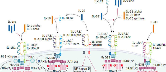
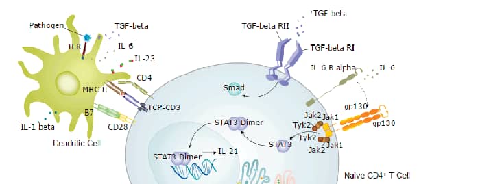
IL-1 is a name that designates two pleiotropic cytokines, IL-1 alpha (IL-1F1) and IL-1 beta (IL-1F2), which are the products of distinct genes. IL-1 alpha and IL-1 beta are structurally related polypeptides that share approximately 27% amino acid (aa) identity in feline. Both proteins are produced by a wide variety of cells in response to inflammatory agents, infections, or microbial endotoxins. While IL-1 alpha and IL-1 beta are regulated independently, they bind to the same receptor and exert identical biological effects. IL‑1 RI binds directly to IL-1 alpha or IL-1 beta and then associates with the IL-1 R accessory protein (IL-1 R3/IL-1 R AcP) to form a high-affinity receptor complex that is competent for signal transduction. IL-1 RII has high affinity for IL-1 beta but functions as a decoy receptor and negative regulator of IL-1 beta activity. IL-1ra functions as a competitive antagonist by preventing IL-1 alpha and IL-1 beta from interacting with IL-1 RI (1‑4). The feline IL-1 beta cDNA encodes a 267 aa precursor. A 115 aa propeptide is cleaved intracellularly by the cysteine protease IL-1 beta -converting enzyme (Caspase-1/ICE) to generate the active cytokine (5, 6). The 17 kDa mature feline IL-1 beta shares 63%‑78% aa sequence identity with canine, cotton rat, equine, human, mouse, porcine, rat, and rhesus IL-1 beta.

Scientific images may be resized, cropped, or adjusted for brightness or contrast for presentation purposes. These updates are limited to presentation and do not affect the underlying scientific interpretation of the data. Where available, additional source data may be provided upon request.
Key Product Details
Species Reactivity
Applications
Label
Antibody Source
Product Specifications
Immunogen
Ala116-Ser267
Accession # P41687
Specificity
Clonality
Host
Isotype
Endotoxin Level
Scientific Data Images for Feline IL-1 beta/IL-1F2 Antibody
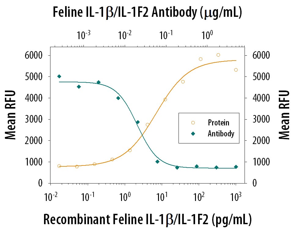
Cell Proliferation Induced by IL‑1 beta /IL‑1F2 and Neutral-ization by Feline IL‑1 beta /IL‑1F2 Antibody.
Recombinant Feline IL-1 beta /IL-1F2 (Catalog # 1796-FL) stimulates proliferation in the the D10.G4.1 mouse helper T cell line in a dose-dependent manner (orange line). Proliferation elicited by Recombinant Feline IL-1 beta /IL-1F2 (50 pg/mL) is neutralized (green line) by increasing concen-trations of Goat Anti-Feline IL-1 beta /IL-1F2 Antigen Affinity-purified Polyclonal Antibody (Catalog # AF1796). The ND50 is typically 0.02-0.08 µg/mL.
Feline IL-1 beta / IL-1F2 ELISA Standard Curve
Recombinant Feline IL‑1 beta /IL‑1F2 (Catalog # 1796-FL) was serially diluted and captured by Goat Anti-Feline IL‑1 beta /IL‑1F2 Antigen Affinity-purified Polyclonal Antibody (Catalog # AF1796) coated on a Clear Polystyrene Microplate (Catalog # DY990). Goat Anti-Feline IL‑1 beta /IL‑1F2 Antigen Affinity-purified Polyclonal Antibody (Catalog # AF1796) was biotinylated and incubated with the protein captured on the plate. Detection of the standard curve was achieved by incubating Streptavidin-HRP (Catalog # DY998)
Feline IL-1 beta / IL-1F2 ELISA Standard Curve
Recombinant Feline IL‑1 beta /IL‑1F2 (Catalog # 1796-FL) was serially diluted and captured by Goat Anti-Feline IL‑1 beta /IL‑1F2 Antigen Affinity-purified Polyclonal Antibody (Catalog # AF1796) coated on a Clear Polystyrene Microplate (Catalog # DY990). Goat Anti-Feline IL‑1 beta /IL‑1F2 Antigen Affinity-purified Polyclonal Antibody (Catalog # AF1796) was biotinylated and incubated with the protein captured on the plate. Detection of the standard curve was achieved by incubating Streptavidin-HRP (Catalog # DY998)Applications for Feline IL-1 beta/IL-1F2 Antibody
Immunocytochemistry
Sample: Immersion fixed feline peripheral blood mononuclear cells
Western Blot
Sample: Recombinant Feline IL‑1 beta /IL‑1F2 (Catalog # 1796-FL)
Neutralization
Feline IL-1 beta /IL-1F2 Sandwich Immunoassay
Reviewed Applications
Read 1 review rated 4 using AF1796 in the following applications:
Formulation, Preparation, and Storage
Purification
Reconstitution
Reconstitute at 0.2 mg/mL in sterile PBS. For liquid material, refer to CoA for concentration.
Formulation
Shipping
Stability & Storage
- 12 months from date of receipt, -20 to -70 °C as supplied.
- 1 month, 2 to 8 °C under sterile conditions after reconstitution.
- 6 months, -20 to -70 °C under sterile conditions after reconstitution.
Calculators
Background: IL-1 beta/IL-1F2
References
- Allan, S.M. et al. (2005) Nat. Rev. Immunol. 5:629.
- Boraschi, D. and A. Tagliabue (2006) Vitam. Horm. 74:229.
- Kornman, K.S. (2006) Am. J. Clin. Nutr. 83:475S.
- Isoda, K. and F. Ohsuzu (2006) J. Atheroscler. Thromb. 13:21.
- Accession # AAA30814.
- Martinon, F. and J. Tschopp (2007) Cell Death Differ. 14:10.
Long Name
Alternate Names
Entrez Gene IDs
Gene Symbol
UniProt
Additional IL-1 beta/IL-1F2 Products
Product Documents for Feline IL-1 beta/IL-1F2 Antibody
Certificate of Analysis
To download a Certificate of Analysis, please enter a lot or batch number in the search box below.
Note: Certificate of Analysis not available for kit components.
Product Specific Notices for Feline IL-1 beta/IL-1F2 Antibody
For research use only
Related Research Areas
Customer Reviews for Feline IL-1 beta/IL-1F2 Antibody (1)
Have you used Feline IL-1 beta/IL-1F2 Antibody?
Submit a review and receive an Amazon gift card!
$25/€18/£15/$25CAN/¥2500 for a review with an image
$10/€7/£6/$10CAN/¥1110 for a review without an image
Submit a review
Customer Images

-
Application: Immunohistochemistry-FrozenSample Tested: brain and spinal cordSpecies: FelineVerified Customer | Posted 11/16/2018Image was captured using AxioVision fluorescent microscope. IL1B on feline section is highlighted as red while DAPI nuclear counter staining shows blue signal.AF1796 antibody was used at 1:200 dilution.

There are no reviews that match your criteria.
Protocols
Find general support by application which include: protocols, troubleshooting, illustrated assays, videos and webinars.
- Appropriate Fixation of IHC/ICC Samples
- Cellular Response to Hypoxia Protocols
- ClariTSA™ Fluorophore Kits
- Detection & Visualization of Antibody Binding
- ICC Cell Smear Protocol for Suspension Cells
- ICC Immunocytochemistry Protocol Videos
- ICC for Adherent Cells
- Immunocytochemistry (ICC) Protocol
- Immunocytochemistry Troubleshooting
- Immunofluorescence of Organoids Embedded in Cultrex Basement Membrane Extract
- Immunohistochemistry (IHC) and Immunocytochemistry (ICC) Protocols
- Preparing Samples for IHC/ICC Experiments
- Preventing Non-Specific Staining (Non-Specific Binding)
- Primary Antibody Selection & Optimization
- Protocol for VisUCyte™ HRP Polymer Detection Reagent
- Protocol for the Fluorescent ICC Staining of Cell Smears - Graphic
- Protocol for the Fluorescent ICC Staining of Cultured Cells on Coverslips - Graphic
- Protocol for the Preparation and Fluorescent ICC Staining of Cells on Coverslips
- Protocol for the Preparation and Fluorescent ICC Staining of Non-adherent Cells
- Protocol for the Preparation and Fluorescent ICC Staining of Stem Cells on Coverslips
- Protocol for the Preparation of a Cell Smear for Non-adherent Cell ICC - Graphic
- R&D Systems Quality Control Western Blot Protocol
- TUNEL and Active Caspase-3 Detection by IHC/ICC Protocol
- The Importance of IHC/ICC Controls
- Troubleshooting Guide: Western Blot Figures
- Western Blot Conditions
- Western Blot Protocol
- Western Blot Protocol for Cell Lysates
- Western Blot Troubleshooting
- Western Blot Troubleshooting Guide
- View all Protocols, Troubleshooting, Illustrated assays and Webinars
Associated Pathways