GABA Antibody - Azide and BSA Free
Novus Biologicals | Catalog # NBP2-43558
Loading...
Key Product Details
Species Reactivity
Validated:
All Species
Cited:
Human, Rat
Applications
Validated:
Immunohistochemistry, Immunohistochemistry-Frozen, Immunohistochemistry Whole-Mount, Immunocytochemistry/ Immunofluorescence, Dot Blot
Cited:
Immunohistochemistry, Immunocytochemistry/ Immunofluorescence
Label
Unconjugated
Antibody Source
Polyclonal Rabbit IgG
Format
Azide and BSA Free
Loading...
Product Specifications
Immunogen
The immunogen used to generate this antibody corresponds to GABA
Reactivity Notes
Species independent
Clonality
Polyclonal
Host
Rabbit
Isotype
IgG
Scientific Data Images for GABA Antibody - Azide and BSA Free
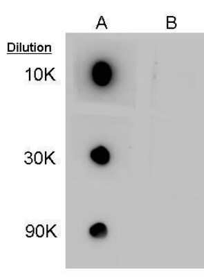
Dot Blot: GABA Antibody [NBP2-43558] - Analysis of 100ng of GABA-conjugated BSA, using GABA Antibody at 1:10000, 1:30000, and 1:90000. A: GABA-conjugated BSA. B: Normal BSA.

Immunocytochemistry/Immunofluorescence: GABA Antibody [NBP2-43558] -
Immunocytochemistry/Immunofluorescence: GABA Antibody [NBP2-43558] - DIV9 rat E18 primary cortical neuron cells were fixed in 4% paraformaldehyde at RT for 15 min.Green: GABA stained by GABA antibody diluted at 1:250. Red: Tau, a axon marker, stained by Tau antibody [287] diluted at 1:500. Blue: Fluoroshield with DAPI.
Immunohistochemistry-Frozen: GABA Antibody [NBP2-43558] -
GABA antibody detects GABA by immunohistochemical analysis.Sample: Frozen-sectioned mouse eye.
Green: GABA stained by GABA antibody (NBP2-43558) diluted at 1:250.
Red: beta Tubulin 3/ Tuj1 antibody [GT11710] diluted at 1:500.
Blue: Fluoroshield with DAPI.
Applications for GABA Antibody - Azide and BSA Free
Application
Recommended Usage
Dot Blot
Assay dependent
Immunocytochemistry/ Immunofluorescence
1:100-1:1000
Immunohistochemistry
1:100-1:1000
Immunohistochemistry Whole-Mount
Assay dependent
Immunohistochemistry-Frozen
1:100-1:1000
Formulation, Preparation, and Storage
Purification
Antigen Affinity-purified
Formulation
PBS, 20% Glycerol
Format
Azide and BSA Free
Preservative
0.025% Proclin 300
Concentration
Concentrations vary lot to lot. See vial label for concentration. If unlisted please contact technical services.
Shipping
The product is shipped with polar packs. Upon receipt, store it immediately at the temperature recommended below.
Stability & Storage
Aliquot and store at -20C or -80C. Avoid freeze-thaw cycles.
Background: GABA
Alternate Names
y-Aminobutyric acid
Additional GABA Products
Product Documents for GABA Antibody - Azide and BSA Free
Certificate of Analysis
To download a Certificate of Analysis, please enter a lot or batch number in the search box below.
Product Specific Notices for GABA Antibody - Azide and BSA Free
This product is for research use only and is not approved for use in humans or in clinical diagnosis. Primary Antibodies are guaranteed for 1 year from date of receipt.
⚠ WARNING: This product can expose you to chemicals including mercury, which is known to the State of California to cause reproductive toxicity with developmental effects. For more information go to www.P65Warnings.ca.gov.Related Research Areas
Citations for GABA Antibody - Azide and BSA Free
 Powered by Bioz
Customer Reviews for GABA Antibody - Azide and BSA Free
There are currently no reviews for this product. Be the first to review GABA Antibody - Azide and BSA Free and earn rewards!
Have you used GABA Antibody - Azide and BSA Free?
Submit a review and receive an Amazon gift card!
$25/€18/£15/$25CAN/¥2500 Yen for a review with an image
$10/€7/£6/$10CAN/¥1110 Yen for a review without an image
Submit a review
Protocols
Find general support by application which include: protocols, troubleshooting, illustrated assays, videos and webinars.
- Antigen Retrieval Protocol (PIER)
- Antigen Retrieval for Frozen Sections Protocol
- Appropriate Fixation of IHC/ICC Samples
- Cellular Response to Hypoxia Protocols
- Chromogenic IHC Staining of Formalin-Fixed Paraffin-Embedded (FFPE) Tissue Protocol
- Chromogenic Immunohistochemistry Staining of Frozen Tissue
- ClariTSA™ Fluorophore Kits
- Detection & Visualization of Antibody Binding
- Fluorescent IHC Staining of Frozen Tissue Protocol
- Graphic Protocol for Heat-induced Epitope Retrieval
- Graphic Protocol for the Preparation and Fluorescent IHC Staining of Frozen Tissue Sections
- Graphic Protocol for the Preparation and Fluorescent IHC Staining of Paraffin-embedded Tissue Sections
- Graphic Protocol for the Preparation of Gelatin-coated Slides for Histological Tissue Sections
- ICC Cell Smear Protocol for Suspension Cells
- ICC Immunocytochemistry Protocol Videos
- ICC for Adherent Cells
- IHC Sample Preparation (Frozen sections vs Paraffin)
- Immunocytochemistry (ICC) Protocol
- Immunocytochemistry Troubleshooting
- Immunofluorescence of Organoids Embedded in Cultrex Basement Membrane Extract
- Immunofluorescent IHC Staining of Formalin-Fixed Paraffin-Embedded (FFPE) Tissue Protocol
- Immunohistochemistry (IHC) and Immunocytochemistry (ICC) Protocols
- Immunohistochemistry Frozen Troubleshooting
- Immunohistochemistry Paraffin Troubleshooting
- Preparing Samples for IHC/ICC Experiments
- Preventing Non-Specific Staining (Non-Specific Binding)
- Primary Antibody Selection & Optimization
- Protocol for Heat-Induced Epitope Retrieval (HIER)
- Protocol for Making a 4% Formaldehyde Solution in PBS
- Protocol for VisUCyte™ HRP Polymer Detection Reagent
- Protocol for the Fluorescent ICC Staining of Cell Smears - Graphic
- Protocol for the Fluorescent ICC Staining of Cultured Cells on Coverslips - Graphic
- Protocol for the Preparation & Fixation of Cells on Coverslips
- Protocol for the Preparation and Chromogenic IHC Staining of Frozen Tissue Sections
- Protocol for the Preparation and Chromogenic IHC Staining of Frozen Tissue Sections - Graphic
- Protocol for the Preparation and Chromogenic IHC Staining of Paraffin-embedded Tissue Sections
- Protocol for the Preparation and Chromogenic IHC Staining of Paraffin-embedded Tissue Sections - Graphic
- Protocol for the Preparation and Fluorescent ICC Staining of Cells on Coverslips
- Protocol for the Preparation and Fluorescent ICC Staining of Non-adherent Cells
- Protocol for the Preparation and Fluorescent ICC Staining of Stem Cells on Coverslips
- Protocol for the Preparation and Fluorescent IHC Staining of Frozen Tissue Sections
- Protocol for the Preparation and Fluorescent IHC Staining of Paraffin-embedded Tissue Sections
- Protocol for the Preparation of Gelatin-coated Slides for Histological Tissue Sections
- Protocol for the Preparation of a Cell Smear for Non-adherent Cell ICC - Graphic
- TUNEL and Active Caspase-3 Detection by IHC/ICC Protocol
- The Importance of IHC/ICC Controls
- Troubleshooting Guide: Immunohistochemistry
- View all Protocols, Troubleshooting, Illustrated assays and Webinars
Loading...