GLUT9 Antibody - BSA Free
Novus Biologicals | Catalog # NBP1-05054
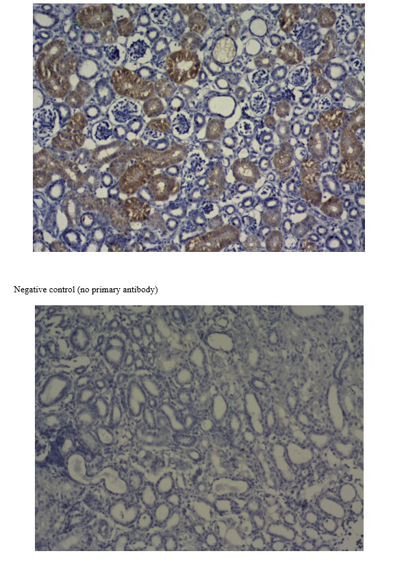
![Immunohistochemistry: GLUT9 Antibody - BSA Free [NBP1-05054]](https://resources.rndsystems.com/images/products/GLUT9-Antibody-Immunohistochemistry-NBP1-05054-img0006.jpg "Immunohistochemistry: GLUT9 Antibody - BSA Free [NBP1-05054]")
Key Product Details
Species Reactivity
Validated:
Human, Mouse
Cited:
Mouse
Applications
Validated:
Immunohistochemistry, Immunohistochemistry-Paraffin, Western Blot, Immunocytochemistry/ Immunofluorescence
Cited:
Western Blot, Immunocytochemistry/ Immunofluorescence
Label
Unconjugated
Antibody Source
Polyclonal Rabbit IgG
Format
BSA Free
Loading...
Product Specifications
Immunogen
Synthetic peptide made to an internal portion of human GLUT9 (within residues 500-540). [Swiss-Prot# Q9NRM0]
Localization
Membrane; Multi-pass membrane protein.
Clonality
Polyclonal
Host
Rabbit
Isotype
IgG
Theoretical MW
55 kDa.
Disclaimer note: The observed molecular weight of the protein may vary from the listed predicted molecular weight due to post translational modifications, post translation cleavages, relative charges, and other experimental factors.
Disclaimer note: The observed molecular weight of the protein may vary from the listed predicted molecular weight due to post translational modifications, post translation cleavages, relative charges, and other experimental factors.
Scientific Data Images for GLUT9 Antibody - BSA Free
Immunohistochemistry: GLUT9 Antibody - BSA Free [NBP1-05054]
GLUT9-Antibody-Immunohistochemistry-NBP1-05054-img0006.jpg![Immunohistochemistry: GLUT9 Antibody - BSA Free [NBP1-05054]](https://resources.rndsystems.com/images/products/GLUT9-Antibody-Immunohistochemistry-NBP1-05054-img0007.jpg "Immunohistochemistry: GLUT9 Antibody - BSA Free [NBP1-05054]")
Immunohistochemistry: GLUT9 Antibody - BSA Free [NBP1-05054]
GLUT9-Antibody-Immunohistochemistry-NBP1-05054-img0007.jpg![Western Blot: GLUT9 AntibodyBSA Free [NBP1-05054]](https://resources.rndsystems.com/images/products/GLUT9-Antibody-Western-Blot-NBP1-05054-img0002.jpg "Western Blot: GLUT9 AntibodyBSA Free [NBP1-05054]")
Western Blot: GLUT9 AntibodyBSA Free [NBP1-05054]
Western Blot: GLUT9 Antibody [NBP1-05054] - Detection of GLUT9 in human kidney membrane.![Immunocytochemistry/ Immunofluorescence: GLUT9 Antibody - BSA Free [NBP1-05054]](https://resources.rndsystems.com/images/products/GLUT9-Antibody-Immunocytochemistry-Immunofluorescence-NBP1-05054-img0005.jpg "Immunocytochemistry/ Immunofluorescence: GLUT9 Antibody - BSA Free [NBP1-05054]")
Immunocytochemistry/ Immunofluorescence: GLUT9 Antibody - BSA Free [NBP1-05054]
Immunocytochemistry/Immunofluorescence: GLUT9 Antibody [NBP1-05054] - HeLa cells were fixed for 10 minutes using 10% formalin and then permeabilized for 5 minutes using 1X TBS + 0.5% Triton-X100. The cells were incubated with anti-GLUT9 at a 1:200 dilution overnight at 4C and detected with an anti-rabbit Dylight 488 (Green) at a 1:500 dilution. Alpha tubulin (DM1A) NB100-690 was used as a co-stain at a 1:1000 dilution and detected with an anti-mouse Dylight 550 (Red) at a 1:500 dilution. Nuclei were counterstained with DAPI (Blue). Cells were imaged using a 40X objective.![Immunohistochemistry-Paraffin: GLUT9 Antibody - BSA Free [NBP1-05054]](https://resources.rndsystems.com/images/products/GLUT9-Antibody-Immunohistochemistry-Paraffin-NBP1-05054-img0004.jpg "Immunohistochemistry-Paraffin: GLUT9 Antibody - BSA Free [NBP1-05054]")
Immunohistochemistry-Paraffin: GLUT9 Antibody - BSA Free [NBP1-05054]
Immunohistochemistry-Paraffin: GLUT9 Antibody [NBP1-05054] - IHC-P analysis of mouse kidney section using GLUT9 antibody at 1:200 dilution. This antibody generated an expected cytoplasmic staining in tubular cells with a more staining towards membranes in some cells but no immunopositivity in Bowmans capsules.
Immunohistochemistry: GLUT9 Antibody - BSA Free [NBP1-05054] -
Immunohistochemistry: GLUT9 Antibody - BSA Free [NBP1-05054] - Immunofluorescence staining of GLUT9 in PFA-fixed murine brain sections. Frozen sections of paraformaldehyde-fixed wild-type murine brain were used for immunofluorescence staining. a, b Antigen absorption test. Immunofluorescence staining of the ependymal wall of the dorsal third ventricle using a anti-GLUT9 antibody & b antigen-preabsorbed antibody. Scale bar 100 µm. c, d Immunofluorescence staining of GLUT9 (magenta), acetylated-tubulin (Ac-Tubulin, green) & DAPI (blue) on ependymal cells. Scale bar 10 µm. e–g Immunofluorescence staining of GLUT9 (magenta) & NeuN (green) showing co-localization in neurons. Scale bar 10 µm. D3V, dorsal third ventricle; DAPI, 4′,6-diamidino-2-phenylindole; NeuN, neuronal nucleus marker Image collected & cropped by CiteAb from the following publication (https://fluidsbarrierscns.biomedcentral.com/articles/10.1186/s12987-016…), licensed under a CC-BY license. Not internally tested by Novus Biologicals.
Immunohistochemistry: GLUT9 Antibody - BSA Free [NBP1-05054] -
Immunohistochemistry: GLUT9 Antibody - BSA Free [NBP1-05054] - Immunofluorescence staining of ABCG2 in methanol/acetone-fixed & methacarn-fixed murine brain. Fresh frozen sections of the dorsal third ventricle were prepared from wild-type (WT) & ABCG2 knockout (KO) mice & post-fixed with methanol & acetone. Immunofluorescence staining of ABCG2 was seen in choroid plexus & capillaries in sections from a WT but not in b ABCG2 KO mouse. Scale bar 100 µm. c Immunofluorescence staining of ABCG2 (magenta) & GLUT9 (green) showing ABCG2 in the choroid plexus & GLUT9 in the ependyma. Scale bar 100 µm. d–f Immunofluorescence staining of d ABCG2 & e GLUT9 using methacarn-fixed paraffin section of capillaries showing co-localization in f. Blue indicates DAPI-stained nucleus of the brain capillary endothelial cell. Scale bar 10 µm. CP, choroid plexus; D3V, dorsal third ventricle Image collected & cropped by CiteAb from the following publication (https://fluidsbarrierscns.biomedcentral.com/articles/10.1186/s12987-016…), licensed under a CC-BY license. Not internally tested by Novus Biologicals.Applications for GLUT9 Antibody - BSA Free
Application
Recommended Usage
Immunocytochemistry/ Immunofluorescence
1:200
Immunohistochemistry
1:200
Immunohistochemistry-Paraffin
1:200
Western Blot
0.5 ug/ml
Application Notes
This GLUT9 antibody is useful for Western blot, where a band is seen ~ 55 kDa. The immunogen of this product corresponds to a cytoplasmic domain of GLUT9 protein and in immunostaining assays, cytoplasmic-membrane staining may be expected for this product. The observed molecular weight of the protein may vary from the listed predicted molecular weight due to post translational modifications, post translation cleavages, relative charges, and other experimental factors.
Reviewed Applications
Read 1 review rated 5 using NBP1-05054 in the following applications:
Formulation, Preparation, and Storage
Purification
Immunogen affinity purified
Formulation
PBS
Format
BSA Free
Preservative
0.02% Sodium Azide
Concentration
1 mg/ml
Shipping
The product is shipped with polar packs. Upon receipt, store it immediately at the temperature recommended below.
Stability & Storage
Store at 4C short term. Aliquot and store at -20C long term. Avoid freeze-thaw cycles.
Background: GLUT9
Alternate Names
facilitated glucose transporter member 9, GLUT-9, GLUT9Glut9, GLUTX, solute carrier family 2 (facilitated glucose transporter), member 9, UAQTL2, urate voltage-driven efflux transporter 1
Gene Symbol
SLC2A9
Additional GLUT9 Products
Product Documents for GLUT9 Antibody - BSA Free
Certificate of Analysis
To download a Certificate of Analysis, please enter a lot or batch number in the search box below.
Product Specific Notices for GLUT9 Antibody - BSA Free
This product is for research use only and is not approved for use in humans or in clinical diagnosis. Primary Antibodies are guaranteed for 1 year from date of receipt.
Citations for GLUT9 Antibody - BSA Free
 Powered by Bioz
Customer Reviews for GLUT9 Antibody - BSA Free (1)
5 out of 5
1 Customer Rating
Have you used GLUT9 Antibody - BSA Free?
Submit a review and receive an Amazon gift card!
$25/€18/£15/$25CAN/¥2500 Yen for a review with an image
$10/€7/£6/$10CAN/¥1110 Yen for a review without an image
Submit a review
Customer Images
Showing
1
-
1 的
1 review
Showing All
Filter By:
-
Application: Immunohistochemistry-ParaffinSample Tested: Goose TissueSpecies: GooseVerified Customer | Posted 09/22/2017

There are no reviews that match your criteria.
Protocols
View specific protocols for GLUT9 Antibody - BSA Free (NBP1-05054):
GLUT9 Antibody:
Western Blot Protocol
1. Perform SDS-PAGE (4-12% MOPS) on samples to be analyzed, loading 30 ug of total protein per lane.
2. Transfer proteins to Nitrocellulose according to the instructions provided by the manufacturer of the transfer
apparatus.
3. Rinse membrane with dH2O and then stain the blot using Ponceau S for 1-2 minutes to access the transfer of proteins onto the nitrocellulose membrane. Rinse the blot in water to remove excess stain and mark the lane locations and locations of molecular weight markers using a pencil.
4. Rinse the blot in TBS for approximately 5 minutes.
5. Block the membrane using 5% BSA in TBS + Tween, 1 hour at RT.
6. Rinse the membrane in dH2O and then wash the membrane in wash buffer [TBS + 0.1% Tween] 3 times for 10 minutes each.
7. Dilute the rabbit anti-GLUT9 primary antibody (NBP1-05054) in blocking buffer and incubate 1 hour at room temperature.
8. Rinse the membrane in dH2O and then wash the membrane in wash buffer [TBS + 0.1% Tween] 3 times for 10 minutes each.
9. Apply the diluted rabbit-IgG HRP-conjugated secondary antibody in blocking buffer (as per manufacturers
instructions) and incubate 1 hour at room temperature.
10. Wash the blot in wash buffer [TBS + 0.1% Tween] 3 times for 10 minutes each (this step can be repeated as required to reduce background).
11. Apply the detection reagent of choice in accordance with the manufacturers instructions (Pierce ECL).
Note: Tween-20 can be added to the blocking or antibody dilution buffer at a final concentration of 0.05-0.2%, provided it does not interfere with antibody-antigen binding.
Find general support by application which include: protocols, troubleshooting, illustrated assays, videos and webinars.
- Antigen Retrieval Protocol (PIER)
- Antigen Retrieval for Frozen Sections Protocol
- Appropriate Fixation of IHC/ICC Samples
- Cellular Response to Hypoxia Protocols
- Chromogenic IHC Staining of Formalin-Fixed Paraffin-Embedded (FFPE) Tissue Protocol
- Chromogenic Immunohistochemistry Staining of Frozen Tissue
- ClariTSA™ Fluorophore Kits
- Detection & Visualization of Antibody Binding
- Fluorescent IHC Staining of Frozen Tissue Protocol
- Graphic Protocol for Heat-induced Epitope Retrieval
- Graphic Protocol for the Preparation and Fluorescent IHC Staining of Frozen Tissue Sections
- Graphic Protocol for the Preparation and Fluorescent IHC Staining of Paraffin-embedded Tissue Sections
- Graphic Protocol for the Preparation of Gelatin-coated Slides for Histological Tissue Sections
- ICC Cell Smear Protocol for Suspension Cells
- ICC Immunocytochemistry Protocol Videos
- ICC for Adherent Cells
- IHC Sample Preparation (Frozen sections vs Paraffin)
- Immunocytochemistry (ICC) Protocol
- Immunocytochemistry Troubleshooting
- Immunofluorescence of Organoids Embedded in Cultrex Basement Membrane Extract
- Immunofluorescent IHC Staining of Formalin-Fixed Paraffin-Embedded (FFPE) Tissue Protocol
- Immunohistochemistry (IHC) and Immunocytochemistry (ICC) Protocols
- Immunohistochemistry Frozen Troubleshooting
- Immunohistochemistry Paraffin Troubleshooting
- Preparing Samples for IHC/ICC Experiments
- Preventing Non-Specific Staining (Non-Specific Binding)
- Primary Antibody Selection & Optimization
- Protocol for Heat-Induced Epitope Retrieval (HIER)
- Protocol for Making a 4% Formaldehyde Solution in PBS
- Protocol for VisUCyte™ HRP Polymer Detection Reagent
- Protocol for the Fluorescent ICC Staining of Cell Smears - Graphic
- Protocol for the Fluorescent ICC Staining of Cultured Cells on Coverslips - Graphic
- Protocol for the Preparation & Fixation of Cells on Coverslips
- Protocol for the Preparation and Chromogenic IHC Staining of Frozen Tissue Sections
- Protocol for the Preparation and Chromogenic IHC Staining of Frozen Tissue Sections - Graphic
- Protocol for the Preparation and Chromogenic IHC Staining of Paraffin-embedded Tissue Sections
- Protocol for the Preparation and Chromogenic IHC Staining of Paraffin-embedded Tissue Sections - Graphic
- Protocol for the Preparation and Fluorescent ICC Staining of Cells on Coverslips
- Protocol for the Preparation and Fluorescent ICC Staining of Non-adherent Cells
- Protocol for the Preparation and Fluorescent ICC Staining of Stem Cells on Coverslips
- Protocol for the Preparation and Fluorescent IHC Staining of Frozen Tissue Sections
- Protocol for the Preparation and Fluorescent IHC Staining of Paraffin-embedded Tissue Sections
- Protocol for the Preparation of Gelatin-coated Slides for Histological Tissue Sections
- Protocol for the Preparation of a Cell Smear for Non-adherent Cell ICC - Graphic
- R&D Systems Quality Control Western Blot Protocol
- TUNEL and Active Caspase-3 Detection by IHC/ICC Protocol
- The Importance of IHC/ICC Controls
- Troubleshooting Guide: Immunohistochemistry
- Troubleshooting Guide: Western Blot Figures
- Western Blot Conditions
- Western Blot Protocol
- Western Blot Protocol for Cell Lysates
- Western Blot Troubleshooting
- Western Blot Troubleshooting Guide
- View all Protocols, Troubleshooting, Illustrated assays and Webinars
Loading...