
HCoV-HKU1 Human Coronavirus Spike RBD Antibody (1045307)
R&D Systems | Catalog # MAB10936

Key Product Details
Species Reactivity
HCoV-HKU1
Applications
Immunocytochemistry
Label
Unconjugated
Antibody Source
Monoclonal Mouse IgG2A Clone # 1045307
Loading...
Product Specifications
Immunogen
Human embryonic kidney cell HEK293-derived HCoV-HKU1 Spike RBD
Thr310-Tyr624
Accession # Q5MQD0.1
Thr310-Tyr624
Accession # Q5MQD0.1
Specificity
Detects Human Coronavirus HCoV-HKU1 Spike RBD and 229E in direct ELISAs. No
staining of NL63 HKu1.
Clonality
Monoclonal
Host
Mouse
Isotype
IgG2A
Scientific Data Images for HCoV-HKU1 Human Coronavirus Spike RBD Antibody (1045307)
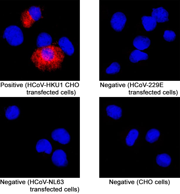
Spike RBD in CHO Chinese hamster ovary cell line transfected with HCoV-HKU1.
Spike RBD was detected in immersion fixed CHO Chinese hamster ovary cell line transfected with HCoV-HKU1 (positive staining), HCoV-229E transfected (negative staining), HCoV-NL63 transfected cell (negative staining), and CHO Chinese hamster ovary cell line (non-transfected, negative staining) using Mouse Anti-HCoV-HKU1 Spike RBD Monoclonal Antibody (Catalog # MAB10936) at 8 µg/mL for 3 hours at room temperature. Cells were stained using the NorthernLights™ 557-conjugated Anti-Mouse IgG Secondary Antibody (red; NL007) and counterstained with DAPI (blue). Specific staining was localized to cell cytoplasm. Staining was performed using our protocol for Fluorescent ICC Staining of Non-adherent Cells.Applications for HCoV-HKU1 Human Coronavirus Spike RBD Antibody (1045307)
Application
Recommended Usage
Immunocytochemistry
8-25 µg/mL
Sample: Immersion fixed CHO Chinese hamster ovary cell line transfected with HCoV-HKU1
Sample: Immersion fixed CHO Chinese hamster ovary cell line transfected with HCoV-HKU1
Formulation, Preparation, and Storage
Purification
Protein A or G purified from hybridoma culture supernatant
Reconstitution
Reconstitute at 0.5 mg/mL in sterile PBS. For liquid material, refer to CoA for concentration.
Loading...
Formulation
Lyophilized from a 0.2 μm filtered solution in PBS with Trehalose. *Small pack size (SP) is supplied either lyophilized or as a 0.2 µm filtered solution in PBS.
Shipping
Lyophilized product is shipped at ambient temperature. Liquid small pack size (-SP) is shipped with polar packs. Upon receipt, store immediately at the temperature recommended below.
Stability & Storage
Use a manual defrost freezer and avoid repeated freeze-thaw cycles.
- 12 months from date of receipt, -20 to -70 °C as supplied.
- 1 month, 2 to 8 °C under sterile conditions after reconstitution.
- 6 months, -20 to -70 °C under sterile conditions after reconstitution.
Calculators
Background: Spike RBD
References
- Woo, P. et al. (2005) J. Virol.79:884.
- Ogimi, C. et al. (2020) J Pediatric Infect Dis Soc doi: 10.1093/jpids/piaa037.
- Qian, Z. et al. (2015) J. Virol. 89:8816.
- Huang, X. et al. (2015) J Virol 89:7202.
- Hulswit, R.J.G. et al. (2019) PNAS 116:2681.
Long Name
Spike Receptor Binding Domain
Gene Symbol
S
UniProt
Additional Spike RBD Products
Product Documents for HCoV-HKU1 Human Coronavirus Spike RBD Antibody (1045307)
Certificate of Analysis
To download a Certificate of Analysis, please enter a lot or batch number in the search box below.
Note: Certificate of Analysis not available for kit components.
Product Specific Notices for HCoV-HKU1 Human Coronavirus Spike RBD Antibody (1045307)
For research use only
Related Research Areas
Citations for HCoV-HKU1 Human Coronavirus Spike RBD Antibody (1045307)
 Powered by Bioz
Customer Reviews for HCoV-HKU1 Human Coronavirus Spike RBD Antibody (1045307)
There are currently no reviews for this product. Be the first to review HCoV-HKU1 Human Coronavirus Spike RBD Antibody (1045307) and earn rewards!
Have you used HCoV-HKU1 Human Coronavirus Spike RBD Antibody (1045307)?
Submit a review and receive an Amazon gift card!
$25/€18/£15/$25CAN/¥2500 for a review with an image
$10/€7/£6/$10CAN/¥1110 for a review without an image
Submit a review
Protocols
Find general support by application which include: protocols, troubleshooting, illustrated assays, videos and webinars.
- Appropriate Fixation of IHC/ICC Samples
- Cellular Response to Hypoxia Protocols
- ClariTSA™ Fluorophore Kits
- Detection & Visualization of Antibody Binding
- ICC Cell Smear Protocol for Suspension Cells
- ICC Immunocytochemistry Protocol Videos
- ICC for Adherent Cells
- Immunocytochemistry (ICC) Protocol
- Immunocytochemistry Troubleshooting
- Immunofluorescence of Organoids Embedded in Cultrex Basement Membrane Extract
- Immunohistochemistry (IHC) and Immunocytochemistry (ICC) Protocols
- Preparing Samples for IHC/ICC Experiments
- Preventing Non-Specific Staining (Non-Specific Binding)
- Primary Antibody Selection & Optimization
- Protocol for VisUCyte™ HRP Polymer Detection Reagent
- Protocol for the Fluorescent ICC Staining of Cell Smears - Graphic
- Protocol for the Fluorescent ICC Staining of Cultured Cells on Coverslips - Graphic
- Protocol for the Preparation and Fluorescent ICC Staining of Cells on Coverslips
- Protocol for the Preparation and Fluorescent ICC Staining of Non-adherent Cells
- Protocol for the Preparation and Fluorescent ICC Staining of Stem Cells on Coverslips
- Protocol for the Preparation of a Cell Smear for Non-adherent Cell ICC - Graphic
- TUNEL and Active Caspase-3 Detection by IHC/ICC Protocol
- The Importance of IHC/ICC Controls
- View all Protocols, Troubleshooting, Illustrated assays and Webinars
Loading...