Histone H2AX [p Ser139] Antibody
Novus Biologicals | Catalog # NB100-2280
Loading...
Key Product Details
Species Reactivity
Validated:
Human, Mouse, Canine
Cited:
Human, Mouse, Canine
Predicted:
Bat (100%), Bovine (100%), Chinese Hamster (100%), Guinea Pig (100%), Monkey (100%), Porcine (100%), Rabbit (100%). Backed by our 100% Guarantee.
Applications
Validated:
Immunohistochemistry, Immunohistochemistry-Paraffin, Immunohistochemistry-Frozen, Western Blot, Immunocytochemistry/ Immunofluorescence, Simple Western
Cited:
Immunohistochemistry-Paraffin, Immunohistochemistry-Frozen, Western Blot, Immunocytochemistry/ Immunofluorescence, IF/IHC
Label
Unconjugated
Antibody Source
Polyclonal Rabbit IgG
Loading...
Product Specifications
Immunogen
This Histone H2AX [p Ser139] Antibody was developed against a synthetic phospho-peptide, which represented a portion of the C-terminus of human histone H2AX surrounding phosphorylated serine 139 (GeneID 3014).
Reactivity Notes
Based on sequence percent identity: Gorilla (100%), Macaque (100%), Canine reactivity reported in scientific literature (PMID: 26991424).
Modification
p Ser139
Marker
DNA Double-strand break marker
Clonality
Polyclonal
Host
Rabbit
Isotype
IgG
Theoretical MW
15 kDa.
Disclaimer note: The observed molecular weight of the protein may vary from the listed predicted molecular weight due to post translational modifications, post translation cleavages, relative charges, and other experimental factors.
Disclaimer note: The observed molecular weight of the protein may vary from the listed predicted molecular weight due to post translational modifications, post translation cleavages, relative charges, and other experimental factors.
Scientific Data Images for Histone H2AX [p Ser139] Antibody
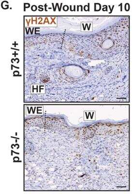
gamma-H2AX-[p-Ser139]-Antibody-Immunohistochemistry-NB100-2280-img0022.jpg

Simple Western: Histone H2AX [p Ser139] Antibody [NB100-2280] - Simple Western lane view shows a specific band for Histone H2AX [p Ser139] in 0.2 mg/ml of Jurkat lysate(s). This experiment was performed under reducing conditions using the 12 - 230 kDa separation system.
gamma-H2AX-[p-Ser139]-Antibody-Immunohistochemistry-NB100-2280-img0021.jpg
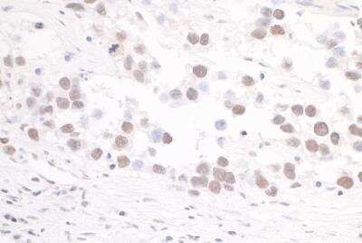
Immunohistochemistry-Paraffin: Histone H2AX [p Ser139] Antibody [NB100-2280] - Detection of human Histone H2AX [p Ser139] antibody by immunohistochemistry. Sample: FFPE section of human seminoma. Antibodies: Affinity purified rabbit Histone H2AX [p Ser139] antibody used at a dilution of 1:500. Detection: DAB.
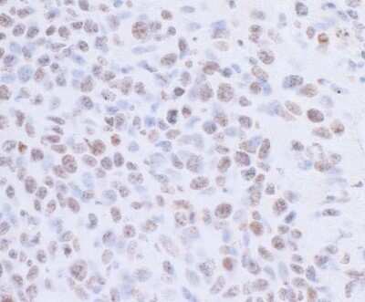
Immunohistochemistry: Histone H2AX [p Ser139] Antibody [NB100-2280] - Detection of Mouse Histone H2AX [p Ser139] by Immunohistochemistry. Sample: FFPE section of mouse colon carcinoma CT26. Antibodies: Affinity purified rabbit Histone H2AX [p Ser139] antibody. Detection: DAB.
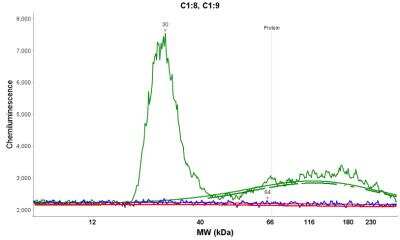
Simple Western: Histone H2AX [p Ser139] Antibody [NB100-2280] - Electropherogram image(s) of corresponding Simple Western lane view. Histone H2AX [p Ser139] antibody was used at 1:20 dilution on Jurkat lysate(s).
![Histone H2AX [p Ser139] Antibody](https://resources.rndsystems.com/images/products/nb100-2280_rabbit-polyclonal-histone-h2ax-p-ser139-antibody-310202415175243.jpg "Immunocytochemistry/ Immunofluorescence: Histone H2AX [p Ser139] Antibody [NB100-2280] -")
Immunocytochemistry/ Immunofluorescence: Histone H2AX [p Ser139] Antibody [NB100-2280] -
Immunocytochemistry/ Immunofluorescence: Histone H2AX [p Ser139] Antibody [NB100-2280] - Treatment with anti-thyroid drug protected RPE & photoreceptors from oxidative damage induced by NaIO3.RPE & retinal oxidative damage were evaluated by immunofluorescence labeling of p-gamma H2AX & 8-OHdG on the RPE whole mounts & retinal sections at 3 days post-NaIO3 injection. a Shown are representative images of p-gamma H2AX immunofluorescence labeling on the RPE whole mounts. b Shown are representative images of p-gamma H2AX & 8-OHdG immunofluorescence labeling on the retinal sections, & corresponding quantitative analysis for p-gamma H2AX labeling. ONL, outer nuclear layer; INL, inner nuclear layer. Data represented the mean ± SEM for 5 mice per group (*p < 0.05). Image collected & cropped by CiteAb from the following publication (https://pubmed.ncbi.nlm.nih.gov/31932580), licensed under a CC-BY license. Not internally tested by Novus Biologicals.![Histone H2AX [p Ser139] Antibody](https://resources.rndsystems.com/images/products/nb100-2280_rabbit-polyclonal-histone-h2ax-p-ser139-antibody-310202415171325.jpg "Immunocytochemistry/ Immunofluorescence: Histone H2AX [p Ser139] Antibody [NB100-2280] -")
Immunocytochemistry/ Immunofluorescence: Histone H2AX [p Ser139] Antibody [NB100-2280] -
Immunocytochemistry/ Immunofluorescence: Histone H2AX [p Ser139] Antibody [NB100-2280] - Treatment with anti-thyroid drug protected RPE & photoreceptors from oxidative damage induced by NaIO3.RPE & retinal oxidative damage were evaluated by immunofluorescence labeling of p-gamma H2AX & 8-OHdG on the RPE whole mounts & retinal sections at 3 days post-NaIO3 injection. a Shown are representative images of p-gamma H2AX immunofluorescence labeling on the RPE whole mounts. b Shown are representative images of p-gamma H2AX & 8-OHdG immunofluorescence labeling on the retinal sections, & corresponding quantitative analysis for p-gamma H2AX labeling. ONL, outer nuclear layer; INL, inner nuclear layer. Data represented the mean ± SEM for 5 mice per group (*p < 0.05). Image collected & cropped by CiteAb from the following publication (https://pubmed.ncbi.nlm.nih.gov/31932580), licensed under a CC-BY license. Not internally tested by Novus Biologicals.![Histone H2AX [p Ser139] Antibody](https://resources.rndsystems.com/images/products/nb100-2280_rabbit-polyclonal-histone-h2ax-p-ser139-antibody-3102024165725.jpg "Immunocytochemistry/ Immunofluorescence: Histone H2AX [p Ser139] Antibody [NB100-2280] -")
Immunocytochemistry/ Immunofluorescence: Histone H2AX [p Ser139] Antibody [NB100-2280] -
Immunocytochemistry/ Immunofluorescence: Histone H2AX [p Ser139] Antibody [NB100-2280] - RES rescued DXR-induced apoptosis through DNA-damage-P63-Caspase3 pathway in mouse oocytes. (A) Representative immunofluorescence images showing the expression of gamma -H2AX in mouse oocytes. Green, gamma -H2AX, Blue, DNA, Bar = 20 μm. (B) The relative immunofluorescence intensity of gamma -H2AX was measured in control, DXR-treated & RES-supplemented oocytes. Experiments were repeated at least 3 times with more than 30 oocytes examined for each group. Data were presented as means ± S.E.M of three independent experiments. **means P < 0.01, *** means P < 0.001. (C) Protein levels of gamma -H2AX, P63 & Active-Caspase3 were examined by Western blotting in control, DXR-treated & RES-supplemented oocytes. GAPDH was used as a loading control. The clean backgrounds for the active-Caspase-3, gamma -H2AX & GAPDH is due to the exposure. Image collected & cropped by CiteAb from the following publication (https://pubmed.ncbi.nlm.nih.gov/32352929), licensed under a CC-BY license. Not internally tested by Novus Biologicals.![Histone H2AX [p Ser139] Antibody](https://resources.rndsystems.com/images/products/nb100-2280_rabbit-polyclonal-histone-h2ax-p-ser139-antibody-310202415541940.jpg "Immunohistochemistry: Histone H2AX [p Ser139] Antibody [NB100-2280] -")
Immunohistochemistry: Histone H2AX [p Ser139] Antibody [NB100-2280] -
Immunohistochemistry: Histone H2AX [p Ser139] Antibody [NB100-2280] - Hepatic activation of FOXO3 induces oxidative damage & Akt activation. a Expression of Bcl2l11 (Bim), Sesn2 & Sesn3 by qPCR in livers from 9-week-old control & FOXO3CAHep transgenic mice (n = 4). b Representative IHC staining for 8-OHdG (bar = 100 μm) & quantification of 8-OHdG-positive cells as a percentage of total hepatocyte cells in livers of patients in area of small cells (SC) & large cells (LC). c Representative IHC staining for phospho-gamma H2AX (bar = 100 μm) & quantification of phospho-gamma H2AX-positive cells as a percentage of total hepatocyte cells in livers of patients in area of small cells & large cells. d Expression of Msh2 by qPCR (upper panel), Western blot analysis for MSH2 (middle panel) & densitometric quantification (lower panel) in livers from 9-week-old control & FOXO3CAHep transgenic mice (n = 4). e Western blot analysis in livers from 9-week-old control & FOXO3CAHep mice for serine-473 phospho-Akt, with Akt & GAPDH as loading control (left panel), as well as densitometric quantification (right panel). f Representative IHC staining for phospho-Akt in livers from 9-week-old control & FOXO3CAHep mice (bar = 100 μm). g Western blot analysis in livers from 9-week-old control & FOXO3CAHep mice for Rictor & GAPDH as loading control (left panel), as well as densitometric quantification (right panel) Image collected & cropped by CiteAb from the following publication (https://pubmed.ncbi.nlm.nih.gov/31488102), licensed under a CC-BY license. Not internally tested by Novus Biologicals.![Histone H2AX [p Ser139] Antibody](https://resources.rndsystems.com/images/products/nb100-2280_rabbit-polyclonal-histone-h2ax-p-ser139-antibody-31020241554196.jpg "Western Blot: Histone H2AX [p Ser139] Antibody [NB100-2280] -")
Western Blot: Histone H2AX [p Ser139] Antibody [NB100-2280] -
Western Blot: Histone H2AX [p Ser139] Antibody [NB100-2280] - RES rescued DXR-induced apoptosis through DNA-damage-P63-Caspase3 pathway in mouse oocytes. (A) Representative immunofluorescence images showing the expression of gamma -H2AX in mouse oocytes. Green, gamma -H2AX, Blue, DNA, Bar = 20 μm. (B) The relative immunofluorescence intensity of gamma -H2AX was measured in control, DXR-treated & RES-supplemented oocytes. Experiments were repeated at least 3 times with more than 30 oocytes examined for each group. Data were presented as means ± S.E.M of three independent experiments. **means P < 0.01, *** means P < 0.001. (C) Protein levels of gamma -H2AX, P63 & Active-Caspase3 were examined by Western blotting in control, DXR-treated & RES-supplemented oocytes. GAPDH was used as a loading control. The clean backgrounds for the active-Caspase-3, gamma -H2AX & GAPDH is due to the exposure. Image collected & cropped by CiteAb from the following publication (https://pubmed.ncbi.nlm.nih.gov/32352929), licensed under a CC-BY license. Not internally tested by Novus Biologicals.![Histone H2AX [p Ser139] Antibody](https://resources.rndsystems.com/images/products/nb100-2280_rabbit-polyclonal-histone-h2ax-p-ser139-antibody-3102024165746.jpg "Immunocytochemistry/ Immunofluorescence: Histone H2AX [p Ser139] Antibody [NB100-2280] -")
Immunocytochemistry/ Immunofluorescence: Histone H2AX [p Ser139] Antibody [NB100-2280] -
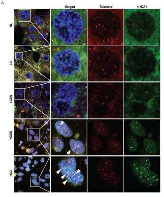
Immunocytochemistry/ Immunofluorescence: Histone H2AX [p Ser139] Antibody [NB100-2280] - Oocyte-specific deletion of Gsk-3 beta disrupted early folliculogenesis in mice. aGsk-3 beta efficiently & specifically deleted in the oocytes in cKO mouse ovary. Immunofluorescence staining for GSK-3 beta (green) & DDX4 (red) on 1 dpp ovary. The nucleus stained by Hoechst (blue). b Control & cKO ovaries at the indicated developmental stages. Oocytes stained w/ DDX4 (green). The nucleus stained by Hoechst (blue). c Statistical analysis showed that the total number of primordial follicle decreased significantly in 7 dpp cKO ovaries (Additional file 8: Individual data values). d Apoptotic cells increased in 1 dpp cKO ovaries compared w/ the control ovaries. TUNEL signals (green) marked apoptotic cells. The nucleus stained by Hoechst (blue). e DSBs persisted in the oocytes of 1 dpp cKO ovaries. The sections stained w/ gamma -H2AX (green) & DDX4 (red). The nucleus stained by Hoechst (blue). f Ectopic RAD51 expression in the oocytes of 1 dpp cKO ovaries. The sections stained w/ RAD51 (green) & DDX4 (red). The nucleus stained by Hoechst (blue). g, h beta -catenin accumulated in the cytoplasm & translocated into nucleus of the oocytes in cKO mice. g The sections stained w/ beta -catenin (green) & DDX4 (red). The nucleus stained by Hoechst (blue). Arrowheads indicated oocytes showing nuclear beta -catenin accumulation. h The statistic analysis demonstrated that the percentage of oocytes showing beta -catenin accumulation per section increased significantly in cKO mice (Additional file 8: Individual data values). The data are presented as mean ± s.d. The asterisk (*) denotes a statistically significant difference between the control & treatment groups. *P < 0.05, **P < 0.01, & ***P < 0.001 (t test). Scale bars, 200 μm Image collected & cropped by CiteAb from the following publication (https://pubmed.ncbi.nlm.nih.gov/30866939), licensed under a CC-BY license. Not internally tested by Novus Biologicals.![Histone H2AX [p Ser139] Antibody](https://resources.rndsystems.com/images/products/nb100-2280_rabbit-polyclonal-histone-h2ax-p-ser139-antibody-31020241612641.jpg "Immunohistochemistry: Histone H2AX [p Ser139] Antibody [NB100-2280] -")
Immunohistochemistry: Histone H2AX [p Ser139] Antibody [NB100-2280] -
Immunohistochemistry: Histone H2AX [p Ser139] Antibody [NB100-2280] - IDO1 inhibitor enhances the efficacy of radiotherapy in vivo. 1 × 10⁵ of SiHa tumorsphere cells were subcutaneously injected to nude mice for tumor growth. After the tumor volume reached 50 mm3, the mice were divided into four groups of non-treated (Ctrl), INCB-024360 treated (INCB), radiotherapy (Rx), or INCB-024360 plus radiotherapy (INCB + Rx). For the INCB or INCB + Rx group, mice were injected once with 50 mg/kg INCB-024360 intraperitoneally before radiotherapy. For the Rx or INCB + Rx group, mice received 2 Gy radiation per day for total 10 Gy. Mice were sacrificed at day 30 after the last radiation treatment & the xenografted tumors were taken out for picturing (A) & weighting (B). The expression of Ki-67 or p-gamma H2AXser139 was determined by paraffin section followed by immunohistochemical staining (C). The inserted bars indicated 50 μM. The quantification results were performed by TissueFAX software (D). * p < 0.05; ** p < 0.01. The experiments were repeated two times & data from one experiment were presented. Image collected & cropped by CiteAb from the following publication (https://pubmed.ncbi.nlm.nih.gov/32545442), licensed under a CC-BY license. Not internally tested by Novus Biologicals.![Histone H2AX [p Ser139] Antibody](https://resources.rndsystems.com/images/products/nb100-2280_rabbit-polyclonal-histone-h2ax-p-ser139-antibody-31020241612651.jpg "Immunocytochemistry/ Immunofluorescence: Histone H2AX [p Ser139] Antibody [NB100-2280] -")
Immunocytochemistry/ Immunofluorescence: Histone H2AX [p Ser139] Antibody [NB100-2280] -
Immunocytochemistry/ Immunofluorescence: Histone H2AX [p Ser139] Antibody [NB100-2280] - Premature upregulation of TAp63 in GSK-3 beta -inhibited ovary. a Expression pattern of gamma -H2AX in fetal & neonatal mouse ovary in vivo. Mouse ovaries from 13.5 dpc, 15.5 dpc, 17.5 dpc, & 1 dpp immunostained for gamma -H2AX (green) & DDX4 (red). Nucleus stained by Hoechst (blue). gamma -H2AX displayed intensive expression in germ cell nucleus from 15.5 to 17.5 dpc. b Expression pattern of TAp63 in fetal & neonatal mouse ovary in vivo. Mouse ovaries from 13.5 dpc, 15.5 dpc, 17.5 dpc, 18.5 dpc, & 1 dpp immunostained for TAp63 (green) & DDX4 (red). Nucleus stained by Hoechst (blue). TAp63 protein located w/in somatic cells in fetal ovary & began to express in germ cell nucleus from 18.5 dpc afterward. c–e TAp63 expression upregulated in fetal ovary & displayed premature localization w/in oocyte nucleus following GSK-3 beta inhibition. Before examination, ovaries of 14.5 dpc cultured in vitro w/ DMSO or BIO for 3 days. c qRT-PCR analysis of TAp63 mRNA level following GSK-3 beta inhibition (normalized to beta -actin) (Additional file 8: Individual data values). d WB analysis of TAp63 protein level following GSK-3 beta inhibition. GAPDH used as internal control. e Immunofluorescence staining for TAp63 (green) & DDX4 (red). The nucleus stained by Hoechst (blue). TAp63 showed premature expression w/in oocyte nucleus following GSK-3 beta inhibition (arrowhead). f qRT-PCR results showed that relative mRNA expression level of p21, Bad, & Noxa increased significantly in GSK-3 beta -inhibiting ovaries (Additional file 8: Individual data values). data are presented as mean ± s.d. The asterisk (*) denotes a statistically significant difference between the control & treatment groups. *P < 0.05, **P < 0.01, & ***P < 0.001 (t test). Scale bars, 200 μm Image collected & cropped by CiteAb from the following publication (https://pubmed.ncbi.nlm.nih.gov/30866939), licensed under a CC-BY license. Not internally tested by Novus Biologicals.Applications for Histone H2AX [p Ser139] Antibody
Application
Recommended Usage
Immunocytochemistry/ Immunofluorescence
1:100 - 1:500
Immunohistochemistry-Paraffin
1:100-1:500
Simple Western
1:20
Western Blot
1:100-1:2000
Application Notes
Epitope exposure is recommended. Epitope exposure with citrate buffer will enhance staining. Likely to work with frozen sections. Use in WB reported in scientific literature ( PMID 24415760). Use in IHC-Frozen reported in scientific literature (PMID 26577699).
In Simple Western only 10 - 15 uL of the recommended dilution is used per data point.
See Simple Western Antibody Database for Simple Western validation: Tested in Jurkat lysate, separated by Size, antibody dilution of 1:20, apparent MW was 30 kDa. Separated by Size-Wes, Sally Sue/Peggy Sue.
In Simple Western only 10 - 15 uL of the recommended dilution is used per data point.
See Simple Western Antibody Database for Simple Western validation: Tested in Jurkat lysate, separated by Size, antibody dilution of 1:20, apparent MW was 30 kDa. Separated by Size-Wes, Sally Sue/Peggy Sue.
Reviewed Applications
Read 1 review rated 5 using NB100-2280 in the following applications:
Formulation, Preparation, and Storage
Purification
Immunogen affinity purified
Formulation
TBS and 0.1% BSA
Preservative
0.09% Sodium Azide
Concentration
0.1 mg/ml
Shipping
The product is shipped with polar packs. Upon receipt, store it immediately at the temperature recommended below.
Stability & Storage
Store at 4C. Do not freeze.
Background: Histone H2AX
References
1. Palla, V. V., Karaolanis, G., Katafigiotis, I., Anastasiou, I., Patapis, P., Dimitroulis, D., & Perrea, D. (2017). gamma-H2AX: Can it be established as a classical cancer prognostic factor?. Tumour biology : the journal of the International Society for Oncodevelopmental Biology and Medicine. https://doi.org/10.1177/1010428317695931
2. Kuo, L. J., & Yang, L. X. (2008). Gamma-H2AX - a novel biomarker for DNA double-strand breaks. In vivo (Athens, Greece).
3. Kinner, A., Wu, W., Staudt, C., & Iliakis, G. (2008). Gamma-H2AX in recognition and signaling of DNA double-strand breaks in the context of chromatin. Nucleic acids research. https://doi.org/10.1093/nar/gkn550
4. Redon, C. E., Weyemi, U., Parekh, P. R., Huang, D., Burrell, A. S., & Bonner, W. M. (2012). gamma-H2AX and other histone post-translational modifications in the clinic. Biochimica et biophysica acta. https://doi.org/10.1016/j.bbagrm.2012.02.021
5. H2AX: Uniprot (P16104)
Additional Histone H2AX Products
Product Documents for Histone H2AX [p Ser139] Antibody
Certificate of Analysis
To download a Certificate of Analysis, please enter a lot or batch number in the search box below.
Product Specific Notices for Histone H2AX [p Ser139] Antibody
Licensed to Novus Biologicals LLC under U.S. Patent Nos. 6,362,317 and 6,884,873.
This product is for research use only and is not approved for use in humans or in clinical diagnosis. Primary Antibodies are guaranteed for 1 year from date of receipt.
Related Research Areas
Citations for Histone H2AX [p Ser139] Antibody
 Powered by Bioz
Customer Reviews for Histone H2AX [p Ser139] Antibody (1)
5 out of 5
1 Customer Rating
Have you used Histone H2AX [p Ser139] Antibody?
Submit a review and receive an Amazon gift card!
$25/€18/£15/$25CAN/¥2500 Yen for a review with an image
$10/€7/£6/$10CAN/¥1110 Yen for a review without an image
Submit a review
Customer Images
![Histone H2AX [p Ser139] Antibody NB100-2280](https://resources.rndsystems.com/images/reviews/Immunohistochemisty-Paraffin__NB100-2280_21811.png)
Showing
1
-
1 的
1 review
Showing All
Filter By:
-
Application: Immunohistochemistry-ParaffinSample Tested: Ramos lymphomaSpecies: HumanVerified Customer | Posted 10/06/2015Lymphoma treated with DNA damaging agent
![Histone H2AX [p Ser139] Antibody NB100-2280](data:image/png;base64,R0lGODlhAQABAAD/ACwAAAAAAQABAAACADs=)
There are no reviews that match your criteria.
Protocols
Find general support by application which include: protocols, troubleshooting, illustrated assays, videos and webinars.
- Antigen Retrieval Protocol (PIER)
- Antigen Retrieval for Frozen Sections Protocol
- Appropriate Fixation of IHC/ICC Samples
- Cellular Response to Hypoxia Protocols
- Chromogenic IHC Staining of Formalin-Fixed Paraffin-Embedded (FFPE) Tissue Protocol
- Chromogenic Immunohistochemistry Staining of Frozen Tissue
- ClariTSA™ Fluorophore Kits
- Detection & Visualization of Antibody Binding
- Fluorescent IHC Staining of Frozen Tissue Protocol
- Graphic Protocol for Heat-induced Epitope Retrieval
- Graphic Protocol for the Preparation and Fluorescent IHC Staining of Frozen Tissue Sections
- Graphic Protocol for the Preparation and Fluorescent IHC Staining of Paraffin-embedded Tissue Sections
- Graphic Protocol for the Preparation of Gelatin-coated Slides for Histological Tissue Sections
- ICC Cell Smear Protocol for Suspension Cells
- ICC Immunocytochemistry Protocol Videos
- ICC for Adherent Cells
- IHC Sample Preparation (Frozen sections vs Paraffin)
- Immunocytochemistry (ICC) Protocol
- Immunocytochemistry Troubleshooting
- Immunofluorescence of Organoids Embedded in Cultrex Basement Membrane Extract
- Immunofluorescent IHC Staining of Formalin-Fixed Paraffin-Embedded (FFPE) Tissue Protocol
- Immunohistochemistry (IHC) and Immunocytochemistry (ICC) Protocols
- Immunohistochemistry Frozen Troubleshooting
- Immunohistochemistry Paraffin Troubleshooting
- Preparing Samples for IHC/ICC Experiments
- Preventing Non-Specific Staining (Non-Specific Binding)
- Primary Antibody Selection & Optimization
- Protocol for Heat-Induced Epitope Retrieval (HIER)
- Protocol for Making a 4% Formaldehyde Solution in PBS
- Protocol for VisUCyte™ HRP Polymer Detection Reagent
- Protocol for the Fluorescent ICC Staining of Cell Smears - Graphic
- Protocol for the Fluorescent ICC Staining of Cultured Cells on Coverslips - Graphic
- Protocol for the Preparation & Fixation of Cells on Coverslips
- Protocol for the Preparation and Chromogenic IHC Staining of Frozen Tissue Sections
- Protocol for the Preparation and Chromogenic IHC Staining of Frozen Tissue Sections - Graphic
- Protocol for the Preparation and Chromogenic IHC Staining of Paraffin-embedded Tissue Sections
- Protocol for the Preparation and Chromogenic IHC Staining of Paraffin-embedded Tissue Sections - Graphic
- Protocol for the Preparation and Fluorescent ICC Staining of Cells on Coverslips
- Protocol for the Preparation and Fluorescent ICC Staining of Non-adherent Cells
- Protocol for the Preparation and Fluorescent ICC Staining of Stem Cells on Coverslips
- Protocol for the Preparation and Fluorescent IHC Staining of Frozen Tissue Sections
- Protocol for the Preparation and Fluorescent IHC Staining of Paraffin-embedded Tissue Sections
- Protocol for the Preparation of Gelatin-coated Slides for Histological Tissue Sections
- Protocol for the Preparation of a Cell Smear for Non-adherent Cell ICC - Graphic
- R&D Systems Quality Control Western Blot Protocol
- TUNEL and Active Caspase-3 Detection by IHC/ICC Protocol
- The Importance of IHC/ICC Controls
- Troubleshooting Guide: Immunohistochemistry
- Troubleshooting Guide: Western Blot Figures
- Western Blot Conditions
- Western Blot Protocol
- Western Blot Protocol for Cell Lysates
- Western Blot Troubleshooting
- Western Blot Troubleshooting Guide
- View all Protocols, Troubleshooting, Illustrated assays and Webinars
FAQs for Histone H2AX [p Ser139] Antibody
Showing
1
-
1 的
1 FAQ
Showing All
-
Q: Could you direct me where to find secondary antibodies for NB100-2280 that would be appropriate for certain applications?
A: By clicking on the "Support & Research" tab on NB100-2280's product page under the "Product Details" display, you will find a link to view all anti-Rabbit secondary antibodies. This will lead you to a new page that will allow you to filter products by application.
Loading...