
ARX (Aristaless-Related homeoboX protein) is a 56-62 kDa member of the aristaless subfamily, paired homeobox family of proteins. In the nervous system, it is expressed in embryonic telencephalon plus adult cortical GABAergic neurons and differentiating neuronal stem cells; in the pancreas, it promotes the differentiation of alpha -cells at the expense of beta -cells. Human ARX is a 562 amino acid (aa) transcriptional repressor. It contains an octapeptide motif that binds Groucho proteins (aa 27‑34), an NLS (aa 82-89), one Ala-rich region (aa 100-155), an acidic/Glu-rich region (aa 224-253), a DNA-binding homeodomain with an embedded NLS (aa 328‑387), one Pro-rich segment that binds CtBP1 (aa 395-459) and a C-terminal Aristaless domain (aa 530-543). There are mutations that show poly-Ala expansions in the N-terminus (aa 115-155), often associated with developmental abnormalities. Over aa 1-100, human ARX shares 97% aa identity with mouse ARX.

Scientific images may be resized, cropped, or adjusted for brightness or contrast for presentation purposes. These updates are limited to presentation and do not affect the underlying scientific interpretation of the data. Where available, additional source data may be provided upon request.
Key Product Details
Species Reactivity
Validated:
Cited:
Applications
Validated:
Cited:
Label
Antibody Source
Product Specifications
Immunogen
Ser2-Ala100
Accession # Q96QS3
Specificity
Clonality
Host
Isotype
Scientific Data Images for Human ARX Antibody
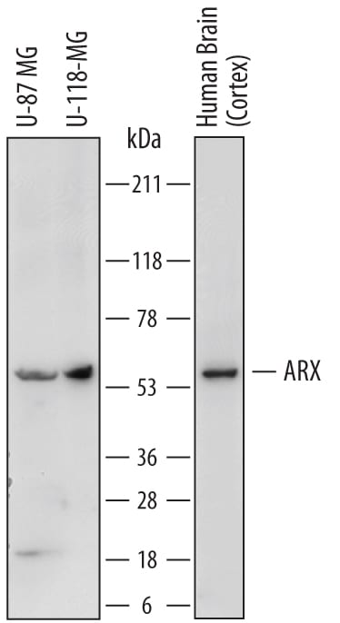
Detection of Human ARX by Western Blot.
Western blot shows lysates of U-87 MG human glioblastoma/astrocytoma cell line, U-118-MG human glioblastoma/astrocytoma cell line, and human brain (cortex) tissue. PVDF membrane was probed with 2 µg/mL of Sheep Anti-Human ARX Antigen Affinity-purified Polyclonal Antibody (Catalog # AF7068) followed by HRP-conjugated Anti-Sheep IgG Secondary Antibody (Catalog # HAF016). A specific band was detected for ARX at approximately 58 kDa (as indicated). This experiment was conducted under reducing conditions and using Immunoblot Buffer Group 1.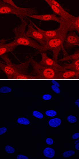
ARX in U‑87 MG Human Cell Line.
ARX was detected in immersion fixed U-87 MG human glioblastoma/astrocytoma cell line using Sheep Anti-Human ARX Antigen Affinity-purified Polyclonal Antibody (Catalog # AF7068) at 10 µg/mL for 3 hours at room temperature. Cells were stained using the NorthernLights™ 557-conjugated Anti-Sheep IgG Secondary Antibody (red, upper panel; Catalog # NL010) and counterstained with DAPI (blue, lower panel). Specific staining was localized to nuclei. View our protocol for Fluorescent ICC Staining of Cells on Coverslips.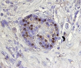
ARX in Human Pancreas.
ARX was detected in immersion fixed paraffin-embedded sections of human pancreas using Sheep Anti-Human ARX Antigen Affinity-purified Polyclonal Antibody (Catalog # AF7068) at 15 µg/mL overnight at 4 °C. Tissue was stained using the Anti-Sheep HRP-DAB Cell & Tissue Staining Kit (brown; Catalog # CTS019) and counterstained with hematoxylin (blue). Specific staining was localized to nuclei of pancreatic islets. View our protocol for Chromogenic IHC Staining of Paraffin-embedded Tissue Sections.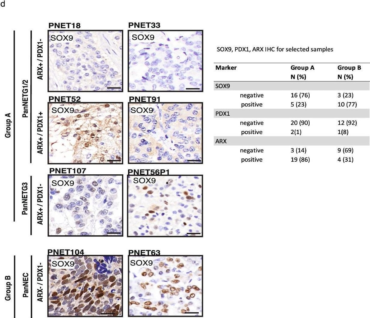
Detection of ARX by Immunohistochemistry
Cell-of-origin analysis using normal cell type methylation profiles and SOX9 in PanNEN subgroups indicate exocrine lineage for Group B tumors. a (i) Phylo-epigenetic analysis of PanNEN tumors and normal pancreatic cell types. Pearson distance between the samples computed using differentially methylated CpGs between normal alpha -, beta -, ductal and acinar cells (n = 46,500, adj. p value < 0.01 and | delta beta | > 0.2) and neighbor-joining tree estimation. (ii) IHC of outliers PNET4 and PNET 60, which were re-assessed and removed from phylo-epigenetic analysis (i). (iii) New phylo-epigenetic analysis without PNET4 and PNET60. b Euclidean distance between each cell type was computed and correlation matrix of the distances is displayed. Heatmap depicts the distance between a given normal cell-type pair. c Boxplot representing distribution of the proportion of atlas signatures of alpha -, beta -, ductal and acinar cells (each main box) in the subgroups and PDACs; each dot depicts the proportion of atlas signatures of the respective cell type in a given sample. Two-sample Wilcoxon test. d IHC of SOX9. Representative images for each subgroup; scale bar: 20μm. Table depicts the total IHC score for Group A and Group B samples Image collected and cropped by CiteAb from the following open publication (https://pubmed.ncbi.nlm.nih.gov/35227293), licensed under a CC-BY license. Not internally tested by R&D Systems.Applications for Human ARX Antibody
Immunocytochemistry
Sample: Immersion fixed U‑87 MG human glioblastoma/astrocytoma cell line
Immunohistochemistry
Sample: Immersion fixed paraffin-embedded sections of human pancreas
Western Blot
Sample: U‑87 MG human glioblastoma/astrocytoma cell line, U‑118‑MG human glioblastoma/astrocytoma cell line, and human brain (cortex) tissue
Formulation, Preparation, and Storage
Purification
Reconstitution
Sterile PBS to a final concentration of 0.2 mg/mL. For liquid material, refer to CoA for concentration.
Formulation
Shipping
Stability & Storage
- 12 months from date of receipt, -20 to -70 °C as supplied.
- 1 month, 2 to 8 °C under sterile conditions after reconstitution.
- 6 months, -20 to -70 °C under sterile conditions after reconstitution.
Calculators
Background: ARX
Long Name
Alternate Names
Gene Symbol
UniProt
Additional ARX Products
Product Documents for Human ARX Antibody
Certificate of Analysis
To download a Certificate of Analysis, please enter a lot or batch number in the search box below.
Note: Certificate of Analysis not available for kit components.
Product Specific Notices for Human ARX Antibody
For research use only
Related Research Areas
Citations for Human ARX Antibody
 Powered by Bioz
Customer Reviews for Human ARX Antibody
There are currently no reviews for this product. Be the first to review Human ARX Antibody and earn rewards!
Have you used Human ARX Antibody?
Submit a review and receive an Amazon gift card!
$25/€18/£15/$25CAN/¥2500 for a review with an image
$10/€7/£6/$10CAN/¥1110 for a review without an image
Submit a review
Protocols
Find general support by application which include: protocols, troubleshooting, illustrated assays, videos and webinars.
- Antigen Retrieval Protocol (PIER)
- Antigen Retrieval for Frozen Sections Protocol
- Appropriate Fixation of IHC/ICC Samples
- Cellular Response to Hypoxia Protocols
- Chromogenic IHC Staining of Formalin-Fixed Paraffin-Embedded (FFPE) Tissue Protocol
- Chromogenic Immunohistochemistry Staining of Frozen Tissue
- ClariTSA™ Fluorophore Kits
- Detection & Visualization of Antibody Binding
- Fluorescent IHC Staining of Frozen Tissue Protocol
- Graphic Protocol for Heat-induced Epitope Retrieval
- Graphic Protocol for the Preparation and Fluorescent IHC Staining of Frozen Tissue Sections
- Graphic Protocol for the Preparation and Fluorescent IHC Staining of Paraffin-embedded Tissue Sections
- Graphic Protocol for the Preparation of Gelatin-coated Slides for Histological Tissue Sections
- ICC Cell Smear Protocol for Suspension Cells
- ICC Immunocytochemistry Protocol Videos
- ICC for Adherent Cells
- IHC Sample Preparation (Frozen sections vs Paraffin)
- Immunocytochemistry (ICC) Protocol
- Immunocytochemistry Troubleshooting
- Immunofluorescence of Organoids Embedded in Cultrex Basement Membrane Extract
- Immunofluorescent IHC Staining of Formalin-Fixed Paraffin-Embedded (FFPE) Tissue Protocol
- Immunohistochemistry (IHC) and Immunocytochemistry (ICC) Protocols
- Immunohistochemistry Frozen Troubleshooting
- Immunohistochemistry Paraffin Troubleshooting
- Preparing Samples for IHC/ICC Experiments
- Preventing Non-Specific Staining (Non-Specific Binding)
- Primary Antibody Selection & Optimization
- Protocol for Heat-Induced Epitope Retrieval (HIER)
- Protocol for Making a 4% Formaldehyde Solution in PBS
- Protocol for VisUCyte™ HRP Polymer Detection Reagent
- Protocol for the Fluorescent ICC Staining of Cell Smears - Graphic
- Protocol for the Fluorescent ICC Staining of Cultured Cells on Coverslips - Graphic
- Protocol for the Preparation & Fixation of Cells on Coverslips
- Protocol for the Preparation and Chromogenic IHC Staining of Frozen Tissue Sections
- Protocol for the Preparation and Chromogenic IHC Staining of Frozen Tissue Sections - Graphic
- Protocol for the Preparation and Chromogenic IHC Staining of Paraffin-embedded Tissue Sections
- Protocol for the Preparation and Chromogenic IHC Staining of Paraffin-embedded Tissue Sections - Graphic
- Protocol for the Preparation and Fluorescent ICC Staining of Cells on Coverslips
- Protocol for the Preparation and Fluorescent ICC Staining of Non-adherent Cells
- Protocol for the Preparation and Fluorescent ICC Staining of Stem Cells on Coverslips
- Protocol for the Preparation and Fluorescent IHC Staining of Frozen Tissue Sections
- Protocol for the Preparation and Fluorescent IHC Staining of Paraffin-embedded Tissue Sections
- Protocol for the Preparation of Gelatin-coated Slides for Histological Tissue Sections
- Protocol for the Preparation of a Cell Smear for Non-adherent Cell ICC - Graphic
- R&D Systems Quality Control Western Blot Protocol
- TUNEL and Active Caspase-3 Detection by IHC/ICC Protocol
- The Importance of IHC/ICC Controls
- Troubleshooting Guide: Immunohistochemistry
- Troubleshooting Guide: Western Blot Figures
- Western Blot Conditions
- Western Blot Protocol
- Western Blot Protocol for Cell Lysates
- Western Blot Troubleshooting
- Western Blot Troubleshooting Guide
- View all Protocols, Troubleshooting, Illustrated assays and Webinars