
Human C5L2/GPR77 Antibody (650918)
R&D Systems | Catalog # MAB10254


Scientific images may be resized, cropped, or adjusted for brightness or contrast for presentation purposes. These updates are limited to presentation and do not affect the underlying scientific interpretation of the data. Where available, additional source data may be provided upon request.
Key Product Details
Species Reactivity
Validated:
Human
Cited:
Human
Applications
Validated:
Flow Cytometry, CyTOF-ready
Cited:
Flow Cytometry
Label
Unconjugated
Antibody Source
Monoclonal Mouse IgG2B Clone # 650918
Loading...
Product Specifications
Immunogen
NS0 mouse myeloma cell line transfected with human C5L2/GPR77
Met1-Val337
Accession # Q9P296
Met1-Val337
Accession # Q9P296
Specificity
Detects human C5L2/GPR77 in direct ELISAs.
Clonality
Monoclonal
Host
Mouse
Isotype
IgG2B
Scientific Data Images for Human C5L2/GPR77 Antibody (650918)
Detection of C5L2/GPR77 in HEK293 Human Cell Line Transfected with Human C5L2/GPR77 and eGFP by Flow Cytometry.
HEK293 human embryonic kidney cell line transfected with (A) C5L2/GPR77 or (B) irrelevant protein, and eGFP were stained with Mouse Anti-Human C5L2/GPR77 Monoclonal Antibody (Catalog # MAB10254) followed by Allophycocyanin-conjugated Anti-Mouse IgG Secondary Antibody (Catalog # F0101B). Quadrant markers were set based Mouse IgG2B Isotype Control Antibody staining (Catalog # MAB0041, data not shown). View our protocol for Staining Membrane-associated Proteins.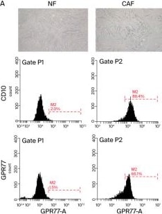
Detection of C5L2/GPR77 by Flow Cytometry
Characterization and functional analysis of CAFs in CRC. (A) Flow cytometric analysis illustrating the identification of fibroblasts isolated from samples from CRC patients using the markers CD10 and GPR77, highlighting the heterogeneity within the CAF population. The experiment was conducted in triplicate to ensure the reliability and reproducibility of the results. (B) Morphological examination of CAFs through light microscopy, depicting their elongated spindle or stellate shapes with thin cytoplasmic extensions and prominent actin stress fibers. The adjacent panels show the immunofluorescent staining results for alpha -SMA and vimentin with a comparison of the expression levels in CAFs and NFs, demonstrating significantly higher expression in CAFs; magnification, ×400. (C) Gene expression microarray data showcasing significant changes in the expression profiles of the proteins, including chemokines and cytokine receptors, secreted by CAFs compared with NFs, with alterations marked as greater-than-five-fold increases. (D) Additional microarray results focusing on distinct clusters of upregulated genes related to tumor invasion and metastasis in CAFs. (E) qRT-PCR results displaying elevated mRNA levels of vimentin, fibroblast activation protein, alpha -SMA, SDF-1, and CXCR4 in CAFs extracted from CRC tissues; these were corroborated by corresponding increases in protein levels, reinforcing the pro-tumorigenic role of these markers in the tumor microenvironment. This experiment was conducted in triplicate to ensure the reliability and reproducibility of the results. (Scale bar: 50 μm). Data are shown as representative images and numerical data are represented as the mean ± SD of each group of cells from three separate experiments. *** p < 0.0001. Image collected and cropped by CiteAb from the following open publication (https://pubmed.ncbi.nlm.nih.gov/39195225), licensed under a CC-BY license. Not internally tested by R&D Systems.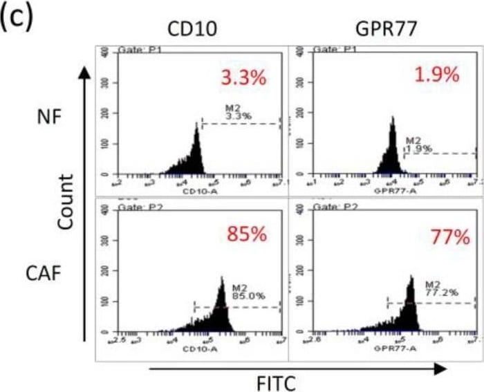
Detection of C5L2/GPR77 by Flow Cytometry
Characterization of CAFs and NFs obtained from clinical surgical tissues from patients with head and neck cancer (HNC) (a) Morphological comparisons between CAFs and NFs from a representative HNC case showed that CAFs (right panel) consisted of more cytoplasmic protrusions than NFs (left panel). Photographs were captured at 40× magnification. (b) Quantitative PCR (left panel) of the culture medium showed a significantly higher expression of vimentin and alpha -SMA in CAFs than in NFs. Western blot analysis (right panel) also demonstrated that levels of vimentin and alpha -SMA were significantly higher in CAFs than in NFs. (c) Flow cytometric analysis of the cell surface markers, CD10 and GPR77, showed a marked increase in their expression in CAFs compared to NFs. (d) A heat map of the gene microarray of NFs and CAFs showed that there were several differences in the expression profile of the secreted genes. The arrow indicates a marked discrepancy of the relative mRNA levels of CCL11 in CAF compared with NF. (e) The RT-PCR (left panel) and ELISA (right panel) analysis showed an increased expression of CCL11 in CAFs compared with that in NFs. (f) Western blot analysis showed that the protein level of CCL11 was higher in CAFs than in NFs in cell lysates. (g) Western blot analysis showed a higher CCL11 expression in CAF-CM compared to NF-CM. The asterisk indicated a significant difference (*: p < 0.05; **: p < 0.01) between experimental and control groups. Image collected and cropped by CiteAb from the following open publication (https://www.mdpi.com/2072-6694/14/13/3141), licensed under a CC-BY license. Not internally tested by R&D Systems.Applications for Human C5L2/GPR77 Antibody (650918)
Application
Recommended Usage
CyTOF-ready
Ready to be labeled using established conjugation methods. No BSA or other carrier proteins that could interfere with conjugation.
Flow Cytometry
0.25 µg/106 cells
Sample: HEK293 Human Cell Line Transfected with Human C5L2/GPR77 and eGFP
Sample: HEK293 Human Cell Line Transfected with Human C5L2/GPR77 and eGFP
Flow Cytometry Panel Builder
Bio-Techne Knows Flow Cytometry
Save time and reduce costly mistakes by quickly finding compatible reagents using the Panel Builder Tool.
Advanced Features
- Spectra Viewer - Custom analysis of spectra from multiple fluorochromes
- Spillover Popups - Visualize the spectra of individual fluorochromes
- Antigen Density Selector - Match fluorochrome brightness with antigen density

Formulation, Preparation, and Storage
Purification
Protein A or G purified from hybridoma culture supernatant
Reconstitution
Reconstitute at 0.5 mg/mL in sterile PBS. For liquid material, refer to CoA for concentration.
Loading...
Formulation
Lyophilized from a 0.2 μm filtered solution in PBS with Trehalose. *Small pack size (SP) is supplied either lyophilized or as a 0.2 µm filtered solution in PBS.
Shipping
Lyophilized product is shipped at ambient temperature. Liquid small pack size (-SP) is shipped with polar packs. Upon receipt, store immediately at the temperature recommended below.
Stability & Storage
Use a manual defrost freezer and avoid repeated freeze-thaw cycles.
- 12 months from date of receipt, -20 to -70 °C as supplied.
- 1 month, 2 to 8 °C under sterile conditions after reconstitution.
- 6 months, -20 to -70 °C under sterile conditions after reconstitution.
Calculators
Background: C5L2/GPR77
Long Name
C5a Anaphylatoxin Chemotactic Receptor C5L2
Alternate Names
GPR77
Gene Symbol
C5AR2
UniProt
Additional C5L2/GPR77 Products
Product Documents for Human C5L2/GPR77 Antibody (650918)
Certificate of Analysis
To download a Certificate of Analysis, please enter a lot or batch number in the search box below.
Note: Certificate of Analysis not available for kit components.
Product Specific Notices for Human C5L2/GPR77 Antibody (650918)
For research use only
Related Research Areas
Citations for Human C5L2/GPR77 Antibody (650918)
 Powered by Bioz
Customer Reviews for Human C5L2/GPR77 Antibody (650918)
There are currently no reviews for this product. Be the first to review Human C5L2/GPR77 Antibody (650918) and earn rewards!
Have you used Human C5L2/GPR77 Antibody (650918)?
Submit a review and receive an Amazon gift card!
$25/€18/£15/$25CAN/¥2500 for a review with an image
$10/€7/£6/$10CAN/¥1110 for a review without an image
Submit a review
Protocols
Find general support by application which include: protocols, troubleshooting, illustrated assays, videos and webinars.
- 7-Amino Actinomycin D (7-AAD) Cell Viability Flow Cytometry Protocol
- Extracellular Membrane Flow Cytometry Protocol
- Flow Cytometry Protocol for Cell Surface Markers
- Flow Cytometry Protocol for Staining Membrane Associated Proteins
- Flow Cytometry Staining Protocols
- Flow Cytometry Troubleshooting Guide
- Intracellular Flow Cytometry Protocol Using Alcohol (Methanol)
- Intracellular Flow Cytometry Protocol Using Detergents
- Intracellular Nuclear Staining Flow Cytometry Protocol Using Detergents
- Intracellular Staining Flow Cytometry Protocol Using Alcohol Permeabilization
- Intracellular Staining Flow Cytometry Protocol Using Detergents to Permeabilize Cells
- Propidium Iodide Cell Viability Flow Cytometry Protocol
- Protocol for Liperfluo
- Protocol for the Characterization of Human Th22 Cells
- Protocol for the Characterization of Human Th9 Cells
- Protocol: Annexin V and PI Staining by Flow Cytometry
- Protocol: Annexin V and PI Staining for Apoptosis by Flow Cytometry
- Troubleshooting Guide: Fluorokine Flow Cytometry Kits
- View all Protocols, Troubleshooting, Illustrated assays and Webinars
Loading...