
Ectonucleoside triphosphate diphosphohydrolase-3 (NTPDase-3), encoded by the ENTPD3 gene and also known as CD39L3, is an integral membrane protein with an extracellular active site (1). Recombinant human NTPDase-3 was expressed as a protein lacking its N- and C-terminal transmembrane domains, resulting in the secretion of the soluble ectodomain. NTPDase-3 hydrolyzes the beta - and gamma -phosphate residues of nucleotides, preferring ATP, ADP, UTP, and UDP as substrates (1). Through its hydrolysis of extracellular nucleotides, NTPDase-3 plays a role in the regulation of purinergic signaling (2). The enzyme is expressed at its highest levels in brain, pancreas, spleen and prostate tissues (3).
Human CD39L3/ENTPD3 Antibody
R&D Systems | Catalog # AF4400

Scientific images may be resized, cropped, or adjusted for brightness or contrast for presentation purposes. These updates are limited to presentation and do not affect the underlying scientific interpretation of the data. Where available, additional source data may be provided upon request.
Key Product Details
Species Reactivity
Validated:
Human
Cited:
Human
Applications
Validated:
Immunohistochemistry, Western Blot
Cited:
Immunohistochemistry
Label
Unconjugated
Antibody Source
Polyclonal Sheep IgG
Loading...
Product Specifications
Immunogen
Mouse myeloma cell line NS0-derived recombinant human CD39L3/ENTPD3
Gln44-Pro485
Accession # O75355
Gln44-Pro485
Accession # O75355
Specificity
Detects human CD39L3/ENTPD3 in direct ELISAs and Western blots. In direct ELISAs, approximately 50% cross-reactivity with recombinant mouse CD39L3 is observed, and less than 2% cross-reactivity with recombinant human (rh) CD39L1 and rhCD39 is observed.
Clonality
Polyclonal
Host
Sheep
Isotype
IgG
Scientific Data Images for Human CD39L3/ENTPD3 Antibody
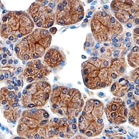
CD39L3/ENTPD3 in Human Salivary Gland.
CD39L3/ENTPD3 was detected in immersion fixed paraffin-embedded sections of human salivary gland using Sheep Anti-Human CD39L3/ENTPD3 Antigen Affinity-purified Polyclonal Antibody (Catalog # AF4400) at 3 µg/mL overnight at 4 °C. Before incubation with the primary antibody, tissue was subjected to heat-induced epitope retrieval using Antigen Retrieval Reagent-Basic (Catalog # CTS013). Tissue was stained using the Anti-Sheep HRP-DAB Cell & Tissue Staining Kit (brown; Catalog # CTS019) and counterstained with hematoxylin (blue). Specific staining was localized to the plasma membranes of epithelial cells. View our protocol for Chromogenic IHC Staining of Paraffin-embedded Tissue Sections.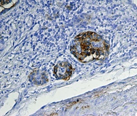
CD39L3/ENTPD3 in Human Pancreas.
CD39L3/ENTPD3 was detected in immersion fixed paraffin-embedded sections of human pancreas using Sheep Anti-Human CD39L3/ENTPD3 Antigen Affinity-purified Polyclonal Antibody (Catalog # AF4400) at 15 µg/mL overnight at 4 °C. Tissue was stained using the Anti-Sheep HRP-DAB Cell & Tissue Staining Kit (brown; Catalog # CTS019) and counterstained with hematoxylin (blue). Specific staining was localized to pancreatic islets. View our protocol for Chromogenic IHC Staining of Paraffin-embedded Tissue Sections.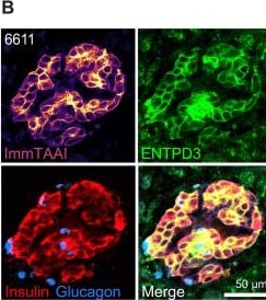
Detection of CD39L3/ENTPD3 by Immunohistochemistry
ImmTAAI binding and labeling of beta cells in pancreas slices is concentration dependent.(A) Dark-field stereomicroscopy image of a pancreas slice showing islets outlined in red. (B) Pancreas slice incubated with CF647-labeled PPI ImmTAAI and subsequently fixed and stained for insulin, glucagon, and ENTPD3, and imaged by confocal. (C) Confocal microscopy images of live pancreas slices incubated with CF647-labeled PPI ImmTAAI at 2, 20, or 200 nM for 1 hour and stained with a labeled primary antibody for cell surface ENTPD3. The off-target CF647-labeled Tel ImmTAAI was used as a control. (D) MFI quantification of confocal microscopy images showing the dose-dependent binding of PPI ImmTAAI to tissue slices. (E) Pearson’s coefficient colocalization analysis of ImmTAAI fluorescent signal with anti-ENTPD3 fluorescent signal. PPI ImmTAAI colocalizes with anti-ENTPD3 well at all three concentrations tested, indicating that beta cell–specific binding is maintained in native tissue. Individual dots represent measurements per islet (means from one donor for Tel ImmTAAI and two donors for PPI ImmTAAI labeled). Statistical differences were determined by two-way analysis of variance (ANOVA) followed by Tukey’s post hoc analysis. ns, not significant; ***P < 0.001, ****P < 0.0001. Image collected and cropped by CiteAb from the following open publication (https://pubmed.ncbi.nlm.nih.gov/41920992), licensed under a CC-BY license. Not internally tested by R&D Systems.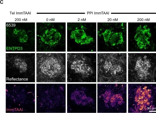
Detection of CD39L3/ENTPD3 by Immunocytochemistry/ Immunofluorescence
ImmTAAI binding and labeling of beta cells in pancreas slices is concentration dependent.(A) Dark-field stereomicroscopy image of a pancreas slice showing islets outlined in red. (B) Pancreas slice incubated with CF647-labeled PPI ImmTAAI and subsequently fixed and stained for insulin, glucagon, and ENTPD3, and imaged by confocal. (C) Confocal microscopy images of live pancreas slices incubated with CF647-labeled PPI ImmTAAI at 2, 20, or 200 nM for 1 hour and stained with a labeled primary antibody for cell surface ENTPD3. The off-target CF647-labeled Tel ImmTAAI was used as a control. (D) MFI quantification of confocal microscopy images showing the dose-dependent binding of PPI ImmTAAI to tissue slices. (E) Pearson’s coefficient colocalization analysis of ImmTAAI fluorescent signal with anti-ENTPD3 fluorescent signal. PPI ImmTAAI colocalizes with anti-ENTPD3 well at all three concentrations tested, indicating that beta cell–specific binding is maintained in native tissue. Individual dots represent measurements per islet (means from one donor for Tel ImmTAAI and two donors for PPI ImmTAAI labeled). Statistical differences were determined by two-way analysis of variance (ANOVA) followed by Tukey’s post hoc analysis. ns, not significant; ***P < 0.001, ****P < 0.0001. Image collected and cropped by CiteAb from the following open publication (https://pubmed.ncbi.nlm.nih.gov/41920992), licensed under a CC-BY license. Not internally tested by R&D Systems.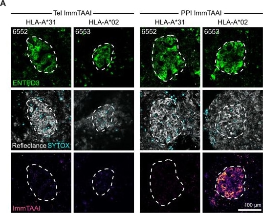
Detection of CD39L3/ENTPD3 by Immunocytochemistry/ Immunofluorescence
ImmTAAI binding is HLA specific in tissue slices.(A) Confocal microscopy images of pancreas slices incubated with 20 nM CF647-labeled PPI ImmTAAI for 2 hours (h) and costained for ENTPD3. No ImmTAAI staining is observed in slices from HLA-A*31 tissue. Islet granularity (reflectance) and viability (SYTOX blue) are also shown. (B) MFI analysis of ImmTAAI binding in HLA-A2–negative and –positive tissue, showing binding only in HLA-A2–positive tissue. (C) Pearson’s coefficient colocalization analysis of ImmTAAI fluorescent signal with anti-ENTPD3 fluorescent signal. ImmTAAI signal colocalizes with ENTPD3 only in HLA-A2–positive tissue. Each dot represents a separate islet from one or two separate slices per condition (n = 1 donor for Tel ImmTAAI in HLA-A*31 tissue, n = 2 donors for HLA-A2) and is the mean of three to six z-planes taken per islet. (D) Confocal microscopy images of pancreas slices treated with a 5-hour pulse of IFN-gamma (100 IU/ml) and then fixed after 24 hours and stained for insulin and HLA-ABC. (E) MFI analysis of HLA-ABC expression in islet and exocrine regions. Each dot represents a separate islet/exocrine region image from ≥three slices per condition. (F) Live confocal microscopy images of pancreas slices treated with a 5-hour pulse of IFN-gamma (100 IU/ml) and incubated with 20 nM CF647-labeled PPI ImmTAAI at 2, 6, or 18 hours. (G) MFI analysis of ImmTAAI signal at each time point post–IFN-gamma treatment. Each dot represents a separate islet from ≥three slices per condition. NT, no treatment. Statistical differences were determined by two-way ANOVA followed by Tukey’s post hoc analysis. ns, not significant. **P < 0.01, ***P < 0.001, ****P < 0.0001. Image collected and cropped by CiteAb from the following open publication (https://pubmed.ncbi.nlm.nih.gov/41920992), licensed under a CC-BY license. Not internally tested by R&D Systems.Applications for Human CD39L3/ENTPD3 Antibody
Application
Recommended Usage
Immunohistochemistry
5-15 µg/mL
Sample: Immersion fixed paraffin-embedded sections of human salivary gland and human pancreas
Sample: Immersion fixed paraffin-embedded sections of human salivary gland and human pancreas
Western Blot
0.1 µg/mL
Sample: Recombinant Human CD39L3/ENTPD3 (Catalog # 4400-EN)
Sample: Recombinant Human CD39L3/ENTPD3 (Catalog # 4400-EN)
Formulation, Preparation, and Storage
Purification
Antigen Affinity-purified
Reconstitution
Sterile PBS to a final concentration of 0.2 mg/mL. For liquid material, refer to CoA for concentration.
Loading...
Formulation
Lyophilized from a 0.2 μm filtered solution in PBS with Trehalose. *Small pack size (SP) is supplied either lyophilized or as a 0.2 µm filtered solution in PBS.
Shipping
Lyophilized product is shipped at ambient temperature. Liquid small pack size (-SP) is shipped with polar packs. Upon receipt, store immediately at the temperature recommended below.
Stability & Storage
Use a manual defrost freezer and avoid repeated freeze-thaw cycles.
- 12 months from date of receipt, -20 to -70 °C as supplied.
- 1 month, 2 to 8 °C under sterile conditions after reconstitution.
- 6 months, -20 to -70 °C under sterile conditions after reconstitution.
Calculators
Background: CD39L3/ENTPD3
References
- Lavoie, E.G. et al. (2004) Biochem. Pharmacol. 67:1917.
- Crawford, P.A. et al. (2007) Arch. Biochem. Biophys. 457:7.
- Chadwick, B.P. and A.M. Frischauf (1998) Genomics 50:357.
Long Name
Ecto-Nucleoside Triphosphate Diphosphohydrolase 3
Alternate Names
ATPDase, CD39L3, ENTPD3, HB6, NTPDase-3
Gene Symbol
ENTPD3
UniProt
Additional CD39L3/ENTPD3 Products
Product Documents for Human CD39L3/ENTPD3 Antibody
Certificate of Analysis
To download a Certificate of Analysis, please enter a lot or batch number in the search box below.
Note: Certificate of Analysis not available for kit components.
Product Specific Notices for Human CD39L3/ENTPD3 Antibody
For research use only
Citations for Human CD39L3/ENTPD3 Antibody
 Powered by Bioz
Customer Reviews for Human CD39L3/ENTPD3 Antibody
There are currently no reviews for this product. Be the first to review Human CD39L3/ENTPD3 Antibody and earn rewards!
Have you used Human CD39L3/ENTPD3 Antibody?
Submit a review and receive an Amazon gift card!
$25/€18/£15/$25CAN/¥2500 for a review with an image
$10/€7/£6/$10CAN/¥1110 for a review without an image
Submit a review
Protocols
Find general support by application which include: protocols, troubleshooting, illustrated assays, videos and webinars.
- Antigen Retrieval Protocol (PIER)
- Antigen Retrieval for Frozen Sections Protocol
- Appropriate Fixation of IHC/ICC Samples
- Cellular Response to Hypoxia Protocols
- Chromogenic IHC Staining of Formalin-Fixed Paraffin-Embedded (FFPE) Tissue Protocol
- Chromogenic Immunohistochemistry Staining of Frozen Tissue
- ClariTSA™ Fluorophore Kits
- Detection & Visualization of Antibody Binding
- Fluorescent IHC Staining of Frozen Tissue Protocol
- Graphic Protocol for Heat-induced Epitope Retrieval
- Graphic Protocol for the Preparation and Fluorescent IHC Staining of Frozen Tissue Sections
- Graphic Protocol for the Preparation and Fluorescent IHC Staining of Paraffin-embedded Tissue Sections
- Graphic Protocol for the Preparation of Gelatin-coated Slides for Histological Tissue Sections
- IHC Sample Preparation (Frozen sections vs Paraffin)
- Immunofluorescent IHC Staining of Formalin-Fixed Paraffin-Embedded (FFPE) Tissue Protocol
- Immunohistochemistry (IHC) and Immunocytochemistry (ICC) Protocols
- Immunohistochemistry Frozen Troubleshooting
- Immunohistochemistry Paraffin Troubleshooting
- Preparing Samples for IHC/ICC Experiments
- Preventing Non-Specific Staining (Non-Specific Binding)
- Primary Antibody Selection & Optimization
- Protocol for Heat-Induced Epitope Retrieval (HIER)
- Protocol for Making a 4% Formaldehyde Solution in PBS
- Protocol for VisUCyte™ HRP Polymer Detection Reagent
- Protocol for the Preparation & Fixation of Cells on Coverslips
- Protocol for the Preparation and Chromogenic IHC Staining of Frozen Tissue Sections
- Protocol for the Preparation and Chromogenic IHC Staining of Frozen Tissue Sections - Graphic
- Protocol for the Preparation and Chromogenic IHC Staining of Paraffin-embedded Tissue Sections
- Protocol for the Preparation and Chromogenic IHC Staining of Paraffin-embedded Tissue Sections - Graphic
- Protocol for the Preparation and Fluorescent IHC Staining of Frozen Tissue Sections
- Protocol for the Preparation and Fluorescent IHC Staining of Paraffin-embedded Tissue Sections
- Protocol for the Preparation of Gelatin-coated Slides for Histological Tissue Sections
- R&D Systems Quality Control Western Blot Protocol
- TUNEL and Active Caspase-3 Detection by IHC/ICC Protocol
- The Importance of IHC/ICC Controls
- Troubleshooting Guide: Immunohistochemistry
- Troubleshooting Guide: Western Blot Figures
- Western Blot Conditions
- Western Blot Protocol
- Western Blot Protocol for Cell Lysates
- Western Blot Troubleshooting
- Western Blot Troubleshooting Guide
- View all Protocols, Troubleshooting, Illustrated assays and Webinars
Loading...