CEACAM-7 (carcinoembryonic antigen cell adhesion molecule 7; also CGM2) is a 40‑45 kDa member of the CEACAM subfamily of the CEA family of proteins. It has a restricted expression pattern, being found on epithelium of the colon, appendix and pancreatic ducts. It is markedly down‑regulated in colon carcinoma. Mature human CEACAM-7 is a 209 amino acid (aa), GPI-linked glycoprotein. It contains one V-type (aa 36‑142) and one C2-type (aa 146‑233) Ig-like domain, plus a 21 aa C‑terminal propeptide. One alternate splice form exists that shows a deletion of aa 143‑235. No definitive rodent CEACAM-7 has been reported.

Key Product Details
Species Reactivity
Human
Applications
Immunohistochemistry, Western Blot
Label
Unconjugated
Antibody Source
Polyclonal Sheep IgG
Loading...
Product Specifications
Immunogen
E. coli-derived recombinant human CEACAM-7
Asn37-Phe142
Accession # Q14002
Asn37-Phe142
Accession # Q14002
Specificity
Detects human CEACAM-7 in direct ELISAs and Western blots. In direct ELISAs and Western blots, less than 15% cross-reactivity with recombinant human (rh) CEACAM-3, rhCEACAM-8, and rhCEACAM-1 is observed and less than 2% cross-reactivity with rhCEACAM-6 and rhCEACAM-5 is observed.
Clonality
Polyclonal
Host
Sheep
Isotype
IgG
Scientific Data Images for Human CEACAM-7 Antibody
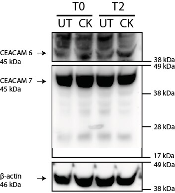
Detection of Human CEACAM‑7 by Western Blot.
Western blot shows lysates of LNCaP human prostate cancer cell line, HeLa human cervical epithelial carcinoma cell line and human liver tissue. PVDF membrane was probed with 1 µg/mL of Sheep Anti-Human CEACAM-7 Antigen Affinity-purified Polyclonal Antibody (Catalog # AF4478) followed by HRP-conjugated Anti-Sheep IgG Secondary Antibody (Catalog # HAF016). A specific band was detected for CEACAM-7 at approximately 45 kDa (as indicated). This experiment was conducted under reducing conditions and using Immunoblot Buffer Group 8.
CEACAM‑7 in Human Colon Cancer Tissue.
CEACAM-7 was detected in immersion fixed paraffin-embedded sections of human colon cancer tissue using Sheep Anti-Human CEACAM-7 Antigen Affinity-purified Polyclonal Antibody (Catalog # AF4478) at 3 µg/mL overnight at 4 °C. Tissue was stained using the Anti-Sheep HRP-DAB Cell & Tissue Staining Kit (brown; Catalog # CTS019) and counterstained with hematoxylin (blue). Specific staining was localized to cell surfaces of cancer cells. View our protocol for Chromogenic IHC Staining of Paraffin-embedded Tissue Sections.Applications for Human CEACAM-7 Antibody
Application
Recommended Usage
Immunohistochemistry
3-15 µg/mL
Sample: Immersion fixed paraffin-embedded sections of human colon cancer tissue
Sample: Immersion fixed paraffin-embedded sections of human colon cancer tissue
Western Blot
1 µg/mL
Sample: LNCaP human prostate cancer cell line, HeLa human cervical epithelial carcinoma cell line and human liver tissue
Sample: LNCaP human prostate cancer cell line, HeLa human cervical epithelial carcinoma cell line and human liver tissue
Reviewed Applications
Read 1 review rated 5 using AF4478 in the following applications:
Formulation, Preparation, and Storage
Purification
Antigen Affinity-purified
Reconstitution
Reconstitute at 0.2 mg/mL in sterile PBS. For liquid material, refer to CoA for concentration.
Loading...
Formulation
Lyophilized from a 0.2 μm filtered solution in PBS with Trehalose. *Small pack size (SP) is supplied either lyophilized or as a 0.2 µm filtered solution in PBS.
Shipping
Lyophilized product is shipped at ambient temperature. Liquid small pack size (-SP) is shipped with polar packs. Upon receipt, store immediately at the temperature recommended below.
Stability & Storage
Use a manual defrost freezer and avoid repeated freeze-thaw cycles.
- 12 months from date of receipt, -20 to -70 °C as supplied.
- 1 month, 2 to 8 °C under sterile conditions after reconstitution.
- 6 months, -20 to -70 °C under sterile conditions after reconstitution.
Calculators
Background: CEACAM-7
Long Name
Carcinoembryonic Antigen-related Cell Adhesion Molecule 7
Alternate Names
CEACAM7, CGM2
Entrez Gene IDs
1087 (Human)
Gene Symbol
CEACAM7
UniProt
Additional CEACAM-7 Products
Product Documents for Human CEACAM-7 Antibody
Certificate of Analysis
To download a Certificate of Analysis, please enter a lot or batch number in the search box below.
Note: Certificate of Analysis not available for kit components.
Product Specific Notices for Human CEACAM-7 Antibody
For research use only
Related Research Areas
Citations for Human CEACAM-7 Antibody
 Powered by Bioz
Customer Reviews for Human CEACAM-7 Antibody (1)
5 out of 5
1 Customer Rating
Have you used Human CEACAM-7 Antibody?
Submit a review and receive an Amazon gift card!
$25/€18/£15/$25CAN/¥2500 Yen for a review with an image
$10/€7/£6/$10CAN/¥1110 Yen for a review without an image
Submit a review
Customer Images
Showing
1
-
1 的
1 review
Showing All
Filter By:
-
Application: Western BlotSample Tested: HT-29 human colon adenocarcinoma cell lineSpecies: HumanVerified Customer | Posted 08/09/2021

There are no reviews that match your criteria.
Protocols
Find general support by application which include: protocols, troubleshooting, illustrated assays, videos and webinars.
- Antigen Retrieval Protocol (PIER)
- Antigen Retrieval for Frozen Sections Protocol
- Appropriate Fixation of IHC/ICC Samples
- Cellular Response to Hypoxia Protocols
- Chromogenic IHC Staining of Formalin-Fixed Paraffin-Embedded (FFPE) Tissue Protocol
- Chromogenic Immunohistochemistry Staining of Frozen Tissue
- ClariTSA™ Fluorophore Kits
- Detection & Visualization of Antibody Binding
- Fluorescent IHC Staining of Frozen Tissue Protocol
- Graphic Protocol for Heat-induced Epitope Retrieval
- Graphic Protocol for the Preparation and Fluorescent IHC Staining of Frozen Tissue Sections
- Graphic Protocol for the Preparation and Fluorescent IHC Staining of Paraffin-embedded Tissue Sections
- Graphic Protocol for the Preparation of Gelatin-coated Slides for Histological Tissue Sections
- IHC Sample Preparation (Frozen sections vs Paraffin)
- Immunofluorescent IHC Staining of Formalin-Fixed Paraffin-Embedded (FFPE) Tissue Protocol
- Immunohistochemistry (IHC) and Immunocytochemistry (ICC) Protocols
- Immunohistochemistry Frozen Troubleshooting
- Immunohistochemistry Paraffin Troubleshooting
- Preparing Samples for IHC/ICC Experiments
- Preventing Non-Specific Staining (Non-Specific Binding)
- Primary Antibody Selection & Optimization
- Protocol for Heat-Induced Epitope Retrieval (HIER)
- Protocol for Making a 4% Formaldehyde Solution in PBS
- Protocol for VisUCyte™ HRP Polymer Detection Reagent
- Protocol for the Preparation & Fixation of Cells on Coverslips
- Protocol for the Preparation and Chromogenic IHC Staining of Frozen Tissue Sections
- Protocol for the Preparation and Chromogenic IHC Staining of Frozen Tissue Sections - Graphic
- Protocol for the Preparation and Chromogenic IHC Staining of Paraffin-embedded Tissue Sections
- Protocol for the Preparation and Chromogenic IHC Staining of Paraffin-embedded Tissue Sections - Graphic
- Protocol for the Preparation and Fluorescent IHC Staining of Frozen Tissue Sections
- Protocol for the Preparation and Fluorescent IHC Staining of Paraffin-embedded Tissue Sections
- Protocol for the Preparation of Gelatin-coated Slides for Histological Tissue Sections
- R&D Systems Quality Control Western Blot Protocol
- TUNEL and Active Caspase-3 Detection by IHC/ICC Protocol
- The Importance of IHC/ICC Controls
- Troubleshooting Guide: Immunohistochemistry
- Troubleshooting Guide: Western Blot Figures
- Western Blot Conditions
- Western Blot Protocol
- Western Blot Protocol for Cell Lysates
- Western Blot Troubleshooting
- Western Blot Troubleshooting Guide
- View all Protocols, Troubleshooting, Illustrated assays and Webinars
Loading...