Complement Factor H is a 155 kDa glycoprotein that provides critical negative regulation to the alternative pathway of complement cascade. It is secreted by Kupffer cells, hepatocytes, vascular endothelial cells, and platelets, and circulates in the serum at high concentration (1). Complement Factor H is composed of 20 SCRs (short consensus repeats), each of which consists of approximately 60 amino acids with four invariant Cys residues (2). Alternate splicing generates an isoform that is truncated following SCR7. Complement Factor H interacts with cell surface polyanions including heparin and sialoglycoproteins (3-6), and immobilized Complement Factor H supports the CD11b/CD18 integrin-dependent adhesion of neutrophils (7). It prevents local complement activation by sequestering complement component C3b, accelerating the decay of C3 and C5 convertases, and functions as a cofactor for the C3b inactivator, Factor I (1, 3, 6, 8). The recombinant protein expressed here corresponds to SCR15-20, which encompass the primary binding sites for heparin and C3b, as well as for the peptide hormone adrenomedullin (4, 9‑11). Within SCR15-20, human Complement Factor H shares 60% and 63% amino acid sequence identity with mouse and rat Complement Factor H, respectively. Dozens of mutations clustered in SCR15-20 are associated with atypical hemolytic uremic syndrome, a disorder characterized by anemia, thrombocytopenia, and renal failure (12). Binding of Complement Factor H to tumor cell-associated dentin matrix protein 1, bone sialoprotein, or osteopontin results in the protection of that cell from complement-mediated lysis (13, 14). A variety of pathogenic microbes also express Complement Factor H binding molecules that interfere with immune clearance of the infection (15).
Human Complement Factor H Antibody
R&D Systems | Catalog # AF4779

Key Product Details
Species Reactivity
Validated:
Human
Cited:
Human
Applications
Validated:
Immunohistochemistry, Western Blot
Cited:
Immunohistochemistry, Western Blot
Label
Unconjugated
Antibody Source
Polyclonal Goat IgG
Loading...
Product Specifications
Immunogen
Mouse myeloma cell line NS0-derived recombinant human Complement Factor H
Ser860-Arg1231
Accession # P08603
Ser860-Arg1231
Accession # P08603
Specificity
Detects human Complement Factor H in direct ELISAs and Western blots. In direct ELISAs, less than 5% cross‑reactivity with recombinant mouse Complement Factor H is observed.
Clonality
Polyclonal
Host
Goat
Isotype
IgG
Scientific Data Images for Human Complement Factor H Antibody
Complement Factor H in Human Liver.
Complement Factor H was detected in immersion fixed paraffin-embedded sections of human liver using Goat Anti-Human Complement Factor H Antigen Affinity-purified Polyclonal Antibody (Catalog # AF4779) at 10 µg/mL overnight at 4 °C. Before incubation with the primary antibody tissue was subjected to heat-induced epitope retrieval using Antigen Retrieval Reagent-Basic (Catalog # CTS013). Tissue was stained using the Anti-Goat HRP-DAB Cell & Tissue Staining Kit (brown; Catalog # CTS008) and counterstained with hematoxylin (blue). View our protocol for Chromogenic IHC Staining of Paraffin-embedded Tissue Sections.Applications for Human Complement Factor H Antibody
Application
Recommended Usage
Immunohistochemistry
5-15 µg/mL
Sample: Immersion fixed paraffin-embedded sections of human liver subjected to Antigen Retrieval Reagent-Basic (Catalog # CTS013)
Sample: Immersion fixed paraffin-embedded sections of human liver subjected to Antigen Retrieval Reagent-Basic (Catalog # CTS013)
Western Blot
0.1 µg/mL
Sample: Recombinant Human Complement Factor H aa 860-1231 (Catalog # 4779-FH)
Sample: Recombinant Human Complement Factor H aa 860-1231 (Catalog # 4779-FH)
Reviewed Applications
Read 2 reviews rated 5 using AF4779 in the following applications:
Formulation, Preparation, and Storage
Purification
Antigen Affinity-purified
Reconstitution
Reconstitute at 0.2 mg/mL in sterile PBS. For liquid material, refer to CoA for concentration.
Loading...
Formulation
Lyophilized from a 0.2 μm filtered solution in PBS with Trehalose. *Small pack size (SP) is supplied either lyophilized or as a 0.2 µm filtered solution in PBS.
Shipping
Lyophilized product is shipped at ambient temperature. Liquid small pack size (-SP) is shipped with polar packs. Upon receipt, store immediately at the temperature recommended below.
Stability & Storage
Use a manual defrost freezer and avoid repeated freeze-thaw cycles.
- 12 months from date of receipt, -20 to -70 °C as supplied.
- 1 month, 2 to 8 °C under sterile conditions after reconstitution.
- 6 months, -20 to -70 °C under sterile conditions after reconstitution.
Calculators
Background: Complement Factor H
References
- Schmidt, C.Q. et al. (2008) Clin. Exp. Immunol. 151:14.
- Ripoche, J. et al. (1988) Biochem. J. 249:593.
- Meri, S. and M.K. Pangburn (1990) Proc. Natl. Acad. Sci. USA 87:3982.
- Jokiranta, T.S. et al. (2005) Am. J. Pathol. 167:1173.
- Blackmore, T.K. et al. (1998) J. Immunol. 160:3342.
- Hellwage, J. et al. (2002) J. Immunol. 169:6935.
- DiScipio, R.G. et al. (1998) J. Immunol. 160:4057.
- Sharma, A.K. and M.K. Pangburn (1996) Proc. Natl. Acad. Sci. USA 93:10996.
- Oppermann, M. et al. (2006) Clin. Exp. Immunol. 144:342.
- Pangburn, M.K. et al. (2000) J. Immunol. 164:4742.
- Martinez, A. et al. (2003) Hypertens. Res. 26:S55.
- de Cordoba, S.R. and E.G. de Jorge (2008) Clin. Exp. Immunol. 151:1.
- Jain, A. et al. (2002) J. Biol. Chem. 277:13700.
- Fedarko, N.S. et al. (2000) J. Biol. Chem. 275:16666.
- Kraiczy, P. and R. Wurzner (2006) Mol. Immunol. 43:31.
Alternate Names
ARMD4, ARMS1, beta-1-H-globulin, CFH, CFHL3, FH, FHL1, HF1, HF2, HUS
Gene Symbol
CFH
UniProt
Additional Complement Factor H Products
Product Documents for Human Complement Factor H Antibody
Certificate of Analysis
To download a Certificate of Analysis, please enter a lot or batch number in the search box below.
Note: Certificate of Analysis not available for kit components.
Product Specific Notices for Human Complement Factor H Antibody
For research use only
Related Research Areas
Citations for Human Complement Factor H Antibody
 Powered by Bioz
Customer Reviews for Human Complement Factor H Antibody (2)
5 out of 5
2 Customer Ratings
Have you used Human Complement Factor H Antibody?
Submit a review and receive an Amazon gift card!
$25/€18/£15/$25CAN/¥2500 Yen for a review with an image
$10/€7/£6/$10CAN/¥1110 Yen for a review without an image
Submit a review
Customer Images

Showing
1
-
2 的
2 reviews
Showing All
Filter By:
-
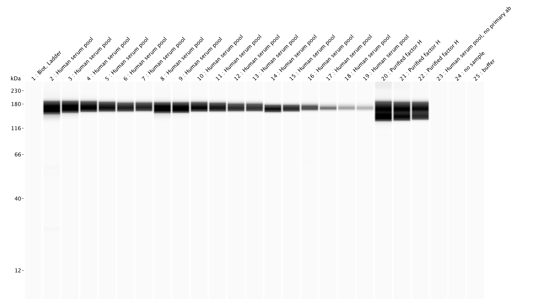
Application: Simple WesternSample Tested: Human serum and Purified proteinSpecies: HumanVerified Customer | Posted 09/25/2019Input samples were dilution series of human serum pool or purified complement factor H. The AF4779 antibody was tested at three different concentrations; 10, 5 and 1 ug/ml (left to right). Samples were analysed by Simple Western under reducing conditions and the 12-230 kDa separation module.

-
Application: Simple WesternSample Tested: Serum and purified complement factor HSpecies: HumanVerified Customer | Posted 09/06/2019Input samples were dilution series of human serum pool or purified complement factor H. The AF4779 antibody was tested at three different concentrations; 10, 5 and 1 ug/ml (left to right). Samples were analysed by Simple Western under reducing conditions and the 12-230 kDa separation module.
There are no reviews that match your criteria.
Protocols
Find general support by application which include: protocols, troubleshooting, illustrated assays, videos and webinars.
- Antigen Retrieval Protocol (PIER)
- Antigen Retrieval for Frozen Sections Protocol
- Appropriate Fixation of IHC/ICC Samples
- Cellular Response to Hypoxia Protocols
- Chromogenic IHC Staining of Formalin-Fixed Paraffin-Embedded (FFPE) Tissue Protocol
- Chromogenic Immunohistochemistry Staining of Frozen Tissue
- ClariTSA™ Fluorophore Kits
- Detection & Visualization of Antibody Binding
- Fluorescent IHC Staining of Frozen Tissue Protocol
- Graphic Protocol for Heat-induced Epitope Retrieval
- Graphic Protocol for the Preparation and Fluorescent IHC Staining of Frozen Tissue Sections
- Graphic Protocol for the Preparation and Fluorescent IHC Staining of Paraffin-embedded Tissue Sections
- Graphic Protocol for the Preparation of Gelatin-coated Slides for Histological Tissue Sections
- IHC Sample Preparation (Frozen sections vs Paraffin)
- Immunofluorescent IHC Staining of Formalin-Fixed Paraffin-Embedded (FFPE) Tissue Protocol
- Immunohistochemistry (IHC) and Immunocytochemistry (ICC) Protocols
- Immunohistochemistry Frozen Troubleshooting
- Immunohistochemistry Paraffin Troubleshooting
- Preparing Samples for IHC/ICC Experiments
- Preventing Non-Specific Staining (Non-Specific Binding)
- Primary Antibody Selection & Optimization
- Protocol for Heat-Induced Epitope Retrieval (HIER)
- Protocol for Making a 4% Formaldehyde Solution in PBS
- Protocol for VisUCyte™ HRP Polymer Detection Reagent
- Protocol for the Preparation & Fixation of Cells on Coverslips
- Protocol for the Preparation and Chromogenic IHC Staining of Frozen Tissue Sections
- Protocol for the Preparation and Chromogenic IHC Staining of Frozen Tissue Sections - Graphic
- Protocol for the Preparation and Chromogenic IHC Staining of Paraffin-embedded Tissue Sections
- Protocol for the Preparation and Chromogenic IHC Staining of Paraffin-embedded Tissue Sections - Graphic
- Protocol for the Preparation and Fluorescent IHC Staining of Frozen Tissue Sections
- Protocol for the Preparation and Fluorescent IHC Staining of Paraffin-embedded Tissue Sections
- Protocol for the Preparation of Gelatin-coated Slides for Histological Tissue Sections
- R&D Systems Quality Control Western Blot Protocol
- TUNEL and Active Caspase-3 Detection by IHC/ICC Protocol
- The Importance of IHC/ICC Controls
- Troubleshooting Guide: Immunohistochemistry
- Troubleshooting Guide: Western Blot Figures
- Western Blot Conditions
- Western Blot Protocol
- Western Blot Protocol for Cell Lysates
- Western Blot Troubleshooting
- Western Blot Troubleshooting Guide
- View all Protocols, Troubleshooting, Illustrated assays and Webinars
Loading...