
Human CXCR4 Antibody (44708)
R&D Systems | Catalog # MAB171

Scientific images may be resized, cropped, or adjusted for brightness or contrast for presentation purposes. These updates are limited to presentation and do not affect the underlying scientific interpretation of the data. Where available, additional source data may be provided upon request.
Key Product Details
Validated by
Biological Validation
Species Reactivity
Validated:
Human
Cited:
Human, Xenograft
Applications
Validated:
Neutralization
Cited:
Immunohistochemistry, Immunohistochemistry-Paraffin, Immunohistochemistry-Frozen, Western Blot, Neutralization, Flow Cytometry, Immunocytochemistry, Binding Assay, FCCS
Label
Unconjugated
Antibody Source
Monoclonal Mouse IgG2A Clone # 44708
Loading...
Product Specifications
Immunogen
3T3 cells transfected with human CXCR4
Specificity
Reacts specifically with human and non-human cells expressing human CXCR4 (fusin) as detected by flow cytometry. It will also react with cells expressing feline CXCR4 but not rat CXCR4. This antibody does not cross-react with other chemokine receptors.
Clonality
Monoclonal
Host
Mouse
Isotype
IgG2A
Endotoxin Level
<0.10 EU per 1 μg of the antibody by the LAL method.
Scientific Data Images for Human CXCR4 Antibody (44708)
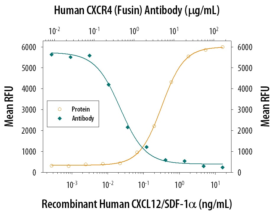
Chemotaxis Induced by CXCL12/SDF‑1 alpha and Neutralization by Human CXCR4 Antibody.
Recombinant Human/Feline/Rhesus Macaque CXCL12/SDF-1a (Catalog # 350-NS) chemoattracts the BaF3 mouse pro-B cell line transfected with human CXCR4 in a dose-dependent manner (orange line). The amount of cells that migrated through to the lower chemotaxis chamber was measured by Resazurin (Catalog # AR002). Chemotaxis elicited by Recombinant Human/Feline/Rhesus Macaque CXCL12/SDF-1a (1 ng/mL) is neutralized (green line) by increasing concentrations of Human CXCR4 Monoclonal Antibody (Catalog # MAB171). The ND50 is typically 0.2-1.2 µg/mL.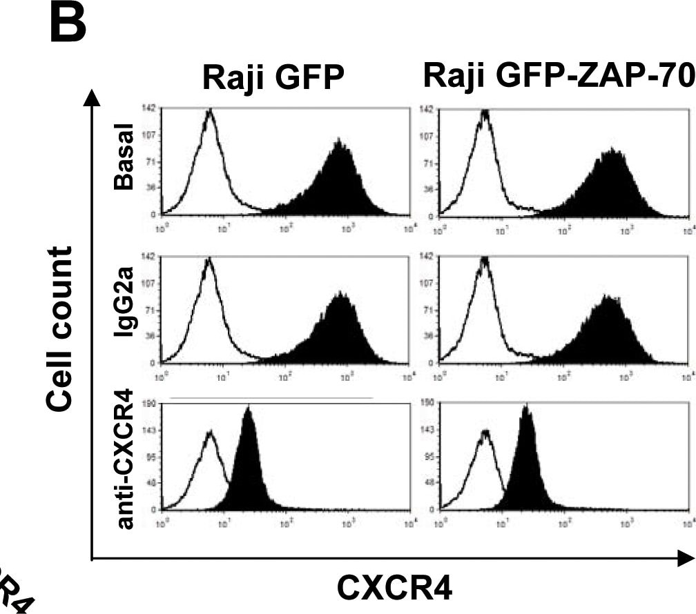
Detection of Mouse CXCR4 by Flow Cytometry
Anti-CXCR4 antibody reduces CXCR4 surface expression, and blocks migration toward CXCL12 and BMSCs.(A) Relative MFIR of CXCR4 surface staining after incubation of Raji transfectants with 100 µg/mL of anti-CXCR4 antibody for 30 minutes. (B) Flow cytometry histograms showing the reduction in CXCR4 expression after incubation with neutralizing anti-CXCR4 antibody. White histograms represent negative unstained controls. (C) CXCR4-blocked cells were stimulated with 100 ng/mL of CXCL12 for 5 minutes and activation of Akt and ERK1/2 proteins analyzed by western blotting. (D) CXCR4-blocked cells were subjected to migration assay for 4 hours at 37°C in 5% CO2 toward 100 ng/mL of CXCL12 (left panel) or the stromal MS-5 cell line (right panel). Cells in the lower chamber were counted by flow cytometry under a defined flow rate for 5 minutes. Number of migrated cells treated with isotypic control was considered 100%. Results are shown as the mean ± SEM of 4 independent experiments. (E) Cell viability was measured by Annexin V-PI staining in cells incubated with 100 µg/mL of anti-CXCR4 or isotypic control for 48 hours. Results are shown as the mean ± SEM of 4 independent experiments Image collected and cropped by CiteAb from the following publication (https://dx.plos.org/10.1371/journal.pone.0081221), licensed under a CC-BY license. Not internally tested by R&D Systems.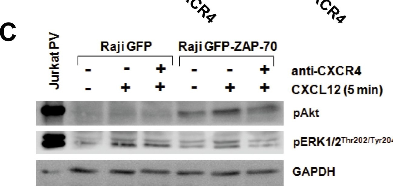
Detection of Mouse CXCR4 by Western Blot
Anti-CXCR4 antibody reduces CXCR4 surface expression, and blocks migration toward CXCL12 and BMSCs.(A) Relative MFIR of CXCR4 surface staining after incubation of Raji transfectants with 100 µg/mL of anti-CXCR4 antibody for 30 minutes. (B) Flow cytometry histograms showing the reduction in CXCR4 expression after incubation with neutralizing anti-CXCR4 antibody. White histograms represent negative unstained controls. (C) CXCR4-blocked cells were stimulated with 100 ng/mL of CXCL12 for 5 minutes and activation of Akt and ERK1/2 proteins analyzed by western blotting. (D) CXCR4-blocked cells were subjected to migration assay for 4 hours at 37°C in 5% CO2 toward 100 ng/mL of CXCL12 (left panel) or the stromal MS-5 cell line (right panel). Cells in the lower chamber were counted by flow cytometry under a defined flow rate for 5 minutes. Number of migrated cells treated with isotypic control was considered 100%. Results are shown as the mean ± SEM of 4 independent experiments. (E) Cell viability was measured by Annexin V-PI staining in cells incubated with 100 µg/mL of anti-CXCR4 or isotypic control for 48 hours. Results are shown as the mean ± SEM of 4 independent experiments Image collected and cropped by CiteAb from the following publication (https://dx.plos.org/10.1371/journal.pone.0081221), licensed under a CC-BY license. Not internally tested by R&D Systems.Applications for Human CXCR4 Antibody (44708)
Application
Recommended Usage
Neutralization
Measured by its ability to neutralize CXCL12/SDF‑1 alpha -induced chemotaxis in the BaF3 mouse pro‑B cell line transfected with human CXCR4. The Neutralization Dose (ND50) is typically 0.2-1.2 µg/mL in the presence of 1 ng/mL Recombinant Human/Feline/Rhesus Macaque CXCL12/SDF‑1 alpha.
Formulation, Preparation, and Storage
Purification
Protein A or G purified from ascites
Reconstitution
Reconstitute at 0.5 mg/mL in sterile PBS. For liquid material, refer to CoA for concentration.
Loading...
Formulation
Lyophilized from a 0.2 μm filtered solution in PBS with Trehalose. *Small pack size (SP) is supplied either lyophilized or as a 0.2 µm filtered solution in PBS.
Shipping
Lyophilized product is shipped at ambient temperature. Liquid small pack size (-SP) is shipped with polar packs. Upon receipt, store immediately at the temperature recommended below.
Stability & Storage
Use a manual defrost freezer and avoid repeated freeze-thaw cycles.
- 12 months from date of receipt, -20 to -70 °C as supplied.
- 1 month, 2 to 8 °C under sterile conditions after reconstitution.
- 6 months, -20 to -70 °C under sterile conditions after reconstitution.
Calculators
Background: CXCR4
References
- Orsini, M.J. et al. (1999) J. Biol. Chem. 274:31076.
- Zagzag, D. et al. (2005) Cancer Res. 65:6178.
- Speetjens, F.M. et al. (2009) Cancer Microenvironment 2:1.
- Wang, L. et al. (2009) Oncology Reports 22:1333.
- Amara, S. et al. (2015) Cancer Biomark. 15:869.
Long Name
C-X-C Motif Chemokine Receptor 4
Alternate Names
CD184, D2S201E, FB22, Fusin, HM89, HSY3RR, LAP-3, LAP3, LCR1, LESTR, NPY3R, NPYRL, NPYY3R, WHIMS
Gene Symbol
CXCR4
Additional CXCR4 Products
Product Documents for Human CXCR4 Antibody (44708)
Certificate of Analysis
To download a Certificate of Analysis, please enter a lot or batch number in the search box below.
Note: Certificate of Analysis not available for kit components.
Product Specific Notices for Human CXCR4 Antibody (44708)
For research use only
Related Research Areas
Citations for Human CXCR4 Antibody (44708)
 Powered by Bioz
Customer Reviews for Human CXCR4 Antibody (44708)
There are currently no reviews for this product. Be the first to review Human CXCR4 Antibody (44708) and earn rewards!
Have you used Human CXCR4 Antibody (44708)?
Submit a review and receive an Amazon gift card!
$25/€18/£15/$25CAN/¥2500 for a review with an image
$10/€7/£6/$10CAN/¥1110 for a review without an image
Submit a review


