
Extracellular matrix protein-1 (ECM-1) is an 85 kDa, secreted glycoprotein important in connective tissue organization (1‑3). Of three identified splice variants the 540 amino acid (aa) form, ECM-1a, is the most widely expressed, with the highest expression in the placenta and heart (2). ECM-1b (415 aa) is found only in tonsil and associated with suprabasal keratinocytes (2, 4). Since ECM-1b expression is differentiation-dependent, a role in terminal keratinocyte differentiation has been suggested (4). ECM-1c (559 aa) accounts for approximately 15% of skin ECM-1 (5). Human ECM-1a contains a 19 aa signal peptide and a 521 aa secreted portion that includes an N-terminal proline-rich, cysteine-free region, two tandem repeat domains, and a C-terminal domain. There are six repeats of a CC(X7 ‑10)C motif (x = any aa) within the tandem repeat and C‑terminal domains. These motifs are involved in ligand binding to members of the albumin family, and are expected to form two (in ECM-1b) or three (in ECM-1a) “double loop” structures (2). Mature human ECM-1a shows 69%, 71%, 72%, and 76% aa identity with corresponding isoforms of mouse, rat, canine, and bovine ECM-1, respectively. ECM-1 is over-expressed in many malignant epithelial tumors and has demonstrated angiogenic activity (6, 7). A variety of ECM-1 mutations, mainly within the first tandem repeat, are considered causative of lipoid proteinosis, a condition showing thickened and irregular extracellular matrix within connective tissue (8). In the autoimmune condition lichen sclerosis, auto-antibodies mainly recognize the second tandem repeat or the C-terminus of ECM-1 (9). These domains also bind the extracellular matrix molecules fibulin-1 and perlecan (5, 10). The phenotypes of lipoid proteinosis and lichen sclerosis support a role for ECM-1 as a “biological glue” in the dermis (1).
Human ECM1 Antibody (966231)
R&D Systems | Catalog # MAB39371

Scientific images may be resized, cropped, or adjusted for brightness or contrast for presentation purposes. These updates are limited to presentation and do not affect the underlying scientific interpretation of the data. Where available, additional source data may be provided upon request.
Key Product Details
Species Reactivity
Validated:
Cited:
Applications
Validated:
Cited:
Label
Antibody Source
Product Specifications
Immunogen
Ala20-Glu540
Accession # Q16610
Specificity
Clonality
Host
Isotype
Scientific Data Images for Human ECM1 Antibody (966231)
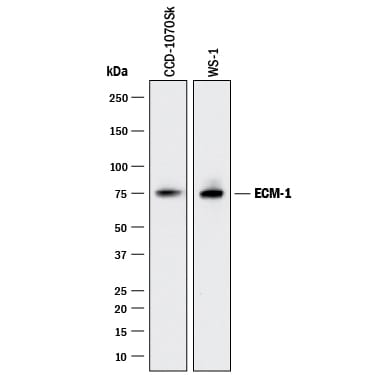
Detection of Human ECM‑1 by Western Blot.
Western blot shows lysates of CCD-1070Sk human foreskin fibroblast cell line and WS-1 human fetal skin fibroblast cell line. PVDF membrane was probed with 2 µg/mL of Mouse Anti-Human ECM-1 Monoclonal Antibody (Catalog # MAB39371) followed by HRP-conjugated Anti-Mouse IgG Secondary Antibody (Catalog # HAF018). A specific band was detected for ECM-1 at approximately 75 kDa (as indicated). This experiment was conducted under reducing conditions and using Immunoblot Buffer Group 3.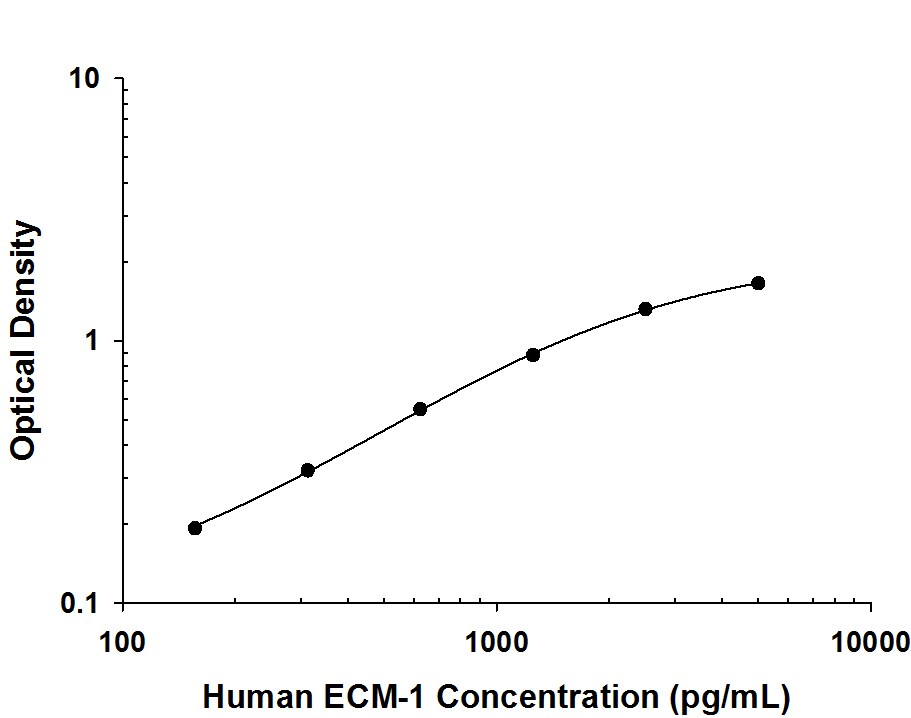
Human ECM‑1 ELISA Standard Curve.
Recombinant human ECM-1 protein was serially diluted 2-fold and captured by Mouse Anti-Human ECM-1 Monoclonal Antibody (Catalog # MAB39371) coated on a Clear Polystyrene Microplate (Catalog # DY990). Mouse Anti-Human ECM-1 Monoclonal Antibody (Catalog # MAB3937) was biotinylated and incubated with the protein captured on the plate. Detection of the standard curve was achieved by incubating Streptavidin-HRP (Catalog # DY998) followed by Substrate Solution (Catalog # DY999) and stopping the enzymatic reaction with Stop Solution (Catalog # DY994).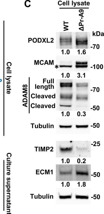
Detection of ECM1 by Western Blot
Knockout (KO) of lnc-FANCI-2 in CaSki cells affects expression of cellular soluble receptors.(A) Dot blots show differentially expressed soluble receptors in delta Pr-A9 cells when compared to those in the wild-type (WT) CaSki cells. A Proteome Profiler Human sReceptor Array was used to examine 105 cellular soluble receptors. Label in red, upregulated receptors; label in blue, downregulated receptors. (B) Quantification of differentially expressed and/or released soluble receptors (A) in delta Pr-A9 cells when compared to those in the WT CaSki cells. Error bars represent standard deviation of replicates in each proteome array. The dashed line indicates the threshold of fold change (FC) with a score above or below the threshold (-/+0.3 FC) being determined as differentially expressed and/or released receptors. (C) Validation of several differentially expressed soluble receptors by immunoblot analysis using specific antibodies. Tubulin served as an internal loading control. *Nonspecific band in MCAM immunoblot.Figure 4—source data 1.Knockout (KO) of lnc-FANCI-2 in CaSki cells affects expression of cellular soluble receptors.Figure 4—source data 2.Knockout (KO) of lnc-FANCI-2 in CaSki cells affects expression of cellular soluble receptors.Knockout (KO) of lnc-FANCI-2 in CaSki cells affects expression of cellular soluble receptors.Knockout (KO) of lnc-FANCI-2 in CaSki cells affects expression of cellular soluble receptors. Image collected and cropped by CiteAb from the following open publication (https://elifesciences.org/articles/102681), licensed under a CC-BY license. Not internally tested by R&D Systems.Applications for Human ECM1 Antibody (966231)
ELISA
This antibody functions as an ELISA capture antibody when paired with Mouse Anti-Human ECM‑1 Monoclonal Antibody (Catalog # MAB3937).
This product is intended for assay development on various assay platforms requiring antibody pairs.
Western Blot
Sample: CCD‑1070Sk human foreskin fibroblast cell line and WS‑1 human fetal skin fibroblast cell line
Formulation, Preparation, and Storage
Purification
Reconstitution
Reconstitute at 0.5 mg/mL in sterile PBS. For liquid material, refer to CoA for concentration.
Formulation
Shipping
Stability & Storage
- 12 months from date of receipt, -20 to -70 °C as supplied.
- 1 month, 2 to 8 °C under sterile conditions after reconstitution.
- 6 months, -20 to -70 °C under sterile conditions after reconstitution.
Calculators
Background: ECM1
References
- Chan, I. (2004) Exp. Dermatol. 29:52.
- Smits, P. et al. (1997) Genomics 45:487.
- Bhalerao, J. et al. (1995) J. Biol. Chem 270:16385.
- Smits, P. et al. (2000) J. Invest. Dermatol. 114:718.
- Mongiat, M. et al. (2003) J. Biol. Chem. 278:17491.
- Han, Z. et al. (2001) FASEB J. 15:988.
- Wang, L. et al. (2003) Cancer Lett. 200:57.
- Hamada, T. et al. (2003) J. Invest. Dermatol. 120:345.
- Oyama, N. et al. (2004) J. Clin. Invest. 113:1550.
- Fujimoto, N. et al. (2005) Biochem. Biophys. Res. Commun. 333:1327.
Long Name
Alternate Names
Gene Symbol
UniProt
Additional ECM1 Products
Product Documents for Human ECM1 Antibody (966231)
Certificate of Analysis
To download a Certificate of Analysis, please enter a lot or batch number in the search box below.
Note: Certificate of Analysis not available for kit components.
Product Specific Notices for Human ECM1 Antibody (966231)
For research use only
Related Research Areas
Citations for Human ECM1 Antibody (966231)
 Powered by Bioz
Customer Reviews for Human ECM1 Antibody (966231)
There are currently no reviews for this product. Be the first to review Human ECM1 Antibody (966231) and earn rewards!
Have you used Human ECM1 Antibody (966231)?
Submit a review and receive an Amazon gift card!
$25/€18/£15/$25CAN/¥2500 for a review with an image
$10/€7/£6/$10CAN/¥1110 for a review without an image
Submit a review
Protocols
Find general support by application which include: protocols, troubleshooting, illustrated assays, videos and webinars.
- Cellular Response to Hypoxia Protocols
- ELISA Sample Preparation & Collection Guide
- ELISA Troubleshooting Guide
- How to Run an R&D Systems DuoSet ELISA
- How to Run an R&D Systems Quantikine ELISA
- How to Run an R&D Systems Quantikine™ QuicKit™ ELISA
- Quantikine HS ELISA Kit Assay Principle, Alkaline Phosphatase
- Quantikine HS ELISA Kit Principle, Streptavidin-HRP Polymer
- R&D Systems Quality Control Western Blot Protocol
- Sandwich ELISA (Colorimetric) – Biotin/Streptavidin Detection Protocol
- Sandwich ELISA (Colorimetric) – Direct Detection Protocol
- Troubleshooting Guide: ELISA
- Troubleshooting Guide: Western Blot Figures
- Western Blot Conditions
- Western Blot Protocol
- Western Blot Protocol for Cell Lysates
- Western Blot Troubleshooting
- Western Blot Troubleshooting Guide
- View all Protocols, Troubleshooting, Illustrated assays and Webinars