
RNF31 (RING [Really INteresting Gene] Finger Protein 31; also HOIL-1-interacting protein/HOIP, and Zn in-between-RING-finger ubiquitin-associated domain protein/ZIBRA) is a cytoplasmic E3 ubiquitin-protein ligase that is found in breast epithelium and multiple cancer types. Although its predicted MW is 102 kDa, it runs anomalously at 95-135 kDa in SDS-Page. Ubiquitin (Ub) chains are typically thought of as 9 kDa additions to Lys residues of target molecules. The activity associated with Ub addition depends upon the location of the attachment, and the monomeric vs. polymeric nature of the chains. Ub can also be added to N-terminal Met residues by an intracellular complex called LUBAC (Linear Ub chain Assembly Complex). This complex is key to NF kappa B pathway activation. Following exposure of cells to cytokines, LUBAC ubiquitinates NEMO, which subsequently induces IKK beta phosphorylation, I kappa B alpha degradation, and NF kappa B translocation into the nucleus with gene activation. The LUBAC complex contains RNF31, HOIL-1L and sharpin, and it is now known that RNF31 is the catalyst for linear Ub chain formation. Human RNF31 is 1072 amino acids (aa) in length. It contains three consecutive RanBP2-type Zn finger domains (aa 299-438), a utilized phosphoryation site at Ser466, one UBA domain (aa 564-615), and two RING-type Zn finger domains (aa 699-747 and 860-909) with an intervening IBR-type Zn finger region (aa 779-841). There are at least two isoform variants, one that contains a 13 aa substitution for aa 1-164, and another that shows a deletion of aa 73-630 coupled to a deletion of aa 833-841. Human RNF31 shares 87% aa sequence identity with mouse RNF31.
Human HOIP/RNF31 Antibody
R&D Systems | Catalog # AF8039

Scientific images may be resized, cropped, or adjusted for brightness or contrast for presentation purposes. These updates are limited to presentation and do not affect the underlying scientific interpretation of the data. Where available, additional source data may be provided upon request.
Key Product Details
Species Reactivity
Validated:
Cited:
Applications
Validated:
Cited:
Label
Antibody Source
Product Specifications
Immunogen
Arg970-Lys1072
Accession # Q96EP0
Specificity
Clonality
Host
Isotype
Scientific Data Images for Human HOIP/RNF31 Antibody
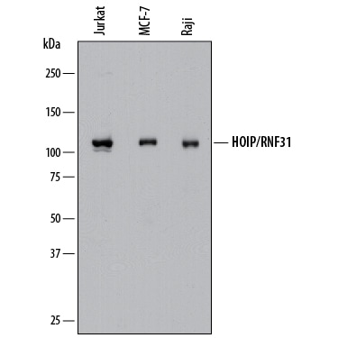
Detection of Human HOIP/RNF31 by Western Blot.
Western blot shows lysates of Jurkat human acute T cell leukemia cell line, MCF-7 human breast cancer cell line, and Raji human Burkitt's lymphoma cell line. PVDF membrane was probed with 1 µg/mL of Sheep Anti-Human HOIP/RNF31 Antigen Affinity-purified Polyclonal Antibody (Catalog # AF8039) followed by HRP-conjugated Anti-Sheep IgG Secondary Antibody (Catalog # HAF016). A specific band was detected for HOIP/RNF31 at approximately 120 kDa (as indicated). This experiment was conducted under reducing conditions and using Immunoblot Buffer Group 1.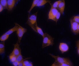
HOIP/RNF31 in MCF‑7 Human Cell Line.
HOIP/RNF31 was detected in immersion fixed MCF-7 human breast cancer cell line using Sheep Anti-Human HOIP/RNF31 Antigen Affinity-purified Polyclonal Antibody (Catalog # AF8039) at 5 µg/mL for 3 hours at room temperature. Cells were stained using the NorthernLights™ 557-conjugated Anti-Sheep IgG Secondary Antibody (red; Catalog # NL010) and counterstained with DAPI (blue). Specific staining was localized to cytoplasm. View our protocol for Fluorescent ICC Staining of Cells on Coverslips.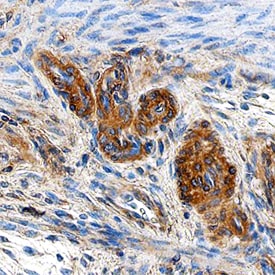
HOIP/RNF31 in Human Uterus.
HOIP/RNF31 was detected in immersion fixed paraffin-embedded sections of human uterus using Sheep Anti-Human HOIP/RNF31 Antigen Affinity-purified Polyclonal Antibody (Catalog # AF8039) at 10 µg/mL overnight at 4 °C. Tissue was stained using the Anti-Sheep HRP-DAB Cell & Tissue Staining Kit (brown; Catalog # CTS019) and counterstained with hematoxylin (blue). Specific staining was localized to cytoplasm in epithelial cells. View our protocol for Chromogenic IHC Staining of Paraffin-embedded Tissue Sections.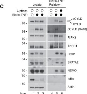
Detection of HOIP/RNF31 by Western Blot
CYLD is phosphorylated at a novel Ser568 site as well as within the phospho-rich patch to activate its activity(A) Qualitative DUB assay of CYLD (aa 436–956) purified from sf9 cells. CYLD was incubated in buffer alone, or with lambda PP as in Figures 1D and 1E. lambda PP was removed and dephosphorylated CYLD was either incubated with buffer alone (−), IKK beta, or the IKK complex (IKK beta + NEMO) prior to DUB assay using Lys63-Ub4 as substrate. For comparison, phosphorylated CYLD from sf9 cells was included (control).(B) as in (A) but using Met1-Ub4 as substrate.(C) Purification and western blot analysis of the TNFR1 complex in U2OS/NOD2 cell lines with subsequent lambda PP treatment. Cells were stimulated with biotin-TNF for 15 min (closed circles) or after lysis (open circles).(D) Purification and western blot analysis of the TNFR1 complex in HCT116 and U937 cells expressing shRNA targeting CYLD or control shRNA. Cells were stimulated with biotin-TNF for the indicated time points.(E and F) Western blot analysis of U2OS/NOD2 cells transfected with siRNA targeting OTULIN or mismatch control (shMM) and treated or not with IKK inhibitors VII + XII (E) or TAK1 inhibitor 5Z-7-oxozeaonol (F) for 30 min before stimulation with L18-MDP for 1 h or TNF for 15 min.(G) Workflow for identification of CYLD phosphorylation sites by mass spectrometry.(H) Schematic representation of identified CYLD phosphorylation sites.(I) Analysis of evolutionary conservation of identified phosphorylation sites. TNF-induced sites are shown in red.(J) Spectral data for identification of the novel S568 site in CYLD.(K) Qualitative DUB assay with CYLD variants purified from sf9 cells. Samples were either incubated in buffer alone or in the presence of lambda PP prior to incubation with Lys63-Ub4 as substrate. Image collected and cropped by CiteAb from the following open publication (https://pubmed.ncbi.nlm.nih.gov/34610306), licensed under a CC-BY license. Not internally tested by R&D Systems.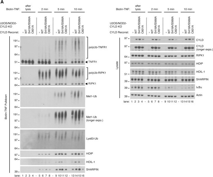
Detection of HOIP/RNF31 by Western Blot
Non-phosphorylation-mediated activity of CYLD retains LUBAC at the TNF-RSC and restores normal Met1-Ub, but not Lys63-Ub, deposition(A) Purification and western blot analysis of the TNFR1 complex in U2OS/NOD2 cell lines stimulated with biotin-TNF for the indicated time points.(B) Schematic model of how Ub-binding via CAP-Gly3 and phosphorylation affects CYLD-regulation of Lys63- and Met1-Ub at receptor complexes. Image collected and cropped by CiteAb from the following open publication (https://pubmed.ncbi.nlm.nih.gov/34610306), licensed under a CC-BY license. Not internally tested by R&D Systems.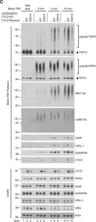
Detection of HOIP/RNF31 by Western Blot
The absence of CYLD activity leads to reduced LUBAC retention at the TNF-RSC as well as increased Lys63-Ub and reduced Met1-Ub on receptor components(A–C) Purification and western blot analysis of the TNFR1 complex in THP1 cells with shRNA knockdown of CYLD or mismatch control (A) and U2OS/NOD2 cell lines (B and C) stimulated with biotin-TNF as indicated.(D) UbiCRest analysis of TNFR1 complexes purified as in (B). Samples were incubated or not with OTULIN and/or OTUD1 as indicated and analyzed by western blotting. Image collected and cropped by CiteAb from the following open publication (https://pubmed.ncbi.nlm.nih.gov/34610306), licensed under a CC-BY license. Not internally tested by R&D Systems.Applications for Human HOIP/RNF31 Antibody
Immunocytochemistry
Sample: Immersion fixed MCF‑7 human breast cancer cell line
Immunohistochemistry
Sample: Immersion fixed paraffin-embedded sections of human uterus
Western Blot
Sample: Jurkat human acute T cell leukemia cell line, MCF‑7 human breast cancer cell line, and Raji human Burkitt's lymphoma cell line
Formulation, Preparation, and Storage
Purification
Reconstitution
Sterile PBS to a final concentration of 0.2 mg/mL. For liquid material, refer to CoA for concentration.
Formulation
Shipping
Stability & Storage
- 12 months from date of receipt, -20 to -70 °C as supplied.
- 1 month, 2 to 8 °C under sterile conditions after reconstitution.
- 6 months, -20 to -70 °C under sterile conditions after reconstitution.
Calculators
Background: HOIP/RNF31
Long Name
Alternate Names
Gene Symbol
UniProt
Additional HOIP/RNF31 Products
Product Documents for Human HOIP/RNF31 Antibody
Certificate of Analysis
To download a Certificate of Analysis, please enter a lot or batch number in the search box below.
Note: Certificate of Analysis not available for kit components.
Product Specific Notices for Human HOIP/RNF31 Antibody
For research use only
Related Research Areas
Citations for Human HOIP/RNF31 Antibody
 Powered by Bioz
Customer Reviews for Human HOIP/RNF31 Antibody
There are currently no reviews for this product. Be the first to review Human HOIP/RNF31 Antibody and earn rewards!
Have you used Human HOIP/RNF31 Antibody?
Submit a review and receive an Amazon gift card!
$25/€18/£15/$25CAN/¥2500 for a review with an image
$10/€7/£6/$10CAN/¥1110 for a review without an image
Submit a review
Protocols
Find general support by application which include: protocols, troubleshooting, illustrated assays, videos and webinars.
- Antigen Retrieval Protocol (PIER)
- Antigen Retrieval for Frozen Sections Protocol
- Appropriate Fixation of IHC/ICC Samples
- Cellular Response to Hypoxia Protocols
- Chromogenic IHC Staining of Formalin-Fixed Paraffin-Embedded (FFPE) Tissue Protocol
- Chromogenic Immunohistochemistry Staining of Frozen Tissue
- ClariTSA™ Fluorophore Kits
- Detection & Visualization of Antibody Binding
- Fluorescent IHC Staining of Frozen Tissue Protocol
- Graphic Protocol for Heat-induced Epitope Retrieval
- Graphic Protocol for the Preparation and Fluorescent IHC Staining of Frozen Tissue Sections
- Graphic Protocol for the Preparation and Fluorescent IHC Staining of Paraffin-embedded Tissue Sections
- Graphic Protocol for the Preparation of Gelatin-coated Slides for Histological Tissue Sections
- ICC Cell Smear Protocol for Suspension Cells
- ICC Immunocytochemistry Protocol Videos
- ICC for Adherent Cells
- IHC Sample Preparation (Frozen sections vs Paraffin)
- Immunocytochemistry (ICC) Protocol
- Immunocytochemistry Troubleshooting
- Immunofluorescence of Organoids Embedded in Cultrex Basement Membrane Extract
- Immunofluorescent IHC Staining of Formalin-Fixed Paraffin-Embedded (FFPE) Tissue Protocol
- Immunohistochemistry (IHC) and Immunocytochemistry (ICC) Protocols
- Immunohistochemistry Frozen Troubleshooting
- Immunohistochemistry Paraffin Troubleshooting
- Preparing Samples for IHC/ICC Experiments
- Preventing Non-Specific Staining (Non-Specific Binding)
- Primary Antibody Selection & Optimization
- Protocol for Heat-Induced Epitope Retrieval (HIER)
- Protocol for Making a 4% Formaldehyde Solution in PBS
- Protocol for VisUCyte™ HRP Polymer Detection Reagent
- Protocol for the Fluorescent ICC Staining of Cell Smears - Graphic
- Protocol for the Fluorescent ICC Staining of Cultured Cells on Coverslips - Graphic
- Protocol for the Preparation & Fixation of Cells on Coverslips
- Protocol for the Preparation and Chromogenic IHC Staining of Frozen Tissue Sections
- Protocol for the Preparation and Chromogenic IHC Staining of Frozen Tissue Sections - Graphic
- Protocol for the Preparation and Chromogenic IHC Staining of Paraffin-embedded Tissue Sections
- Protocol for the Preparation and Chromogenic IHC Staining of Paraffin-embedded Tissue Sections - Graphic
- Protocol for the Preparation and Fluorescent ICC Staining of Cells on Coverslips
- Protocol for the Preparation and Fluorescent ICC Staining of Non-adherent Cells
- Protocol for the Preparation and Fluorescent ICC Staining of Stem Cells on Coverslips
- Protocol for the Preparation and Fluorescent IHC Staining of Frozen Tissue Sections
- Protocol for the Preparation and Fluorescent IHC Staining of Paraffin-embedded Tissue Sections
- Protocol for the Preparation of Gelatin-coated Slides for Histological Tissue Sections
- Protocol for the Preparation of a Cell Smear for Non-adherent Cell ICC - Graphic
- R&D Systems Quality Control Western Blot Protocol
- TUNEL and Active Caspase-3 Detection by IHC/ICC Protocol
- The Importance of IHC/ICC Controls
- Troubleshooting Guide: Immunohistochemistry
- Troubleshooting Guide: Western Blot Figures
- Western Blot Conditions
- Western Blot Protocol
- Western Blot Protocol for Cell Lysates
- Western Blot Troubleshooting
- Western Blot Troubleshooting Guide
- View all Protocols, Troubleshooting, Illustrated assays and Webinars
Associated Pathways
