Interleukin-22 (IL-22), also known as IL-10-related T cell-derived inducible factor (IL-TIF) was initially identified as a gene induced by IL-9 in mouse T cells and mast cells. Human IL-22 cDNA encodes a 179 amino acid (aa) residue protein with a putative 33 aa signal peptide that is cleaved to generate a 147 aa mature protein that shares approximately 79% and 22% aa sequence identity with mouse IL-22 and human IL-10, respectively. The human IL-22 gene is localized to chromosome 12q15. Although it exists as a single copy gene in human and in many mouse strains, the mouse IL-22 gene is duplicated in some mouse strains including C57B1/6, FVB and 129. The two mouse genes designated IL-TIF alpha and IL-TIF beta, share greater than 98% sequence homology in their coding region. IL-22 has been shown to activate STAT1 and STAT3 in several hepatoma cell lines and upregulate the production of acute phase proteins. IL-22 is produced by normal T cells upon anti-CD3 stimulation in humans. Mouse IL-22 expression is also induced in various organs upon lipopolysaccharide injection, suggesting that IL-22 may be involved in inflammatory responses. The functional IL-22 receptor complex consists of two receptor subunits, IL-22 R (previously an orphan receptor named CRF2-9) and IL-10R beta (previously known as CRF2-4), belonging to the class II cytokine receptor family.

Key Product Details
Species Reactivity
Validated:
Human
Cited:
Human
Applications
Validated:
Neutralization, Intracellular Staining by Flow Cytometry
Cited:
Immunohistochemistry-Paraffin, Neutralization, Flow Cytometry
Label
Unconjugated
Antibody Source
Monoclonal Mouse IgG1 Clone # 142928
Loading...
Product Specifications
Immunogen
E. coli-derived recombinant human IL-22
Ala34-Ile179
Accession # Q9GZX6
Ala34-Ile179
Accession # Q9GZX6
Specificity
Detects human IL-22 in direct ELISAs. In direct ELISAs, this antibody shows 100% cross-reactivity with recombinant mouse IL‑22 and recombinant rat IL‑22 and no cross-reactivity with recombinant human IL‑10.
Clonality
Monoclonal
Host
Mouse
Isotype
IgG1
Endotoxin Level
<0.10 EU per 1 μg of the antibody by the LAL method.
Scientific Data Images for Human IL-22 Antibody (142928)
Detection of IL‑22 in PBMC treated with 20ng/mL PMA and 1ug/mL Calcium iono for 2 hours followed by 1ug/mL brefeldin A for 4 hours.
PBMC treated with 20ng/mL PMA and 1ug/mL Calcium iono for 2 hours followed by 1ug/mL brefeldin A for 4 hours were stained with Mouse Anti-Human CD4 Fluorescein‑conjugated Monoclonal Antibody (Catalog # FAB3791F) and either (A) Mouse Anti-Human IL‑22 Monoclonal Antibody (Catalog # MAB7821) or (B) isotype control antibody (Catalog # MAB002) followed by Allophycocyanin-conjugated Anti-Mouse IgG Secondary Antibody (Catalog # F0101B). To facilitate intracellular staining, cells were fixed with Flow Cytometry Fixation Buffer (Catalog # FC004) and permeabilized with Flow Cytometry Permeabilization/Wash Buffer I (Catalog # FC005). View our protocol for Staining Intracellular Molecules.
IL‑10 Secretion Induced by IL‑22 and Neutralization by Human IL-22 Antibody.
Recombinant Human IL-22 (Catalog # 782-IL) stimulates IL-10 secretion in the COLO 205 human colorectal adeno-carcinoma cell line in a dose-dependent manner (orange line), as measured by the Human IL-10 DuoSet ELISA Development Kit (Catalog # DY217B). IL-10 secretion elicited by Recombinant Human IL-22 (1 ng/mL) is neutralized (green line) by increasing concentrations of Human IL-22 Monoclonal Antibody (Catalog # MAB7821). The ND50 is typically 1-5 µg/mL.
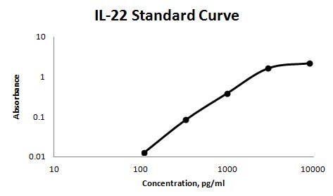
Human IL-22 ELISA Standard Curve
Recombinant Human IL‑22 (Catalog # 782-IL) was serially diluted and captured by Mouse Anti-Human IL‑22 Monoclonal Antibody (Catalog # MAB7822) coated on a Clear Polystyrene Microplate (Catalog # DY990). Mouse Anti-Human IL‑22 Monoclonal Antibody (Catalog # MAB7821) was biotinylated and incubated with the protein captured on the plate. Detection of the standard curve was achieved by incubating Streptavidin-HRP (Catalog # DY998)Applications for Human IL-22 Antibody (142928)
Application
Recommended Usage
Intracellular Staining by Flow Cytometry
0.25 µg/106 cells
Sample: PBMC treated with 20ng/mL PMA and 1ug/mL calcium iono for 2 hours followed by brefeldin A for 4 hours
Sample: PBMC treated with 20ng/mL PMA and 1ug/mL calcium iono for 2 hours followed by brefeldin A for 4 hours
Neutralization
Measured by its ability to neutralize IL‑22-induced IL‑10 secretion in the COLO 205 human colorectal adenocarcinoma cell line. Nagalakshmi, M.L. et al. (2004) Intl. Immunopharmacol. 4:679. The Neutralization Dose (ND50) is typically 1-5 µg/mL in the presence of 1 ng/mL Recombinant Human IL‑22.
Reviewed Applications
Read 1 review rated 5 using MAB7821 in the following applications:
Flow Cytometry Panel Builder
Bio-Techne Knows Flow Cytometry
Save time and reduce costly mistakes by quickly finding compatible reagents using the Panel Builder Tool.
Advanced Features
- Spectra Viewer - Custom analysis of spectra from multiple fluorochromes
- Spillover Popups - Visualize the spectra of individual fluorochromes
- Antigen Density Selector - Match fluorochrome brightness with antigen density

Formulation, Preparation, and Storage
Purification
Protein A or G purified from hybridoma culture supernatant
Reconstitution
Reconstitute at 0.5 mg/mL in sterile PBS. For liquid material, refer to CoA for concentration.
Loading...
Formulation
Lyophilized from a 0.2 μm filtered solution in PBS with Trehalose. See Certificate of Analysis for details.
*Small pack size (-SP) is supplied either lyophilized or as a 0.2 µm filtered solution in PBS.
*Small pack size (-SP) is supplied either lyophilized or as a 0.2 µm filtered solution in PBS.
Shipping
Lyophilized product is shipped at ambient temperature. Liquid small pack size (-SP) is shipped with polar packs. Upon receipt, store immediately at the temperature recommended below.
Stability & Storage
Use a manual defrost freezer and avoid repeated freeze-thaw cycles.
- 12 months from date of receipt, -20 to -70 °C as supplied.
- 1 month, 2 to 8 °C under sterile conditions after reconstitution.
- 6 months, -20 to -70 °C under sterile conditions after reconstitution.
Calculators
Background: IL-22
References
- Dumoutier, L. et al. (2000) J. Immunol. 164:1814.
- Xie, M-H. et al. (2000) J. Biol. Chem. 275:31335.
- Dumoutier, L. et al. (2000) Proc. Natl. Acad. Sci. USA 97:10144.
- Kotenko, S.V. et al. (2001) J. Biol. Chem. 276:2725.
Long Name
Interleukin 22
Alternate Names
IL-TIF, IL22
Gene Symbol
IL22
UniProt
Additional IL-22 Products
Product Documents for Human IL-22 Antibody (142928)
Certificate of Analysis
To download a Certificate of Analysis, please enter a lot or batch number in the search box below.
Note: Certificate of Analysis not available for kit components.
Product Specific Notices for Human IL-22 Antibody (142928)
For research use only
Related Research Areas
Citations for Human IL-22 Antibody (142928)
 Powered by Bioz
Customer Reviews for Human IL-22 Antibody (142928) (1)
5 out of 5
1 Customer Rating
Have you used Human IL-22 Antibody (142928)?
Submit a review and receive an Amazon gift card!
$25/€18/£15/$25CAN/¥2500 Yen for a review with an image
$10/€7/£6/$10CAN/¥1110 Yen for a review without an image
Submit a review
Customer Images
Showing
1
-
1 的
1 review
Showing All
Filter By:
-
Application: ELISASample Tested: Serum and PlasmaSpecies: HumanVerified Customer | Posted 11/27/2017MAB7821 was used as the solid phase antibody, BAM1359 was used as the detection antibody. 1259-RN was used as the immunoassay standard. Sensitivity of the assay was around 100 pg/ml.

There are no reviews that match your criteria.
Protocols
Find general support by application which include: protocols, troubleshooting, illustrated assays, videos and webinars.
- 7-Amino Actinomycin D (7-AAD) Cell Viability Flow Cytometry Protocol
- Extracellular Membrane Flow Cytometry Protocol
- Flow Cytometry Protocol for Cell Surface Markers
- Flow Cytometry Protocol for Staining Membrane Associated Proteins
- Flow Cytometry Staining Protocols
- Flow Cytometry Troubleshooting Guide
- Intracellular Flow Cytometry Protocol Using Alcohol (Methanol)
- Intracellular Flow Cytometry Protocol Using Detergents
- Intracellular Nuclear Staining Flow Cytometry Protocol Using Detergents
- Intracellular Staining Flow Cytometry Protocol Using Alcohol Permeabilization
- Intracellular Staining Flow Cytometry Protocol Using Detergents to Permeabilize Cells
- Propidium Iodide Cell Viability Flow Cytometry Protocol
- Protocol for Liperfluo
- Protocol for the Characterization of Human Th22 Cells
- Protocol for the Characterization of Human Th9 Cells
- Protocol: Annexin V and PI Staining by Flow Cytometry
- Protocol: Annexin V and PI Staining for Apoptosis by Flow Cytometry
- Troubleshooting Guide: Fluorokine Flow Cytometry Kits
- View all Protocols, Troubleshooting, Illustrated assays and Webinars


