Human Jagged 2 is a 131 kDa (predicted) member of the Delta-Serrate-Lag-2 (DSL) family of ligands. This family activates LIN12/Notch proteins and thereby regulates cell fate determination during development (1-5). It is a type 1 transmembrane protein that is synthesized as a 1238 amino acid (aa) precursor. It contains a 23 aa signal sequence, a large 1057 aa extracellular region, a 21 aa transmembrane region, and a short 137 aa cytoplasmic region. The extracellular region contains four potential N-linked glycosylation sites, a DSL domain, 16 EGF-like repeats (many of which are also sites of calcium binding), a von Willebrand factor (vWF) type C domain, and a cysteine-rich region just proximal to the transmembrane segment (2). There are two isoforms for human Jagged 2, named long and short. The short form lacks a splicing variant region (aa 421-461) that is present in the long form of the protein. Human Jagged 2 shares 90% and 87% aa sequence identity with mouse and rat Jagged 2, respectively. During murine embryonic development, Jagged 2 is expressed highest in fetal thymus, epidermis, foregut, dorsal root ganglia, and inner ear (2). In 2 week old mice, the Jagged 2 transcript is most abundant in heart, lung, thymus, skeletal muscle, brain, and testis (2). Functionally, it is suggested that Jagged 2 engages the Notch1 pathway of signal transduction (2). It is involved in the development of the mammalian limb, branchial arches, central and peripheral nervous systems, T cell lineage differentiation, natural killer cells, and the establishment of functional natural killer cell lines (3, 5, 6).

Key Product Details
Species Reactivity
Validated:
Human
Cited:
Human
Applications
Validated:
Immunohistochemistry, Western Blot, Immunocytochemistry
Cited:
Immunohistochemistry-Paraffin, Western Blot, Immunocytochemistry
Label
Unconjugated
Antibody Source
Polyclonal Goat IgG
Loading...
Product Specifications
Immunogen
Mouse myeloma cell line NS0-derived recombinant human Jagged 2
Met27-Asp307
Accession # Q9Y219
Met27-Asp307
Accession # Q9Y219
Specificity
Detects human Jagged 2 in direct ELISAs and Western blots. In Western blots, approximately 35% cross-reactivity with recombinant human Jagged 1 is observed and less than 1% cross-reactivity with recombinant rat Jagged 1 is observed.
Clonality
Polyclonal
Host
Goat
Isotype
IgG
Scientific Data Images for Human Jagged 2 Antibody
Jagged 2 in HUVEC Human Cells.
Jagged 2 was detected in immersion fixed HUVEC human umbilical vein endothelial cells using Goat Anti-Human Jagged 2 Antigen Affinity-purified Polyclonal Antibody (Catalog # AF1726) at 10 µg/mL for 3 hours at room temperature. Cells were stained using the NorthernLights™ 557-conjugated Anti-Goat IgG Secondary Antibody (red; Catalog # NL001) and counterstained with DAPI (blue). View our protocol for Fluorescent ICC Staining of Cells on Coverslips.Applications for Human Jagged 2 Antibody
Application
Recommended Usage
Immunocytochemistry
5-15 µg/mL
Sample: Immersion fixed HUVEC human umbilical vein endothelial cells
Sample: Immersion fixed HUVEC human umbilical vein endothelial cells
Immunohistochemistry
5-15 µg/mL
Sample: Immersion fixed paraffin-embedded sections of human skeletal muscle
Sample: Immersion fixed paraffin-embedded sections of human skeletal muscle
Western Blot
0.1 µg/mL
Sample: Recombinant Human Jagged 2 Fc Chimera (Catalog # 1726-JG)
Sample: Recombinant Human Jagged 2 Fc Chimera (Catalog # 1726-JG)
Reviewed Applications
Read 3 reviews rated 4 using AF1726 in the following applications:
Formulation, Preparation, and Storage
Purification
Antigen Affinity-purified
Reconstitution
Reconstitute at 0.2 mg/mL in sterile PBS. For liquid material, refer to CoA for concentration.
Loading...
Formulation
Lyophilized from a 0.2 μm filtered solution in PBS with Trehalose. *Small pack size (SP) is supplied either lyophilized or as a 0.2 µm filtered solution in PBS.
Shipping
Lyophilized product is shipped at ambient temperature. Liquid small pack size (-SP) is shipped with polar packs. Upon receipt, store immediately at the temperature recommended below.
Stability & Storage
Use a manual defrost freezer and avoid repeated freeze-thaw cycles.
- 12 months from date of receipt, -20 to -70 °C as supplied.
- 1 month, 2 to 8 °C under sterile conditions after reconstitution.
- 6 months, -20 to -70 °C under sterile conditions after reconstitution.
Calculators
Background: Jagged 2
References
- Shawber, C. et al. (1996) Dev. Biol. 180:370.
- Luo, B. et al. (1997) Mol. Cell. Biol. 17:6057.
- Valsecchi, V. et al. (1997) Mech. Dev. 69:203.
- Schickwann, T. et al. (2000) Blood 96:950.
- DeHart, S. et al. (2005) Blood 105:3521.
- de La Coste, A. and A.A. Freitas (2006) Immunol. Lett. 102:1.
Alternate Names
HJ2, JAG2
Gene Symbol
JAG2
UniProt
Additional Jagged 2 Products
Product Documents for Human Jagged 2 Antibody
Certificate of Analysis
To download a Certificate of Analysis, please enter a lot or batch number in the search box below.
Note: Certificate of Analysis not available for kit components.
Product Specific Notices for Human Jagged 2 Antibody
For research use only
Related Research Areas
Citations for Human Jagged 2 Antibody
 Powered by Bioz
Customer Reviews for Human Jagged 2 Antibody (3)
4 out of 5
3 Customer Ratings
Have you used Human Jagged 2 Antibody?
Submit a review and receive an Amazon gift card!
$25/€18/£15/$25CAN/¥2500 Yen for a review with an image
$10/€7/£6/$10CAN/¥1110 Yen for a review without an image
Submit a review
Customer Images
Showing
1
-
3 的
3 reviews
Showing All
Filter By:
-
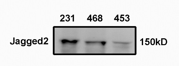
Application: Western BlotSample Tested: Breast cancer cellsSpecies: HumanVerified Customer | Posted 02/16/2020

-
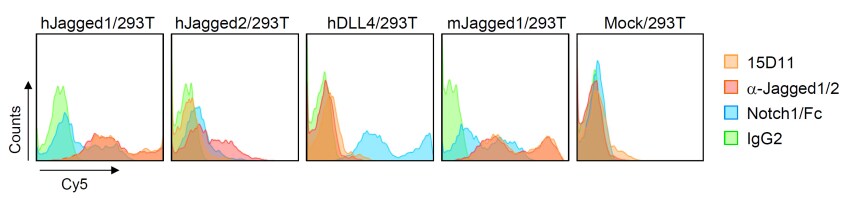
Application: Flow CytometrySample Tested: 293T human embryonic kidney cell lineSpecies: HumanVerified Customer | Posted 11/11/2018See Therapeutic Antibody Targeting Tumor- and Osteoblastic Niche-Derived Jagged1 Sensitizes Bone Metastasis to Chemotherapy Cancer Cell Volume 32, Issue 6, 11 December 2017, Pages 731-747.e6 For experimental details. Note that antibody cross-reacts with hJag1
-
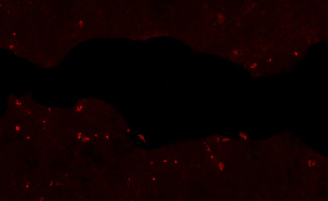
Application: Immunocytochemistry/ImmunofluorescenceSample Tested: Bladder tissueSpecies: HumanVerified Customer | Posted 12/19/2017
There are no reviews that match your criteria.
Protocols
Find general support by application which include: protocols, troubleshooting, illustrated assays, videos and webinars.
- Antigen Retrieval Protocol (PIER)
- Antigen Retrieval for Frozen Sections Protocol
- Appropriate Fixation of IHC/ICC Samples
- Cellular Response to Hypoxia Protocols
- Chromogenic IHC Staining of Formalin-Fixed Paraffin-Embedded (FFPE) Tissue Protocol
- Chromogenic Immunohistochemistry Staining of Frozen Tissue
- ClariTSA™ Fluorophore Kits
- Detection & Visualization of Antibody Binding
- Fluorescent IHC Staining of Frozen Tissue Protocol
- Graphic Protocol for Heat-induced Epitope Retrieval
- Graphic Protocol for the Preparation and Fluorescent IHC Staining of Frozen Tissue Sections
- Graphic Protocol for the Preparation and Fluorescent IHC Staining of Paraffin-embedded Tissue Sections
- Graphic Protocol for the Preparation of Gelatin-coated Slides for Histological Tissue Sections
- ICC Cell Smear Protocol for Suspension Cells
- ICC Immunocytochemistry Protocol Videos
- ICC for Adherent Cells
- IHC Sample Preparation (Frozen sections vs Paraffin)
- Immunocytochemistry (ICC) Protocol
- Immunocytochemistry Troubleshooting
- Immunofluorescence of Organoids Embedded in Cultrex Basement Membrane Extract
- Immunofluorescent IHC Staining of Formalin-Fixed Paraffin-Embedded (FFPE) Tissue Protocol
- Immunohistochemistry (IHC) and Immunocytochemistry (ICC) Protocols
- Immunohistochemistry Frozen Troubleshooting
- Immunohistochemistry Paraffin Troubleshooting
- Preparing Samples for IHC/ICC Experiments
- Preventing Non-Specific Staining (Non-Specific Binding)
- Primary Antibody Selection & Optimization
- Protocol for Heat-Induced Epitope Retrieval (HIER)
- Protocol for Making a 4% Formaldehyde Solution in PBS
- Protocol for VisUCyte™ HRP Polymer Detection Reagent
- Protocol for the Fluorescent ICC Staining of Cell Smears - Graphic
- Protocol for the Fluorescent ICC Staining of Cultured Cells on Coverslips - Graphic
- Protocol for the Preparation & Fixation of Cells on Coverslips
- Protocol for the Preparation and Chromogenic IHC Staining of Frozen Tissue Sections
- Protocol for the Preparation and Chromogenic IHC Staining of Frozen Tissue Sections - Graphic
- Protocol for the Preparation and Chromogenic IHC Staining of Paraffin-embedded Tissue Sections
- Protocol for the Preparation and Chromogenic IHC Staining of Paraffin-embedded Tissue Sections - Graphic
- Protocol for the Preparation and Fluorescent ICC Staining of Cells on Coverslips
- Protocol for the Preparation and Fluorescent ICC Staining of Non-adherent Cells
- Protocol for the Preparation and Fluorescent ICC Staining of Stem Cells on Coverslips
- Protocol for the Preparation and Fluorescent IHC Staining of Frozen Tissue Sections
- Protocol for the Preparation and Fluorescent IHC Staining of Paraffin-embedded Tissue Sections
- Protocol for the Preparation of Gelatin-coated Slides for Histological Tissue Sections
- Protocol for the Preparation of a Cell Smear for Non-adherent Cell ICC - Graphic
- R&D Systems Quality Control Western Blot Protocol
- TUNEL and Active Caspase-3 Detection by IHC/ICC Protocol
- The Importance of IHC/ICC Controls
- Troubleshooting Guide: Immunohistochemistry
- Troubleshooting Guide: Western Blot Figures
- Western Blot Conditions
- Western Blot Protocol
- Western Blot Protocol for Cell Lysates
- Western Blot Troubleshooting
- Western Blot Troubleshooting Guide
- View all Protocols, Troubleshooting, Illustrated assays and Webinars
Loading...
Associated Pathways

