EBF-2 (Early B cell Factor 2; also Mmot1, OLF3 and COE2) is a 62 kDa (predicted) member of the COE family of transcription factors. It is expressed in immature osteoblasts and Purkinje cells, and in the embryo is associated with the migration of postmitotic neuroblasts. In immature osteoblasts, EBF-2 appears to upregulate OPG, suppressing osteoclast formation. And in the developing retina, EBF-2 is found in ganglion, glycinergic Amacrine and horizontal cells, possibly promoting their development over that of photoreceptor cells. Mouse EBF-2 is 575 amino acids (aa) in length. It contains one DNA-binding region with an embedded C5-type zinc‑finger motif (aa 62-238), a dimerization ITP/TIG domain (aa 253-336), and a Pro/Ser-rich transactivation domain (aa 453-534). Although considered an HLH type transcription factor, it does not contain the typical "b", or basic amino acid sequence associated with bHLH factors. EBF-2 both homodimerizes, and heterodimerizes with EBF-1 and -3. There is an alternative start site at Met23. Over aa 407-519, mouse EBF-2 is identical in aa sequence to rat EBF-2 and shares 99% aa identity with human EBF-2.

Key Product Details
Species Reactivity
Validated:
Human, Mouse
Cited:
Human, Mouse, Transgenic Mouse
Applications
Validated:
Immunohistochemistry, Western Blot
Cited:
Immunohistochemistry, Western Blot, Immunocytochemistry, Immunoprecipitation, Chromatin Immunoprecipitation (ChIP)
Label
Unconjugated
Antibody Source
Polyclonal Sheep IgG
Loading...
Product Specifications
Immunogen
E. coli-derived recombinant mouse EBF-2
Arg407-Ser519
Accession # O08792
Arg407-Ser519
Accession # O08792
Specificity
Detects mouse EBF-2 in direct ELISAs. Detects mouse and human EBF-2 in Western blots.
Clonality
Polyclonal
Host
Sheep
Isotype
IgG
Scientific Data Images for EBF-2 Antibody
Detection of Human and Mouse EBF-2 by Western Blot.
Western blot shows lysates of 3T3-L1 mouse embryonic fibroblast adipose-like cell line and HepG2 human hepatocellular carcinoma cell line. PVDF membrane was probed with 1 µg/mL of Sheep Anti-Human/Mouse EBF-2 Antigen Affinity-purified Polyclonal Antibody (Catalog # AF7006) followed by HRP-conjugated Anti-Sheep IgG Secondary Antibody (HAF016). A specific band was detected for EBF-2 at approximately 65 kDa (as indicated). This experiment was conducted under reducing conditions and using Immunoblot Buffer Group 8.
EBF-2 in Mouse Embryo.
EBF-2 was detected in immersion fixed frozen sections of mouse embryo (15 d.p.c.) using Sheep Anti-Mouse EBF-2 Antigen Affinity-purified Polyclonal Antibody (Catalog # AF7006) at 10 µg/mL overnight at 4 °C. Tissue was stained using the Anti-Sheep HRP-DAB Cell & Tissue Staining Kit (brown; CTS019) and counterstained with hematoxylin (blue). Specific staining was localized to developing muscle cells. View our protocol for Chromogenic IHC Staining of Frozen Tissue Sections.Applications for EBF-2 Antibody
Application
Recommended Usage
Immunohistochemistry
5-15 µg/mL
Sample: Immersion fixed frozen sections of mouse embryo (15 d.p.c.)
Sample: Immersion fixed frozen sections of mouse embryo (15 d.p.c.)
Western Blot
1 µg/mL
Sample: 3T3‑L1 mouse embryonic fibroblast adipose-like cell line and HepG2 human hepatocellular carcinoma cell line
Sample: 3T3‑L1 mouse embryonic fibroblast adipose-like cell line and HepG2 human hepatocellular carcinoma cell line
Reviewed Applications
Read 1 review rated 5 using AF7006 in the following applications:
Formulation, Preparation, and Storage
Purification
Antigen Affinity-purified
Reconstitution
Sterile PBS to a final concentration of 0.2 mg/mL. For liquid material, refer to CoA for concentration.
Loading...
Formulation
Lyophilized from a 0.2 μm filtered solution in PBS with Trehalose. *Small pack size (SP) is supplied either lyophilized or as a 0.2 µm filtered solution in PBS.
Shipping
Lyophilized product is shipped at ambient temperature. Liquid small pack size (-SP) is shipped with polar packs. Upon receipt, store immediately at the temperature recommended below.
Stability & Storage
Use a manual defrost freezer and avoid repeated freeze-thaw cycles.
- 12 months from date of receipt, -20 to -70 °C as supplied.
- 1 month, 2 to 8 °C under sterile conditions after reconstitution.
- 6 months, -20 to -70 °C under sterile conditions after reconstitution.
Calculators
Background: EBF-2
Long Name
Early B-cell Factor 2
Alternate Names
COE2, EBF2, Mmot1
Gene Symbol
EBF2
UniProt
Additional EBF-2 Products
Product Documents for EBF-2 Antibody
Certificate of Analysis
To download a Certificate of Analysis, please enter a lot or batch number in the search box below.
Note: Certificate of Analysis not available for kit components.
Product Specific Notices for EBF-2 Antibody
For research use only
Related Research Areas
Citations for EBF-2 Antibody
 Powered by Bioz
Customer Reviews for EBF-2 Antibody (1)
5 out of 5
1 Customer Rating
Have you used EBF-2 Antibody?
Submit a review and receive an Amazon gift card!
$25/€18/£15/$25CAN/¥2500 Yen for a review with an image
$10/€7/£6/$10CAN/¥1110 Yen for a review without an image
Submit a review
Customer Images
Showing
1
-
1 的
1 review
Showing All
Filter By:
-
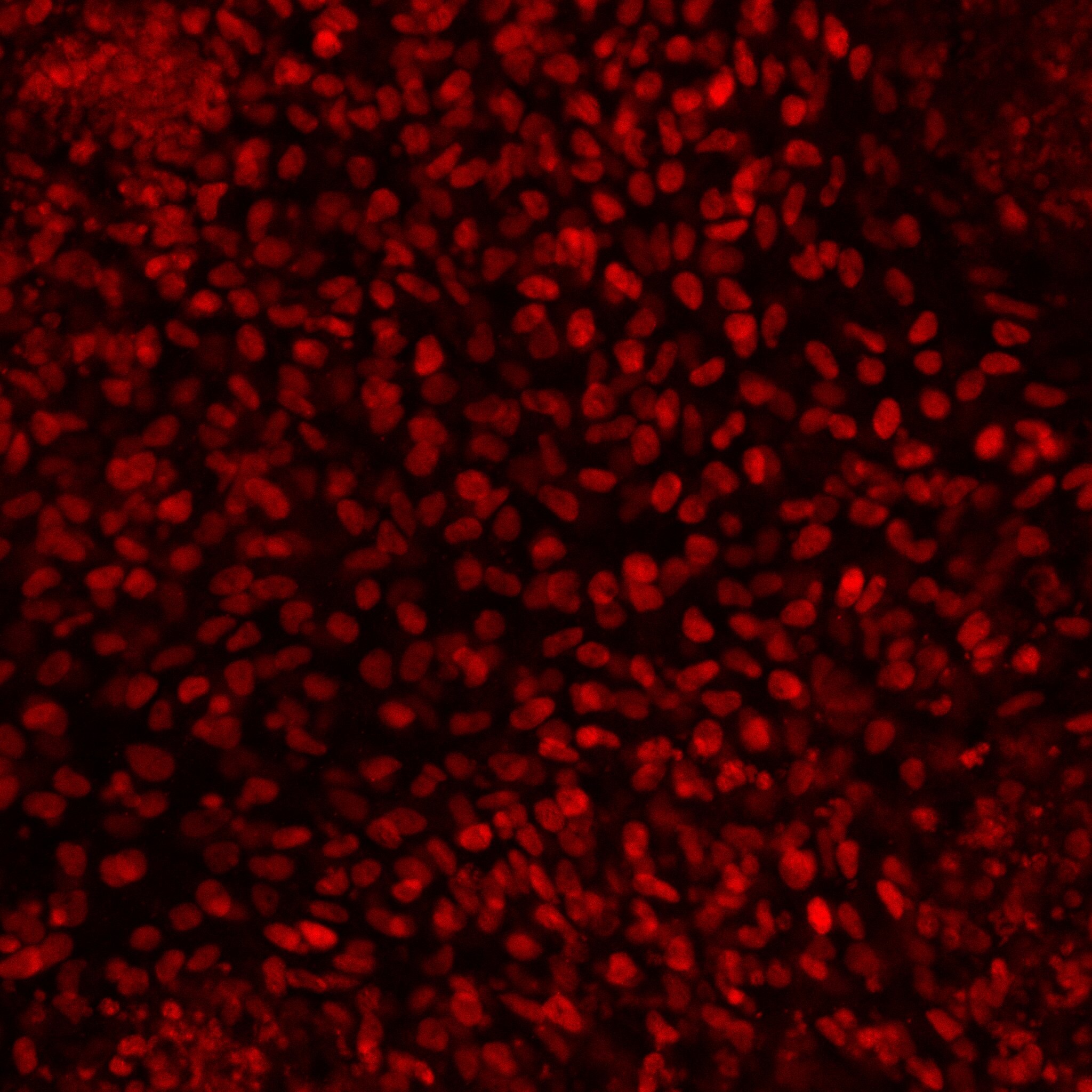
Application: Immunocytochemistry/ImmunofluorescenceSample Tested: Human fibroblastSpecies: HumanVerified Customer | Posted 07/30/2020

There are no reviews that match your criteria.
Protocols
Find general support by application which include: protocols, troubleshooting, illustrated assays, videos and webinars.
- Antigen Retrieval Protocol (PIER)
- Antigen Retrieval for Frozen Sections Protocol
- Appropriate Fixation of IHC/ICC Samples
- Cellular Response to Hypoxia Protocols
- Chromogenic IHC Staining of Formalin-Fixed Paraffin-Embedded (FFPE) Tissue Protocol
- Chromogenic Immunohistochemistry Staining of Frozen Tissue
- ClariTSA™ Fluorophore Kits
- Detection & Visualization of Antibody Binding
- Fluorescent IHC Staining of Frozen Tissue Protocol
- Graphic Protocol for Heat-induced Epitope Retrieval
- Graphic Protocol for the Preparation and Fluorescent IHC Staining of Frozen Tissue Sections
- Graphic Protocol for the Preparation and Fluorescent IHC Staining of Paraffin-embedded Tissue Sections
- Graphic Protocol for the Preparation of Gelatin-coated Slides for Histological Tissue Sections
- IHC Sample Preparation (Frozen sections vs Paraffin)
- Immunofluorescent IHC Staining of Formalin-Fixed Paraffin-Embedded (FFPE) Tissue Protocol
- Immunohistochemistry (IHC) and Immunocytochemistry (ICC) Protocols
- Immunohistochemistry Frozen Troubleshooting
- Immunohistochemistry Paraffin Troubleshooting
- Preparing Samples for IHC/ICC Experiments
- Preventing Non-Specific Staining (Non-Specific Binding)
- Primary Antibody Selection & Optimization
- Protocol for Heat-Induced Epitope Retrieval (HIER)
- Protocol for Making a 4% Formaldehyde Solution in PBS
- Protocol for VisUCyte™ HRP Polymer Detection Reagent
- Protocol for the Preparation & Fixation of Cells on Coverslips
- Protocol for the Preparation and Chromogenic IHC Staining of Frozen Tissue Sections
- Protocol for the Preparation and Chromogenic IHC Staining of Frozen Tissue Sections - Graphic
- Protocol for the Preparation and Chromogenic IHC Staining of Paraffin-embedded Tissue Sections
- Protocol for the Preparation and Chromogenic IHC Staining of Paraffin-embedded Tissue Sections - Graphic
- Protocol for the Preparation and Fluorescent IHC Staining of Frozen Tissue Sections
- Protocol for the Preparation and Fluorescent IHC Staining of Paraffin-embedded Tissue Sections
- Protocol for the Preparation of Gelatin-coated Slides for Histological Tissue Sections
- R&D Systems Quality Control Western Blot Protocol
- TUNEL and Active Caspase-3 Detection by IHC/ICC Protocol
- The Importance of IHC/ICC Controls
- Troubleshooting Guide: Immunohistochemistry
- Troubleshooting Guide: Western Blot Figures
- Western Blot Conditions
- Western Blot Protocol
- Western Blot Protocol for Cell Lysates
- Western Blot Troubleshooting
- Western Blot Troubleshooting Guide
- View all Protocols, Troubleshooting, Illustrated assays and Webinars
Loading...