GFR alpha-2/GDNF R alpha-2 Antibody

R&D Systems | Catalog # AF429

R&D Systems

Key Product Details

Species Reactivity

Validated:

Human, Mouse

Cited:

Human, Mouse, Rat, Transgenic Mouse

Applications

Validated:

Immunohistochemistry, Western Blot, Blockade of Receptor-ligand Interaction, Immunocytochemistry

Cited:

Immunohistochemistry, Immunohistochemistry-Paraffin, Immunohistochemistry-Frozen, Western Blot, Neutralization, Flow Cytometry, Immunocytochemistry

Label

Unconjugated

Antibody Source

Polyclonal Goat IgG
Loading...

Product Specifications

Immunogen

Mouse myeloma cell line NS0-derived recombinant mouse GFR alpha ‑2/GDNF R alpha ‑2
Ser22-Ser441
Accession # Q3UUD8

Specificity

Detects human and mouse GFR alpha ‑2/GDNF R alpha ‑2 in direct ELISAs and Western blots. In direct ELISAs and Western blots, less than

Clonality

Polyclonal

Host

Goat

Isotype

IgG

Endotoxin Level

<0.10 EU per 1 μg of the antibody by the LAL method.

Scientific Data Images for GFR alpha-2/GDNF R alpha-2 Antibody

GFRa-2/GDNF Ra-2 antibody in Mouse Splenocytes by Immunocytochemistry (ICC).

GFR alpha ‑2/GDNF R alpha ‑2 in Mouse Splenocytes.

GFRa-2/GDNF Ra-2 was detected in immersion fixed mouse splenocytes using Goat Anti-Human/Mouse GFRa-2/GDNF Ra-2 Antigen Affinity-purified Polyclonal Antibody (Catalog # AF429) at 5 µg/mL for 3 hours at room temperature. Cells were stained using the NorthernLights™ 557-conjugated Anti-Goat IgG Secondary Antibody (red; Catalog # NL001) and counterstained with DAPI (blue). Specific staining was localized to plasma membrane. View our protocol for Fluorescent ICC Staining of Non-adherent Cells.
GFRa-2/GDNF Ra-2 antibody in Mouse Dorsal Root Ganglion by Immunohistochemistry (IHC-Fr).

GFR alpha ‑2/GDNF R alpha ‑2 in Mouse Dorsal Root Ganglion.

GFRa-2/GDNF Ra-2 was detected in perfusion fixed frozen sections of mouse dorsal root ganglion using Goat Anti-Human/Mouse GFRa-2/GDNF Ra-2 Antigen Affinity-purified Polyclonal Antibody (Catalog # AF429) at 1.7 µg/mL overnight at 4 °C. Tissue was stained using the Anti-Goat HRP-DAB Cell & Tissue Staining Kit (brown; Catalog # CTS008) and counterstained with hematoxylin (blue). Specific staining was localized to plasma membrane of sensory neurons. View our protocol for Chromogenic IHC Staining of Frozen Tissue Sections.
Detection of GFR alpha-2 by Flow Cytometry

Detection of GFR alpha-2 by Flow Cytometry

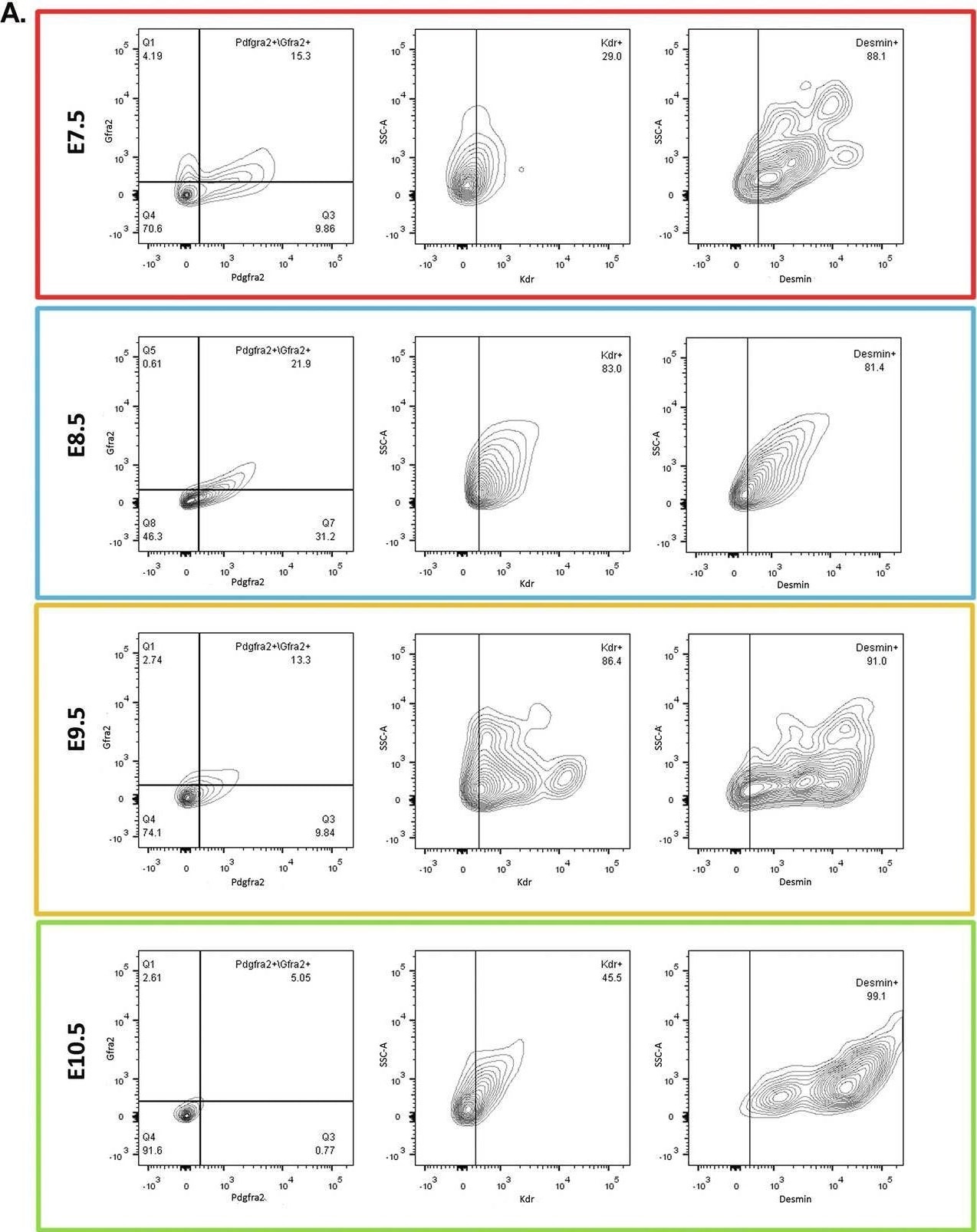
Desmin is expressed in the Pdgfra+/Gfra2+/Kdr+ CPC population and regulates its size.A FACS analyses of desmin expression in Pdgfra+/Gfra2+/Kdr+ CPCs isolated from mouse hearts at embryonic days E7.5 (n = 2), E8.5 (n = 3), E9.5 (n = 3) and E10.5 (n = 2). The first plot presents the cardiac cell population according to Pdgfra+/Gfra2+ expression. The second plot shows the percentage of Kdr+ cells in the Pdgfra+/Gfra2+ population, and the third plot shows the percentage of Pdgfra+/Gfra2+/Kdr+ population that expresses desmin. B FACS plots of CPCs isolated from Des+/+ (Bi and Bii) vs. Des−/− (Biii and Biv) mouse hearts at E8.5, n = 2. The first plot presents the cardiac cell population according to Pdgfra+/Gfra2+ expression. The second plot shows the percentage of Kdr+ cells in the Pdgfra+/Gfra2+ population, that is, the triple-positive Pdgfra+/Gfra2+/Kdr+ CPCs. C FACS plots showing the percentage of MHC (Myh6) in the total cardiac cell population of Des+/+ (Ci) and Des−/− (Cii) of E8.5 mouse embryonic hearts. D Percentage of desmin+ cells in total cardiac cell population; the line with circles represents the percentage of cardiac cells that express desmin during E7.5-E10.5, the line with squares represents the percentage of CPCs that express desmin. CPCs cardiac progenitor cells. Image collected and cropped by CiteAb from the following open publication (https://www.nature.com/articles/s41419-025-08056-3), licensed under a CC-BY license. Not internally tested by R&D Systems.
Detection of GFR alpha-2 by Immunocytochemistry/ Immunofluorescence

Detection of GFR alpha-2 by Immunocytochemistry/ Immunofluorescence

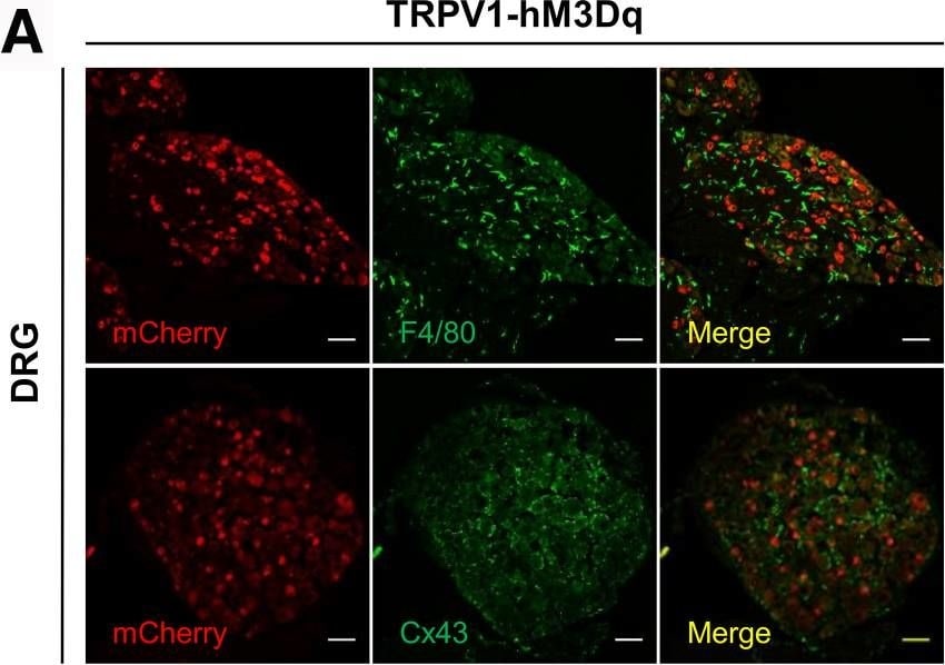
hM3Dq-mCherry is not expressed in non-neuronal cells and in the ENS. The absence of hM3Dq and hM4Di expression in non-neuronal cells and ENS was confirmed by staining with an anti F4/80 (macrophages) and Cx43 (satellite glial cells) antibodies in DRG (A and D, respectively); Iba1 antibody (microglia) in spinal cord (B and E, respectively), and CGRP antibody in the colon (C and F, respectively). G, hM3Dq expression was not detected in the anterior cingulate cortex and the somatosensory cortex in the brain. Scale bars 50 μm. Image collected and cropped by CiteAb from the following open publication (https://pubmed.ncbi.nlm.nih.gov/34954381), licensed under a CC-BY license. Not internally tested by R&D Systems.
Detection of GFR alpha-2 by Flow Cytometry

Detection of GFR alpha-2 by Flow Cytometry

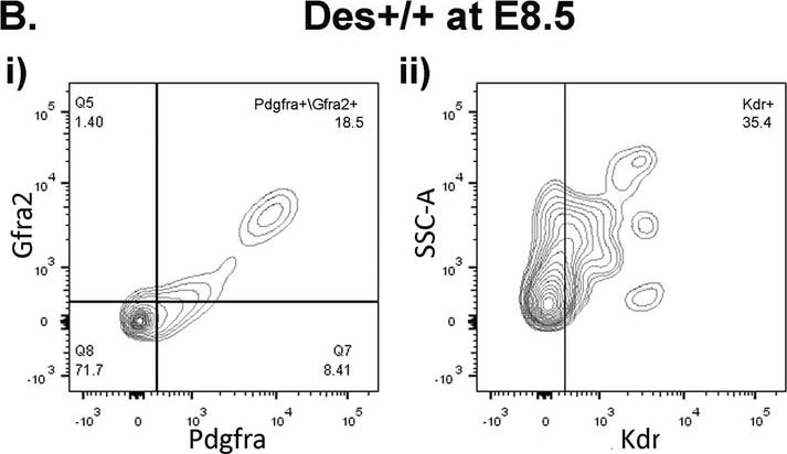
Desmin is expressed in the Pdgfra+/Gfra2+/Kdr+ CPC population and regulates its size.A FACS analyses of desmin expression in Pdgfra+/Gfra2+/Kdr+ CPCs isolated from mouse hearts at embryonic days E7.5 (n = 2), E8.5 (n = 3), E9.5 (n = 3) and E10.5 (n = 2). The first plot presents the cardiac cell population according to Pdgfra+/Gfra2+ expression. The second plot shows the percentage of Kdr+ cells in the Pdgfra+/Gfra2+ population, and the third plot shows the percentage of Pdgfra+/Gfra2+/Kdr+ population that expresses desmin. B FACS plots of CPCs isolated from Des+/+ (Bi and Bii) vs. Des−/− (Biii and Biv) mouse hearts at E8.5, n = 2. The first plot presents the cardiac cell population according to Pdgfra+/Gfra2+ expression. The second plot shows the percentage of Kdr+ cells in the Pdgfra+/Gfra2+ population, that is, the triple-positive Pdgfra+/Gfra2+/Kdr+ CPCs. C FACS plots showing the percentage of MHC (Myh6) in the total cardiac cell population of Des+/+ (Ci) and Des−/− (Cii) of E8.5 mouse embryonic hearts. D Percentage of desmin+ cells in total cardiac cell population; the line with circles represents the percentage of cardiac cells that express desmin during E7.5-E10.5, the line with squares represents the percentage of CPCs that express desmin. CPCs cardiac progenitor cells. Image collected and cropped by CiteAb from the following open publication (https://www.nature.com/articles/s41419-025-08056-3), licensed under a CC-BY license. Not internally tested by R&D Systems.
Detection of GFR alpha-2 by Immunohistochemistry

Detection of GFR alpha-2 by Immunohistochemistry

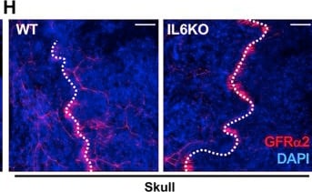
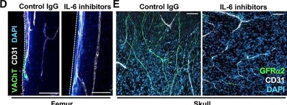
Interleukin-6 promotes postnatal development of skeletal sympathetic cholinergic innervation(A and B) Proximity ligation assay of IL-6 in BM section from postnatal day-3 developing femur. Scale bars, 100 μm (A) and 50 μm (B).(C) Schematic of in vivo IL-6 blockade.(D–F) Immunofluorescence of (D) VAChT+, or (E) GFR alpha 2+ cholinergic nerve fibers (green) in (D) femoral cortical bone and (E) skulls from 6.5-week-old mice following IL-6 blockade, with (F) quantification of fiber area. Dashed lines indicate bone surface.(G–I) Immunofluorescence of (G) VAChT+ or (H) GFR alpha 2+ cholinergic nerve fibers in (G) femoral cortical bone and (H) skulls from male IL-6 KO mice, with (I) quantification of fiber area. Dashed lines indicate cranial sutures.(D, E, G, and H) Scale bars, 100 μm. Nuclei were counterstained with DAPI (blue).(F and I) Data are mean ± SEM, ∗p < 0.05, ∗∗p < 0.01, unpaired two-tailed t test. Image collected and cropped by CiteAb from the following open publication (https://linkinghub.elsevier.com/retrieve/pii/S1934590922000595), licensed under a CC-BY license. Not internally tested by R&D Systems.
Detection of GFR alpha-2 by Immunohistochemistry

Detection of GFR alpha-2 by Immunohistochemistry

Interleukin-6 promotes postnatal development of skeletal sympathetic cholinergic innervation(A and B) Proximity ligation assay of IL-6 in BM section from postnatal day-3 developing femur. Scale bars, 100 μm (A) and 50 μm (B).(C) Schematic of in vivo IL-6 blockade.(D–F) Immunofluorescence of (D) VAChT+, or (E) GFR alpha 2+ cholinergic nerve fibers (green) in (D) femoral cortical bone and (E) skulls from 6.5-week-old mice following IL-6 blockade, with (F) quantification of fiber area. Dashed lines indicate bone surface.(G–I) Immunofluorescence of (G) VAChT+ or (H) GFR alpha 2+ cholinergic nerve fibers in (G) femoral cortical bone and (H) skulls from male IL-6 KO mice, with (I) quantification of fiber area. Dashed lines indicate cranial sutures.(D, E, G, and H) Scale bars, 100 μm. Nuclei were counterstained with DAPI (blue).(F and I) Data are mean ± SEM, ∗p < 0.05, ∗∗p < 0.01, unpaired two-tailed t test. Image collected and cropped by CiteAb from the following open publication (https://linkinghub.elsevier.com/retrieve/pii/S1934590922000595), licensed under a CC-BY license. Not internally tested by R&D Systems.

Applications for GFR alpha-2/GDNF R alpha-2 Antibody

Application
Recommended Usage

Blockade of Receptor-ligand Interaction

In a functional ELISA, 1-3 µg/mL of this antibody will block 50% of the binding of 2 ng/mL of Recombinant Human GDNF (Catalog # 212-GD) to immobilized Recombinant Mouse GFR alpha -2 Fc Chimera (Catalog # 429-FR) coated at 1 µg/mL (100 µL/well). At 15 μg/mL, this antibody will block >90% of the binding.

Immunocytochemistry

5-15 µg/mL
Sample: Immersion fixed mouse splenocytes

Immunohistochemistry

5-15 µg/mL
Sample: Perfusion fixed frozen sections of mouse dorsal root ganglion

Western Blot

0.1 µg/mL
Sample: Recombinant Mouse GFR alpha ‑2/GDNF R alpha ‑2 Fc Chimera (Catalog # 429-FR)

Reviewed Applications

Read 2 reviews rated 5 using AF429 in the following applications:

Formulation, Preparation, and Storage

Purification

Antigen Affinity-purified

Reconstitution

Reconstitute at 0.2 mg/mL in sterile PBS. For liquid material, refer to CoA for concentration.


Loading...

Formulation

Lyophilized from a 0.2 μm filtered solution in PBS with Trehalose. *Small pack size (SP) is supplied either lyophilized or as a 0.2 µm filtered solution in PBS.

Shipping

Lyophilized product is shipped at ambient temperature. Liquid small pack size (-SP) is shipped with polar packs. Upon receipt, store immediately at the temperature recommended below.

Stability & Storage

Use a manual defrost freezer and avoid repeated freeze-thaw cycles.
  • 12 months from date of receipt, -20 to -70 °C as supplied.
  • 1 month, 2 to 8 °C under sterile conditions after reconstitution.
  • 6 months, -20 to -70 °C under sterile conditions after reconstitution.

Calculators

The reconstitution calculator allows you to quickly calculate the volume of a reagent to reconstitute your vial. Simply enter the mass of reagent and the target concentration and the calculator will determine the rest.

=
÷

Background: GFR alpha-2/GDNF R alpha-2

Glial cell line-derived growth factor (GDNF), neurturin (NTN), persephin (PSP) and artemin, distant members of the TGF-beta  superfamily, are neurotrophic factors for a variety of neuronal populations in the central and peripheral nervous systems. The bioactivities of GDNF and NTN are mediated through a receptor complex composed of the non ligand-binding signaling subunit (c-Ret receptor tyrosine kinase) and either of two ligand binding subunits, GDNF receptor alpha -1 (GFR alpha -1, also known as Trn R1) or GFR alpha -2 (also known as Trn R2). GFR alpha -1 and -2 are members of a family of at least four cysteine-rich glycosyl-phosphatidylinositol (GPI)-linked cell surface proteins that share conserved placements of many of their cysteine residues. Binding of GDNF or NTN to membrane-associated GFR alpha -1 or GFR alpha -2 initiates the association with and activation of the Ret tyrosine kinase.

Mouse GFR alpha -2 cDNA encodes a 463 amino acid (aa) residue protein with a putative N-terminal 21 aa residue hydrophobic signal peptide. Like other GPI-linked proteins, rat GFR alpha -2 has a C-terminal hydrophobic region which is preceded by a 3 aa residue (SGS) GPI-binding site. Human GFR alpha -2 shares 96.5% amino acid identity with mouse GFR alpha -2. The expression of the various GFR alpha s are differentially regulated in the central and peripheral nervous system, suggesting complementary roles for the GFR alpha s in mediating the activities of the GDNF family of neurotrophic factors.

References

  1. Thompson, J. et al. (1998) Mol. Cell Neurosci. 11:117.
  2. Trupp, M. et al. (1998) Mol. Cell Neurosci. 11:47.
  3. Baloh, R.H. et al. (1998) Proc. Natl. Acad. Sci. USA 95:5801.
  4. Baloh, R.H. et al. (1998) Neuron 21:1291.

Long Name

Glial Cell line-derived Neurotrophic Factor Receptor alpha 2

Alternate Names

GDNF R alpha-2, GFR alpha2, GFRa-2, GFRA2

Entrez Gene IDs

2675 (Human); 14586 (Mouse)

Gene Symbol

GFRA2

UniProt

Additional GFR alpha-2/GDNF R alpha-2 Products

Product Documents for GFR alpha-2/GDNF R alpha-2 Antibody

Certificate of Analysis

To download a Certificate of Analysis, please enter a lot or batch number in the search box below.

Note: Certificate of Analysis not available for kit components.

Product Specific Notices for GFR alpha-2/GDNF R alpha-2 Antibody

For research use only

Related Research Areas

Citations for GFR alpha-2/GDNF R alpha-2 Antibody

Customer Reviews for GFR alpha-2/GDNF R alpha-2 Antibody (2)

5 out of 5
2 Customer Ratings
5 Stars
100%
4 Stars
0%
3 Stars
0%
2 Stars
0%
1 Stars
0%

Have you used GFR alpha-2/GDNF R alpha-2 Antibody?

Submit a review and receive an Amazon gift card!

$25/€18/£15/$25CAN/¥2500 for a review with an image

$10/€7/£6/$10CAN/¥1110 for a review without an image

Submit a review
Amazon Gift Card

Customer Images


Showing  1 - 2 的 2 reviews Showing All
Filter By:
  • Human/Mouse GFR alpha-2/GDNF R alpha-2 Antibody
    Name: Anonymous
    Application: Western Blot
    Sample Tested: COS-7 cells
    Species: Mouse
    Verified Customer | Posted 07/16/2021
    GFR alpha-2/GDNF R alpha-2 Antibody AF429
  • Human/Mouse GFR alpha-2/GDNF R alpha-2 Antibody
    Name: Anonymous
    Application: Immunocytochemistry/Immunofluorescence
    Sample Tested: microglial cells
    Species: Mouse
    Verified Customer | Posted 05/05/2021
    GFR alpha-2/GDNF R alpha-2 Antibody AF429

There are no reviews that match your criteria.

Showing  1 - 2 的 2 reviews Showing All

Protocols

Find general support by application which include: protocols, troubleshooting, illustrated assays, videos and webinars.

FAQs

No product specific FAQs exist for this product.

View all FAQs for Antibodies
Loading...
