
PRDM16 (PR [PRDI-BF1 and RIZ] domain containing protein 16; also MEL-1) is a 170 kDa member of the PR Domain family of proteins. It is a transcriptional regulator expressed in the embryo, and is reported to participate in the maintenance of both neuronal and hematopoietic progenitor stem cells populations, and to preferentially promote the development of brown fat from adipomyocyte precursors. The generation of brown fat is likely due to suppression of muscle-specific factors. Mouse PRDM16 is 1275 amino acids (aa) in length. It contains one SET domain (aa 85-208) followed by ten C2H2 type Zn finger motifs (aa 230-1030). There are multiple potential isoform variants that likely vary from 150-170 kDa in size. One isoform shows a deletion of aa 1232-1250, a second isoform shows a three aa substitution for aa 1174-1275, and a third isoform possesses an alternative start site at Met21, coupled to a deletion of aa 1196-1133. Over aa 537-688, mouse PRDM16 shares 81% and 95% aa identity with human and rat PRDM16, respectively.

Scientific images may be resized, cropped, or adjusted for brightness or contrast for presentation purposes. These updates are limited to presentation and do not affect the underlying scientific interpretation of the data. Where available, additional source data may be provided upon request.
Key Product Details
Species Reactivity
Validated:
Cited:
Applications
Validated:
Cited:
Label
Antibody Source
Product Specifications
Immunogen
Lys537-Glu688
Accession # A2A935.1
Specificity
Clonality
Host
Isotype
Scientific Data Images for PRDM16/MEL1 Antibody
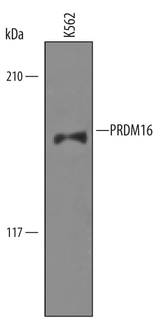
Detection of Human PRDM16 by Western Blot.
Western blot shows lysates of K562 human chronic myelogenous leukemia cell line. PVDF Membrane was probed with 1 µg/mL of Sheep Anti-Human/Mouse PRDM16 Antigen Affinity-purified Polyclonal Antibody (Catalog # AF6295) followed by HRP-conjugated Anti-Sheep IgG Secondary Antibody (Catalog # HAF016). A specific band was detected for PRDM16 at approximately 170 kDa (as indicated). This experiment was conducted under reducing conditions and using Immunoblot Buffer Group 8.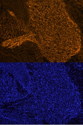
PRDM16 in Mouse Embryo.
PRDM16 was detected in immersion fixed frozen sections of mouse embryo (E13.5) using Sheep Anti-Human/Mouse PRDM16 Antigen Affinity-purified Polyclonal Antibody (Catalog # AF6295) at 10 µg/mL overnight at 4 °C. Tissue was stained using the NorthernLights™ 557-conjugated Anti-Sheep IgG Secondary Antibody (orange, upper panel; Catalog # NL010) and counterstained with DAPI (blue, lower panel). Specific staining was localized to the trigeminal ganglion. View our protocol for Fluorescent IHC Staining of Frozen Tissue Sections.Applications for PRDM16/MEL1 Antibody
Immunohistochemistry
Sample: Immersion fixed frozen sections of mouse embryo (E13.5)
Western Blot
Sample: K562 human chronic myelogenous leukemia cell line and M1 mouse myeloid leukemia cell line
Reviewed Applications
Read 1 review rated 3 using AF6295 in the following applications:
Formulation, Preparation, and Storage
Purification
Reconstitution
Formulation
Shipping
Stability & Storage
- 12 months from date of receipt, -20 to -70 °C as supplied.
- 1 month, 2 to 8 °C under sterile conditions after reconstitution.
- 6 months, -20 to -70 °C under sterile conditions after reconstitution.
Calculators
Background: PRDM16/MEL1
Long Name
Alternate Names
Gene Symbol
UniProt
Additional PRDM16/MEL1 Products
Product Documents for PRDM16/MEL1 Antibody
Certificate of Analysis
To download a Certificate of Analysis, please enter a lot or batch number in the search box below.
Note: Certificate of Analysis not available for kit components.
Product Specific Notices for PRDM16/MEL1 Antibody
For research use only
Citations for PRDM16/MEL1 Antibody
 Powered by Bioz
Customer Reviews for PRDM16/MEL1 Antibody (1)
Have you used PRDM16/MEL1 Antibody?
Submit a review and receive an Amazon gift card!
$25/€18/£15/$25CAN/¥2500 for a review with an image
$10/€7/£6/$10CAN/¥1110 for a review without an image
Submit a review
Customer Images
-
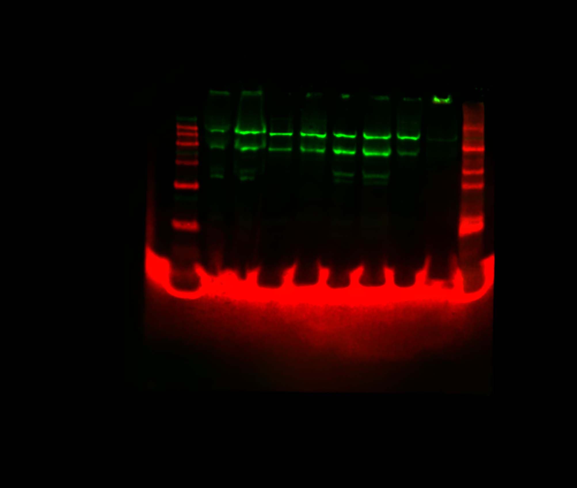
Application: Western BlotSample Tested: Adipose tissue and Brown adipose tissueSpecies: MouseVerified Customer | Posted 03/08/2016Samples were nuclear extracts from brown adipose and subcutaneous adipose tissue (20ug protein loaded). Primary antibody (1:1,000) was detected with HRP-anti sheep (1:10,000) then amplified with Licor streptavidin 800 (1:10,000). Near infrared fluorescence gave much better signal-to-noise than chemiluminescence.

There are no reviews that match your criteria.
Protocols
Find general support by application which include: protocols, troubleshooting, illustrated assays, videos and webinars.
- Antigen Retrieval Protocol (PIER)
- Antigen Retrieval for Frozen Sections Protocol
- Appropriate Fixation of IHC/ICC Samples
- Cellular Response to Hypoxia Protocols
- Chromogenic IHC Staining of Formalin-Fixed Paraffin-Embedded (FFPE) Tissue Protocol
- Chromogenic Immunohistochemistry Staining of Frozen Tissue
- ClariTSA™ Fluorophore Kits
- Detection & Visualization of Antibody Binding
- Fluorescent IHC Staining of Frozen Tissue Protocol
- Graphic Protocol for Heat-induced Epitope Retrieval
- Graphic Protocol for the Preparation and Fluorescent IHC Staining of Frozen Tissue Sections
- Graphic Protocol for the Preparation and Fluorescent IHC Staining of Paraffin-embedded Tissue Sections
- Graphic Protocol for the Preparation of Gelatin-coated Slides for Histological Tissue Sections
- IHC Sample Preparation (Frozen sections vs Paraffin)
- Immunofluorescent IHC Staining of Formalin-Fixed Paraffin-Embedded (FFPE) Tissue Protocol
- Immunohistochemistry (IHC) and Immunocytochemistry (ICC) Protocols
- Immunohistochemistry Frozen Troubleshooting
- Immunohistochemistry Paraffin Troubleshooting
- Preparing Samples for IHC/ICC Experiments
- Preventing Non-Specific Staining (Non-Specific Binding)
- Primary Antibody Selection & Optimization
- Protocol for Heat-Induced Epitope Retrieval (HIER)
- Protocol for Making a 4% Formaldehyde Solution in PBS
- Protocol for VisUCyte™ HRP Polymer Detection Reagent
- Protocol for the Preparation & Fixation of Cells on Coverslips
- Protocol for the Preparation and Chromogenic IHC Staining of Frozen Tissue Sections
- Protocol for the Preparation and Chromogenic IHC Staining of Frozen Tissue Sections - Graphic
- Protocol for the Preparation and Chromogenic IHC Staining of Paraffin-embedded Tissue Sections
- Protocol for the Preparation and Chromogenic IHC Staining of Paraffin-embedded Tissue Sections - Graphic
- Protocol for the Preparation and Fluorescent IHC Staining of Frozen Tissue Sections
- Protocol for the Preparation and Fluorescent IHC Staining of Paraffin-embedded Tissue Sections
- Protocol for the Preparation of Gelatin-coated Slides for Histological Tissue Sections
- R&D Systems Quality Control Western Blot Protocol
- TUNEL and Active Caspase-3 Detection by IHC/ICC Protocol
- The Importance of IHC/ICC Controls
- Troubleshooting Guide: Immunohistochemistry
- Troubleshooting Guide: Western Blot Figures
- Western Blot Conditions
- Western Blot Protocol
- Western Blot Protocol for Cell Lysates
- Western Blot Troubleshooting
- Western Blot Troubleshooting Guide
- View all Protocols, Troubleshooting, Illustrated assays and Webinars