
Caspr2 (CNTNAP2, Contactin-associated protein-like 2 and Cell recognition molecule Caspr2) is a 160-170 kDa glycoprotein, a member of the neurexin family of proteins. It is predominantly expressed in nervous system and likely play a role in the formation of functional distinct domains critical for saltatory conduction of nerve impulses in myelinated nerve fibers. Seems to demarcate the juxtaparanodal region of the axo-glial junction. Caspr2 and closely related molecule Caspr/Paranodin demarcate distinct subdomains in myelinated axons. While contactin-associated protein (Caspr) is present at the paranodal junctions, Caspr2 is precisely colocalized with Shaker-like K+ channels in the juxtaparanodal region. Over aa 29-1262, human CASPR2 shares 94% aa sequence identity with mouse Caspr2.

Scientific images may be resized, cropped, or adjusted for brightness or contrast for presentation purposes. These updates are limited to presentation and do not affect the underlying scientific interpretation of the data. Where available, additional source data may be provided upon request.
Key Product Details
Species Reactivity
Applications
Label
Antibody Source
Product Specifications
Immunogen
Pro29-Ala1262
Accession # Q9UHC6
Specificity
Clonality
Host
Isotype
Scientific Data Images for Caspr2 Antibody
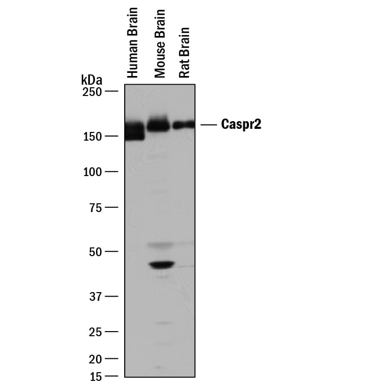
Detection of Human, Mouse, and Rat Caspr2 by Western Blot.
Western blot shows lysates of human brain tissue, mouse brain tissue, and rat brain tissue. PVDF membrane was probed with 2 µg/mL of Sheep Anti-Human/Mouse/Rat Caspr2 Antigen Affinity-purified Polyclonal Antibody (Catalog # AF5145) followed by HRP-conjugated Anti-Sheep IgG Secondary Antibody (Catalog # HAF016). A specific band was detected for Caspr2 at approximately 160-170 kDa (as indicated). This experiment was conducted under reducing conditions and using Immunoblot Buffer Group 1.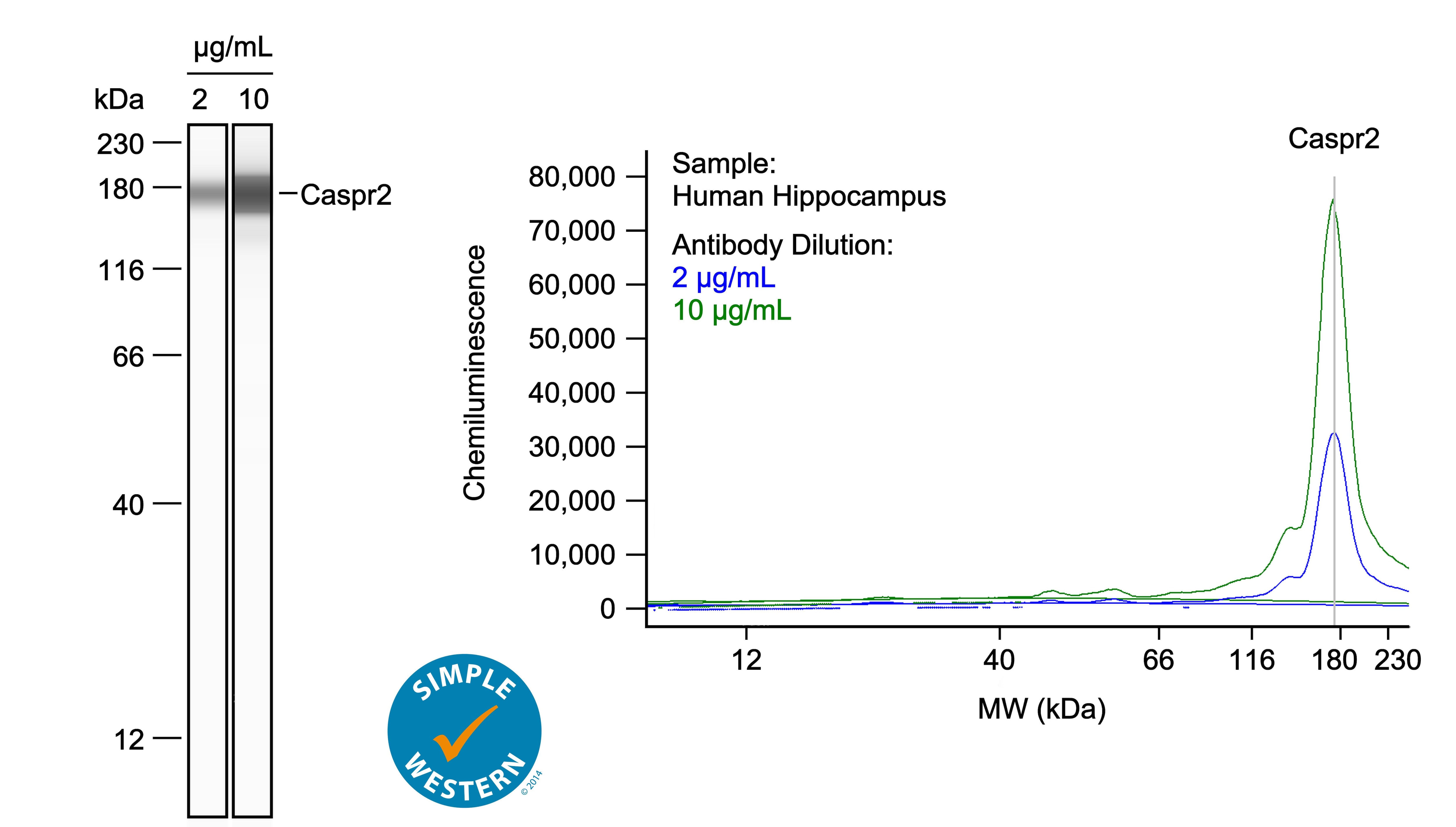
Detection of Human Caspr2 by Simple WesternTM.
Left: Simple Western lane view shows lysates of human hippocampus, loaded at 1 mg/ml. A specific band was detected for Caspr2 at approximately 175 kDa (as indicated) using both 2 µg/ml and 10 µg/ml of Sheep Anti-Human/Mouse/Rat Caspr2 Antigen Affinity-purified Polyclonal Antibody (Catalog # AF5145) followed by 1:50 dilution of HRP-conjugated Anti-Sheep IgG Secondary Antibody (Catalog # HAF016) in Milk-free Antibody Diluent (Catalog # 043-524). This experiment was conducted under reducing conditions and using the 12-230kDa separation system. Right: Simple Western electropherogram showing the same Sheep Anti-Human/Mouse/Rat Caspr2 Antigen Affinity-purified Polyclonal Antibody (Catalog # AF5145) tested at 2 µg/ml (blue line) and 10 µg/ml (green line) in the human hippocampus.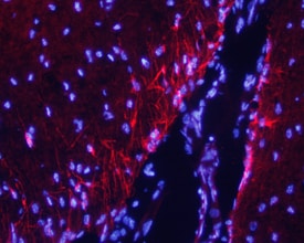
Caspr2 in Mouse Brain.
Caspr2 was detected in perfusion fixed frozen sections of mouse brain using 10 µg/mL Sheep Anti-Human/Mouse/Rat Caspr2 Antigen Affinity-purified Polyclonal Antibody (Catalog # AF5145) overnight at 4 °C. Tissue was stained with the NorthernLights™ 557-conjugated Anti-Sheep IgG Secondary Antibody (red; Catalog # NL010) and counterstained with DAPI (blue). View our protocol for Fluorescent IHC Staining of Frozen Tissue Sections.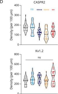
Detection of Caspr2 by Flow Cytometry
Expression of CASPR2 After Long-Term Exposure to CASPR2 aAbs and Structural Integrity of the VGKC Complex(A) Microfluidic chamber (MFC) with embryonic DRGs to separate somatic and axonal compartments and investigate VGKC complex organization after CASPR2 aAb incubation in separated compartments. (B) Exemplary pictures using lattice SIM2 microscopy of soma and axons from DRGs in MFCs treated with healthy control serum. Arrows mark the distance between CASPR2 (magenta) and Kv1.2 (green). (C) Quantification of distance between CASPR2 and Kv1.2 for somata (left) and axons (right) after CASPR2 aAb incubation for 2 hours. Data are shown as mean ± SEM. Somatic and axonal complexes: n = 325–1,465 and n = 1872–3,603, respectively. (D) CASPR2 and Kv1.2 density per 100 µm axon (CASPR2 n = 34–37; Kv1.2 n = 33–37). Data are shown as violin plots with individual values, median = bold line, quartiles = dotted lines. Levels of significance: ns: not significant, *p < 0.05, **p < 0.01, ****p < 0.0001. CASPR2 = Contactin-associated protein 2; DRG = dorsal root ganglia; VGKC complex = voltage-gated potassium channel complex. Image collected and cropped by CiteAb from the following open publication (https://pubmed.ncbi.nlm.nih.gov/40561371), licensed under a CC-BY license. Not internally tested by R&D Systems.Applications for Caspr2 Antibody
Immunohistochemistry
Sample: Perfusion fixed frozen sections of mouse brain
Simple Western
Sample: Human hippocampus tissue
Western Blot
Sample: Human brain tissue, mouse brain tissue, and rat brain tissue
Formulation, Preparation, and Storage
Purification
Reconstitution
Reconstitute at 0.2 mg/mL in sterile PBS. For liquid material, refer to CoA for concentration.
Formulation
Shipping
Stability & Storage
- 12 months from date of receipt, -20 to -70 °C as supplied.
- 1 month, 2 to 8 °C under sterile conditions after reconstitution.
- 6 months, -20 to -70 °C under sterile conditions after reconstitution.
Calculators
Background: Caspr2
Long Name
Alternate Names
Gene Symbol
UniProt
Additional Caspr2 Products
Product Documents for Caspr2 Antibody
Certificate of Analysis
To download a Certificate of Analysis, please enter a lot or batch number in the search box below.
Note: Certificate of Analysis not available for kit components.
Product Specific Notices for Caspr2 Antibody
For research use only
Citations for Caspr2 Antibody
 Powered by Bioz
Customer Reviews for Caspr2 Antibody
There are currently no reviews for this product. Be the first to review Caspr2 Antibody and earn rewards!
Have you used Caspr2 Antibody?
Submit a review and receive an Amazon gift card!
$25/€18/£15/$25CAN/¥2500 for a review with an image
$10/€7/£6/$10CAN/¥1110 for a review without an image
Submit a review
Protocols
Find general support by application which include: protocols, troubleshooting, illustrated assays, videos and webinars.
- Antigen Retrieval Protocol (PIER)
- Antigen Retrieval for Frozen Sections Protocol
- Appropriate Fixation of IHC/ICC Samples
- Cellular Response to Hypoxia Protocols
- Chromogenic IHC Staining of Formalin-Fixed Paraffin-Embedded (FFPE) Tissue Protocol
- Chromogenic Immunohistochemistry Staining of Frozen Tissue
- ClariTSA™ Fluorophore Kits
- Detection & Visualization of Antibody Binding
- Fluorescent IHC Staining of Frozen Tissue Protocol
- Graphic Protocol for Heat-induced Epitope Retrieval
- Graphic Protocol for the Preparation and Fluorescent IHC Staining of Frozen Tissue Sections
- Graphic Protocol for the Preparation and Fluorescent IHC Staining of Paraffin-embedded Tissue Sections
- Graphic Protocol for the Preparation of Gelatin-coated Slides for Histological Tissue Sections
- IHC Sample Preparation (Frozen sections vs Paraffin)
- Immunofluorescent IHC Staining of Formalin-Fixed Paraffin-Embedded (FFPE) Tissue Protocol
- Immunohistochemistry (IHC) and Immunocytochemistry (ICC) Protocols
- Immunohistochemistry Frozen Troubleshooting
- Immunohistochemistry Paraffin Troubleshooting
- Preparing Samples for IHC/ICC Experiments
- Preventing Non-Specific Staining (Non-Specific Binding)
- Primary Antibody Selection & Optimization
- Protocol for Heat-Induced Epitope Retrieval (HIER)
- Protocol for Making a 4% Formaldehyde Solution in PBS
- Protocol for VisUCyte™ HRP Polymer Detection Reagent
- Protocol for the Preparation & Fixation of Cells on Coverslips
- Protocol for the Preparation and Chromogenic IHC Staining of Frozen Tissue Sections
- Protocol for the Preparation and Chromogenic IHC Staining of Frozen Tissue Sections - Graphic
- Protocol for the Preparation and Chromogenic IHC Staining of Paraffin-embedded Tissue Sections
- Protocol for the Preparation and Chromogenic IHC Staining of Paraffin-embedded Tissue Sections - Graphic
- Protocol for the Preparation and Fluorescent IHC Staining of Frozen Tissue Sections
- Protocol for the Preparation and Fluorescent IHC Staining of Paraffin-embedded Tissue Sections
- Protocol for the Preparation of Gelatin-coated Slides for Histological Tissue Sections
- R&D Systems Quality Control Western Blot Protocol
- TUNEL and Active Caspase-3 Detection by IHC/ICC Protocol
- The Importance of IHC/ICC Controls
- Troubleshooting Guide: Immunohistochemistry
- Troubleshooting Guide: Western Blot Figures
- Western Blot Conditions
- Western Blot Protocol
- Western Blot Protocol for Cell Lysates
- Western Blot Troubleshooting
- Western Blot Troubleshooting Guide
- View all Protocols, Troubleshooting, Illustrated assays and Webinars