
Cathepsin X (also known as Cathepsin Z and P) is a cysteine protease of the papain family (1‑5). Compared to other members of the papain family, Cathepsin X has a short proregion and unique insertions. The cysteine residue in the proregion forms a covalent and reversible bond with the active site cysteine residue (6). Acting as a carboxypeptidase, Cathepsin X displays a unique specificity (7‑10). It is ubiquitously expressed in human tissues and conserved in other species such as mouse, nematode and echiuran. The nematode enzyme is apparently involved in molting of third stage larvae (11).
Cathepsin X/Z/P Antibody
R&D Systems | Catalog # AF934


Scientific images may be resized, cropped, or adjusted for brightness or contrast for presentation purposes. These updates are limited to presentation and do not affect the underlying scientific interpretation of the data. Where available, additional source data may be provided upon request.
Key Product Details
Species Reactivity
Validated:
Human, Mouse, Rat
Cited:
Human, Mouse, Transgenic Mouse, Xenograft
Applications
Validated:
Immunohistochemistry, Western Blot, Simple Western, Immunoprecipitation
Cited:
Immunohistochemistry, Immunohistochemistry-Paraffin, Immunohistochemistry-Frozen, Western Blot, Flow Cytometry, Immunocytochemistry, ELISA Capture, ELISA Development
Label
Unconjugated
Antibody Source
Polyclonal Goat IgG
Loading...
Product Specifications
Immunogen
Mouse myeloma cell line NS0-derived recombinant human Cathepsin X/Z/P
Ala21-Val303 (Gly23Val, Ser48Thr)
Accession # Q9UBR2
Ala21-Val303 (Gly23Val, Ser48Thr)
Accession # Q9UBR2
Specificity
Detects human Cathepsin X/Z/P in direct ELISAs and Western blots.
Clonality
Polyclonal
Host
Goat
Isotype
IgG
Scientific Data Images for Cathepsin X/Z/P Antibody
Detection of Human, Mouse, and Rat Cathepsin X/Z/P by Western Blot.
Western blot shows lysates of K562 human chronic myelogenous leukemia cell line, RAW 264.7 mouse monocyte/macrophage cell line, and NR8383 rat alveolar macrophage cell line. PVDF membrane was probed with 1 µg/mL of Goat Anti-Human/ Mouse/Rat Cathepsin X/Z/P Antigen Affinity-purified Polyclonal Antibody (Catalog # AF934) followed by HRP-conjugated Anti-Goat IgG Secondary Antibody (HAF017). A specific band was detected for Cathepsin X/Z/P at approximately 34 kDa (as indicated). This experiment was conducted under reducing conditions and using Immunoblot Buffer Group 1.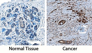
Cathepsin X/Z/P in Human Breast and Breast Cancer Tissue.
Cathepsin X/Z/P was detected in immersion fixed paraffin-embedded sections of normal human breast and breast cancer tissue using Goat Anti-Human/Mouse/Rat Cathepsin X/Z/P Antigen Affinity-purified Polyclonal Antibody (Catalog # AF934) at 3 µg/mL overnight at 4 °C. Tissue was stained using the Anti-Goat HRP-DAB Cell & Tissue Staining Kit (brown; CTS008) and counterstained with hematoxylin (blue). Specific staining was localized to stromal and endothelial cells. View our protocol for Chromogenic IHC Staining of Paraffin-embedded Tissue Sections.
Detection of Human Cathepsin X/Z/P by Simple WesternTM.
Simple Western lane view shows lysates of HeLa human cervical epithelial carcinoma cell line, loaded at 0.2 mg/mL. A specific band was detected for Cathepsin X/Z/P at approximately 36 kDa (as indicated) using 5 µg/mL of Goat Anti-Human/Mouse/Rat Cathepsin X/Z/P Antigen Affinity-purified Polyclonal Antibody (Catalog # AF934) followed by 1:50 dilution of HRP-conjugated Anti-Goat IgG Secondary Antibody (HAF109). This experiment was conducted under reducing conditions and using the 12-230 kDa separation system.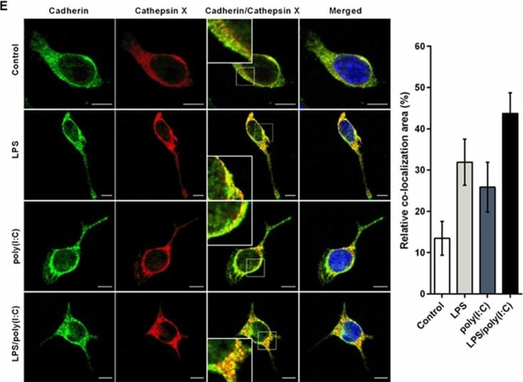
Detection of Cathepsin X/Z/P by Immunocytochemistry/ Immunofluorescence
The effects of simultaneous TLR3/4 activation on cathepsin X expression and activity in microglia. A–D BV2 cells or primary microglia cultures were stimulated with 1 µg/mL LPS and/or 10 µg/mL poly(I:C) in serum-free medium for the times indicated (A, C) or for 24 h (B, D). A, B Cathepsin X protein levels in cell lysates of the control and stimulated cells were measured by ELISA, using a goat anti-cathepsin X capture antibody (AF934) and a mouse anti-cathepsin X detection antibody (3B10). Data are presented as means ± standard deviation of at least two independent experiments, each performed in duplicate. C, D Cathepsin X activity in cell lysates was determined using the cathepsin X-specific substrate Abz-Phe-Glu-Lys(Dnp)-OH. Data are presented as means ± standard deviation of at least two independent experiments, each performed in duplicate. *p ˂ 0.05. E Representative images of double immunofluorescence staining of cadherin (green) and cathepsin X (red) in unstimulated BV2 cells (control) and in BV2 cells stimulated with LPS and/or poly(I:C) for 24 h. Nuclei were counterstained with DAPI (blue). Right: quantification of the relative co-localization areas of cadherin and cathepsin X, presented as means ± standard deviation of pixels (cell numbers ≥ 10). Scale bars: 10 µm. *p ˂ 0.05 Image collected and cropped by CiteAb from the following open publication (https://link.springer.com/10.1007/s12035-021-02694-2), licensed under a CC-BY license. Not internally tested by R&D Systems.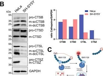
Detection of Cathepsin X/Z/P by Western Blot
Analysis of the endogenous expression and activity of cathepsins in HeLa and SH-SY5Y cells. A Previously published [75] RNA sequencing data from HeLa and SH-SY5Y cells, publicly available through the Human Proteome Atlas portal, show transcripts per million (TPM) for 10 human cathepsins. B Immunoblot and protein expression analysis of endogenous CTSB, CTSD, CTSL and CTSZ expression in HeLa and SH-SY5Y wildtype (WT) cells. The signal intensities of all detected bands with respective CTS-antibodies were normalized to GAPDH. C The quenched cysteine-protease activity-based probe (ABP) BMV-109 contains a fluorophore-quencher pair. Once the ABP binds covalently to the active-site cysteine, the quenching group is released, and the protease is irreversibly labelled, resulting in the emission of fluorescence that can be detected via fluorescent SDS-PAGE. The in-gel fluorescence emission of the scanned SDS-PAGE shows BMV-109-labelled HeLa and SH-SY5Y WT cells treated with or without 50 μM E64D (cysteine-protease inhibitor). Cy5 signal was detected at excitation at 635 nm. Total protein staining with Coomassie Brilliant Blue was used as a loading control. D The immunoblot shows a successful generation of two HeLa CTSB, CTSD, CTSL, CTSZ, and CTSBDLZ KO clones (D) and of SH-SY5Y CTSB, CTSD, CTSL and CTSBDL clones (E) each. F Fluorescent SDS-PAGE of BMV-109-labelled HeLa (F) and SH-SY5Y (G) WT, single KO, and multiple CTS-deficient cells. kDa kilodalton, * indicates non-specific-binding of the antibody, p pro, m mature. sc-single chain, dc-double chain (A, C) “Created with BioRender.com.” Image collected and cropped by CiteAb from the following open publication (https://link.springer.com/10.1007/s00018-024-05274-4), licensed under a CC-BY license. Not internally tested by R&D Systems.Applications for Cathepsin X/Z/P Antibody
Application
Recommended Usage
Immunohistochemistry
3-15 µg/mL
Sample: Immersion fixed paraffin-embedded sections of normal human breast and breast cancer tissue
Sample: Immersion fixed paraffin-embedded sections of normal human breast and breast cancer tissue
Immunoprecipitation
25 µg/mL
Sample: Conditioned cell culture medium spiked with Recombinant Human Cathepsin X/Z/P (Catalog # 934-CY), see our available Western blot detection antibodies
Sample: Conditioned cell culture medium spiked with Recombinant Human Cathepsin X/Z/P (Catalog # 934-CY), see our available Western blot detection antibodies
Simple Western
5 µg/mL
Sample: HeLa human cervical epithelial carcinoma cell line
Sample: HeLa human cervical epithelial carcinoma cell line
Western Blot
1 µg/mL
Sample: K562 human chronic myelogenous leukemia cell line, RAW 264.7 mouse monocyte/macrophage cell line, and NR8383 rat alveolar macrophage cell line
Sample: K562 human chronic myelogenous leukemia cell line, RAW 264.7 mouse monocyte/macrophage cell line, and NR8383 rat alveolar macrophage cell line
Formulation, Preparation, and Storage
Purification
Antigen Affinity-purified
Reconstitution
Reconstitute at 0.2 mg/mL in sterile PBS. For liquid material, refer to CoA for concentration.
Loading...
Formulation
Lyophilized from a 0.2 μm filtered solution in PBS with Trehalose. *Small pack size (SP) is supplied either lyophilized or as a 0.2 µm filtered solution in PBS.
Shipping
Lyophilized product is shipped at ambient temperature. Liquid small pack size (-SP) is shipped with polar packs. Upon receipt, store immediately at the temperature recommended below.
Stability & Storage
Use a manual defrost freezer and avoid repeated freeze-thaw cycles.
- 12 months from date of receipt, -20 to -70 °C as supplied.
- 1 month, 2 to 8 °C under sterile conditions after reconstitution.
- 6 months, -20 to -70 °C under sterile conditions after reconstitution.
Calculators
Background: Cathepsin X/Z/P
References
- Nagler, D.K. and R. Menard (1998) FEBS Lett. 434:135.
- Santamaria, I. et al. (1998) J. Biol. Chem. 273:16816.
- Deussing, J. et al. (2000) Biochim. Biophys. Acta 1491:93.
- Pungercar, J. and G. Ivanovski (2000) Pflugers Arch. Eur. J. Physiol. 439:R116.
- Pungercar, J. et al. (2000) Pflugers Arch. Eur. J. Physiol. 439:R119.
- Sivaraman, J. et al. (2000) J. Mol. Biol. 295:935.
- Menard, R. et al. (2001) Biol. Chem. 382:839.
- Therrien, C. et al. (2001) Biochemistry 40:2702.
- Klemencic, I. et al. (2000) Eur. J. Biochem. 267:5404.
- Guncar, G. et al. (2000) Structure Fold Des. 8:305.
- Lustigman, S. et al. (1996) J. Biol. Chem. 271:30181.
Alternate Names
Cathepsin P, Cathepsin Z, CTSZ
Gene Symbol
CTSZ
UniProt
Additional Cathepsin X/Z/P Products
Product Documents for Cathepsin X/Z/P Antibody
Certificate of Analysis
To download a Certificate of Analysis, please enter a lot or batch number in the search box below.
Note: Certificate of Analysis not available for kit components.
Product Specific Notices for Cathepsin X/Z/P Antibody
For research use only
Related Research Areas
Citations for Cathepsin X/Z/P Antibody
 Powered by Bioz
Customer Reviews for Cathepsin X/Z/P Antibody
There are currently no reviews for this product. Be the first to review Cathepsin X/Z/P Antibody and earn rewards!
Have you used Cathepsin X/Z/P Antibody?
Submit a review and receive an Amazon gift card!
$25/€18/£15/$25CAN/¥2500 for a review with an image
$10/€7/£6/$10CAN/¥1110 for a review without an image
Submit a review
Protocols
Find general support by application which include: protocols, troubleshooting, illustrated assays, videos and webinars.
- Antigen Retrieval Protocol (PIER)
- Antigen Retrieval for Frozen Sections Protocol
- Appropriate Fixation of IHC/ICC Samples
- Cellular Response to Hypoxia Protocols
- Chromogenic IHC Staining of Formalin-Fixed Paraffin-Embedded (FFPE) Tissue Protocol
- Chromogenic Immunohistochemistry Staining of Frozen Tissue
- ClariTSA™ Fluorophore Kits
- Detection & Visualization of Antibody Binding
- Fluorescent IHC Staining of Frozen Tissue Protocol
- Graphic Protocol for Heat-induced Epitope Retrieval
- Graphic Protocol for the Preparation and Fluorescent IHC Staining of Frozen Tissue Sections
- Graphic Protocol for the Preparation and Fluorescent IHC Staining of Paraffin-embedded Tissue Sections
- Graphic Protocol for the Preparation of Gelatin-coated Slides for Histological Tissue Sections
- IHC Sample Preparation (Frozen sections vs Paraffin)
- Immunofluorescent IHC Staining of Formalin-Fixed Paraffin-Embedded (FFPE) Tissue Protocol
- Immunohistochemistry (IHC) and Immunocytochemistry (ICC) Protocols
- Immunohistochemistry Frozen Troubleshooting
- Immunohistochemistry Paraffin Troubleshooting
- Immunoprecipitation Protocol
- Preparing Samples for IHC/ICC Experiments
- Preventing Non-Specific Staining (Non-Specific Binding)
- Primary Antibody Selection & Optimization
- Protocol for Heat-Induced Epitope Retrieval (HIER)
- Protocol for Making a 4% Formaldehyde Solution in PBS
- Protocol for VisUCyte™ HRP Polymer Detection Reagent
- Protocol for the Preparation & Fixation of Cells on Coverslips
- Protocol for the Preparation and Chromogenic IHC Staining of Frozen Tissue Sections
- Protocol for the Preparation and Chromogenic IHC Staining of Frozen Tissue Sections - Graphic
- Protocol for the Preparation and Chromogenic IHC Staining of Paraffin-embedded Tissue Sections
- Protocol for the Preparation and Chromogenic IHC Staining of Paraffin-embedded Tissue Sections - Graphic
- Protocol for the Preparation and Fluorescent IHC Staining of Frozen Tissue Sections
- Protocol for the Preparation and Fluorescent IHC Staining of Paraffin-embedded Tissue Sections
- Protocol for the Preparation of Gelatin-coated Slides for Histological Tissue Sections
- R&D Systems Quality Control Western Blot Protocol
- TUNEL and Active Caspase-3 Detection by IHC/ICC Protocol
- The Importance of IHC/ICC Controls
- Troubleshooting Guide: Immunohistochemistry
- Troubleshooting Guide: Western Blot Figures
- Western Blot Conditions
- Western Blot Protocol
- Western Blot Protocol for Cell Lysates
- Western Blot Troubleshooting
- Western Blot Troubleshooting Guide
- View all Protocols, Troubleshooting, Illustrated assays and Webinars
Loading...