Histone H2AX is a core histone protein that is phosphorylated at S139 in cells exposed to DNA double-strand break-inducing agents, such as ionizing radiation. The S139 phosphorylated H2AX, termed gamma -H2AX, marks the site of DNA double-strand breaks and serves to recruit cell cycle checkpoint and DNA repair factors to the site of damage.

Loading...
Key Product Details
Species Reactivity
Validated:
Human, Mouse, Rat
Cited:
Human, Mouse
Applications
Validated:
Western Blot, Immunocytochemistry
Cited:
Immunohistochemistry, Immunohistochemistry-Paraffin, Immunohistochemistry-Frozen, Western Blot, Immunocytochemistry, IR Fluorescence
Label
Unconjugated
Antibody Source
Monoclonal Mouse IgG2A Clone # 322105
Loading...
Product Specifications
Immunogen
E. coli-derived recombinant human Histone H2AX
Gly3-Ser131
Accession # P16104
Gly3-Ser131
Accession # P16104
Specificity
Detects human, mouse and rat Histone H2AX in Western blots.
Clonality
Monoclonal
Host
Mouse
Isotype
IgG2A
Scientific Data Images for Histone H2AX Antibody (322105)
Detection of Human/Mouse/Rat Histone H2AX by Western Blot.
Western blot shows lysates of HEK293 human embryonic kidney cell line, K562 human chronic myelogenous leukemia cell line, Balb/3T3 mouse embryonic fibroblast cell line, and Nb2-11 rat lymphoma cell line. PVDF membrane was probed with 0.5 µg/mL of Mouse Anti-Human/Mouse/Rat Histone H2AX Monoclonal Antibody (Catalog # MAB3406) followed by HRP-conjugated Anti-Mouse IgG Secondary Antibody (Catalog # HAF007). A specific band was detected for Histone H2AX at approximately 20 kDa (as indicated). This experiment was conducted under reducing conditions and using Immunoblot Buffer Group 1.
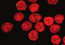
Histone H2AX in HeLa Human Cell Line.
Histone H2AX was detected in immersion fixed HeLa human cervical epithelial carcinoma cell line using Mouse Anti-Human/Mouse/Rat Histone H2AX Monoclonal Antibody (Catalog # MAB3406) at 8 µg/mL for 3 hours at room temperature. Cells were stained using the Northern-Lights™ 557-conjugated Anti-Mouse IgG Secondary Antibody (red; Catalog # NL007) and counterstained with DAPI (blue). Specific staining was localized to nuclei. View our protocol for Fluorescent ICC Staining of Cells on Coverslips.
Detection of Histone H2AX by Western Blot
The NAMPT-dependent NAD+ salvage pathway is necessary for the recovery of NAD+ & induction of apoptosis under weak H2O2 stimulation. A The amounts of endogenous gamma H2AX & NAMPT proteins under 0.7 & 1.1 mM H2O2 stimulation determined by immunoblotting (n = 3). Image collected & cropped by CiteAb from the following open publication (https://pubmed.ncbi.nlm.nih.gov/35410407), licensed under a CC-BY license. Not internally tested by R&D Systems.Applications for Histone H2AX Antibody (322105)
Application
Recommended Usage
Immunocytochemistry
8-25 µg/mL
Sample: Immersion fixed HeLa human cervical epithelial carcinoma cell line
Sample: Immersion fixed HeLa human cervical epithelial carcinoma cell line
Western Blot
0.5 µg/mL
Sample: HEK293 human embryonic kidney cell line, K562 human chronic myelogenous leukemia cell line, Balb/3T3 mouse embryonic fibroblast cell line, and Nb2-11 rat lymphoma cell line
Sample: HEK293 human embryonic kidney cell line, K562 human chronic myelogenous leukemia cell line, Balb/3T3 mouse embryonic fibroblast cell line, and Nb2-11 rat lymphoma cell line
Reviewed Applications
Read 2 reviews rated 4.5 using MAB3406 in the following applications:
Formulation, Preparation, and Storage
Purification
Protein A or G purified from hybridoma culture supernatant
Reconstitution
Reconstitute at 0.5 mg/mL in sterile PBS. For liquid material, refer to CoA for concentration.
Loading...
Formulation
Lyophilized from a 0.2 μm filtered solution in PBS with Trehalose. *Small pack size (SP) is supplied either lyophilized or as a 0.2 µm filtered solution in PBS.
Shipping
Lyophilized product is shipped at ambient temperature. Liquid small pack size (-SP) is shipped with polar packs. Upon receipt, store immediately at the temperature recommended below.
Stability & Storage
Use a manual defrost freezer and avoid repeated freeze-thaw cycles.
- 12 months from date of receipt, -20 to -70 °C as supplied.
- 1 month, 2 to 8 °C under sterile conditions after reconstitution.
- 6 months, -20 to -70 °C under sterile conditions after reconstitution.
Calculators
Background: Histone H2AX
Additional Histone H2AX Products
Product Documents for Histone H2AX Antibody (322105)
Certificate of Analysis
To download a Certificate of Analysis, please enter a lot or batch number in the search box below.
Note: Certificate of Analysis not available for kit components.
Product Specific Notices for Histone H2AX Antibody (322105)
For research use only
Related Research Areas
Citations for Histone H2AX Antibody (322105)
 Powered by Bioz
Customer Reviews for Histone H2AX Antibody (322105) (2)
4.5 out of 5
2 Customer Ratings
Have you used Histone H2AX Antibody (322105)?
Submit a review and receive an Amazon gift card!
$25/€18/£15/$25CAN/¥2500 Yen for a review with an image
$10/€7/£6/$10CAN/¥1110 Yen for a review without an image
Submit a review
Customer Images
Showing
1
-
2 的
2 reviews
Showing All
Filter By:
-
Application: Immunocytochemistry/ImmunofluorescenceSample Tested: SplenocytesSpecies: MouseVerified Customer | Posted 11/04/2021

-
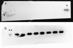
Application: Western BlotSample Tested: Brain (cerebral cortex)Species: MouseVerified Customer | Posted 01/24/201910 micrograms of mouse cortex antibody used at 1/1300 1 second exposition a: membrane after blotting with 15kDa molecular weight (*) b : membrane after blotting with 20kDa molecular weight (**)
There are no reviews that match your criteria.
Protocols
Find general support by application which include: protocols, troubleshooting, illustrated assays, videos and webinars.
- Appropriate Fixation of IHC/ICC Samples
- Cellular Response to Hypoxia Protocols
- ClariTSA™ Fluorophore Kits
- Detection & Visualization of Antibody Binding
- ICC Cell Smear Protocol for Suspension Cells
- ICC Immunocytochemistry Protocol Videos
- ICC for Adherent Cells
- Immunocytochemistry (ICC) Protocol
- Immunocytochemistry Troubleshooting
- Immunofluorescence of Organoids Embedded in Cultrex Basement Membrane Extract
- Immunohistochemistry (IHC) and Immunocytochemistry (ICC) Protocols
- Preparing Samples for IHC/ICC Experiments
- Preventing Non-Specific Staining (Non-Specific Binding)
- Primary Antibody Selection & Optimization
- Protocol for VisUCyte™ HRP Polymer Detection Reagent
- Protocol for the Fluorescent ICC Staining of Cell Smears - Graphic
- Protocol for the Fluorescent ICC Staining of Cultured Cells on Coverslips - Graphic
- Protocol for the Preparation and Fluorescent ICC Staining of Cells on Coverslips
- Protocol for the Preparation and Fluorescent ICC Staining of Non-adherent Cells
- Protocol for the Preparation and Fluorescent ICC Staining of Stem Cells on Coverslips
- Protocol for the Preparation of a Cell Smear for Non-adherent Cell ICC - Graphic
- R&D Systems Quality Control Western Blot Protocol
- TUNEL and Active Caspase-3 Detection by IHC/ICC Protocol
- The Importance of IHC/ICC Controls
- Troubleshooting Guide: Western Blot Figures
- Western Blot Conditions
- Western Blot Protocol
- Western Blot Protocol for Cell Lysates
- Western Blot Troubleshooting
- Western Blot Troubleshooting Guide
- View all Protocols, Troubleshooting, Illustrated assays and Webinars
Loading...