
Key Product Details
Species Reactivity
Human, Mouse
Applications
Immunohistochemistry, Western Blot
Label
Unconjugated
Antibody Source
Monoclonal Mouse IgG1 Clone # 540524
Loading...
Product Specifications
Immunogen
E. coli-derived recombinant human VAMP-1
Met1-Lys96
Accession # P23763
Met1-Lys96
Accession # P23763
Specificity
Detects human and mouse VAMP-1/VAMP-2 in Western blots. This antibody does not react with recombinant VAMP family members 5, 7, and 8.
Clonality
Monoclonal
Host
Mouse
Isotype
IgG1
Scientific Data Images for VAMP-1/VAMP-2 Antibody (540524)
Detection of Human/Mouse VAMP‑1/VAMP‑2 by Western Blot.
Western blot shows lysates of Daudi human Burkitt's lymphoma cell line, HepG2 human hepatocellular carcinoma cell line, L1.2 mouse pro-B cell line, and BaF3 mouse pro-B cell line. PVDF membrane was probed with 0.2 µg/mL of Mouse Anti-Human/Mouse VAMP-1/VAMP-2 Monoclonal Antibody (Catalog # MAB5958) followed by HRP-conjugated Anti-Mouse IgG Secondary Antibody (Catalog # HAF007). A specific band was detected for VAMP-1/VAMP-2 at approximately 18 kDa (as indicated). This experiment was conducted under reducing conditions and using Immunoblot Buffer Group 1.Applications for VAMP-1/VAMP-2 Antibody (540524)
Application
Recommended Usage
Immunohistochemistry
8-25 µg/mL
Sample: Immersion fixed paraffin-embedded sections of human brain (globus pallidus)
Sample: Immersion fixed paraffin-embedded sections of human brain (globus pallidus)
Western Blot
0.2 µg/mL
Sample: Daudi human Burkitt's lymphoma cell line, HepG2 human hepatocellular carcinoma cell line, L1.2 mouse pro-B cell line, and BaF3 mouse pro-B cell line
Sample: Daudi human Burkitt's lymphoma cell line, HepG2 human hepatocellular carcinoma cell line, L1.2 mouse pro-B cell line, and BaF3 mouse pro-B cell line
Reviewed Applications
Read 1 review rated 5 using MAB5958 in the following applications:
Formulation, Preparation, and Storage
Purification
Protein A or G purified from hybridoma culture supernatant
Reconstitution
Reconstitute at 0.5 mg/mL in sterile PBS. For liquid material, refer to CoA for concentration.
Loading...
Formulation
Lyophilized from a 0.2 μm filtered solution in PBS with Trehalose. *Small pack size (SP) is supplied either lyophilized or as a 0.2 µm filtered solution in PBS.
Shipping
Lyophilized product is shipped at ambient temperature. Liquid small pack size (-SP) is shipped with polar packs. Upon receipt, store immediately at the temperature recommended below.
Stability & Storage
Use a manual defrost freezer and avoid repeated freeze-thaw cycles.
- 12 months from date of receipt, -20 to -70 °C as supplied.
- 1 month, 2 to 8 °C under sterile conditions after reconstitution.
- 6 months, -20 to -70 °C under sterile conditions after reconstitution.
Calculators
Background: VAMP-1/VAMP-2
Long Name
Vesicle-Associated Membrane Protein 1/2
Alternate Names
SPAX1, SYB1, VAMP1, VAMP-1, vesicle associated membrane protein 1
Gene Symbol
VAMP1
UniProt
Additional VAMP-1/VAMP-2 Products
Product Documents for VAMP-1/VAMP-2 Antibody (540524)
Certificate of Analysis
To download a Certificate of Analysis, please enter a lot or batch number in the search box below.
Note: Certificate of Analysis not available for kit components.
Product Specific Notices for VAMP-1/VAMP-2 Antibody (540524)
For research use only
Related Research Areas
Customer Reviews for VAMP-1/VAMP-2 Antibody (540524) (1)
5 out of 5
1 Customer Rating
Have you used VAMP-1/VAMP-2 Antibody (540524)?
Submit a review and receive an Amazon gift card!
$25/€18/£15/$25CAN/¥2500 Yen for a review with an image
$10/€7/£6/$10CAN/¥1110 Yen for a review without an image
Submit a review
Customer Images
Showing
1
-
1 的
1 review
Showing All
Filter By:
-
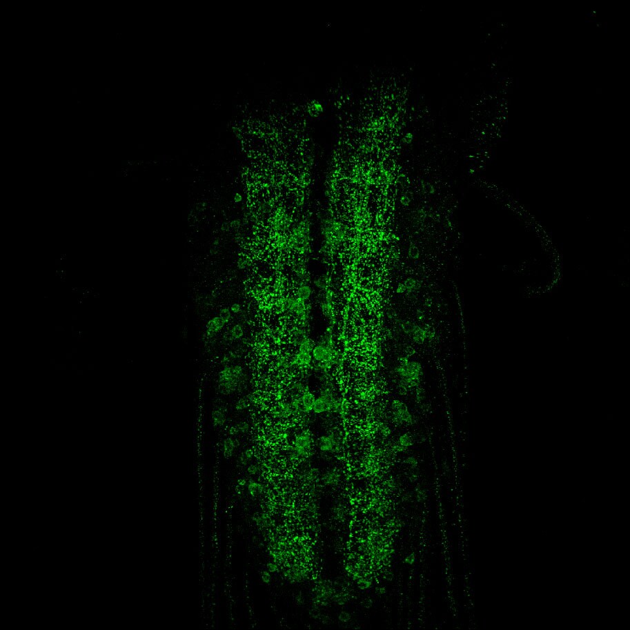
Application: ImmunocytochemistrySample Tested: Larval ventral nerve cord and Larval neuromuscular junctionSpecies: DrosophilaVerified Customer | Posted 02/10/2018Drosophila third instar larval ventral nerve cord.Antibody recognizes Drosophila homolog of VAMP1/2 (neuronal-synaptobrevin) and gives strong staining.

There are no reviews that match your criteria.
Protocols
Find general support by application which include: protocols, troubleshooting, illustrated assays, videos and webinars.
- Antigen Retrieval Protocol (PIER)
- Antigen Retrieval for Frozen Sections Protocol
- Appropriate Fixation of IHC/ICC Samples
- Cellular Response to Hypoxia Protocols
- Chromogenic IHC Staining of Formalin-Fixed Paraffin-Embedded (FFPE) Tissue Protocol
- Chromogenic Immunohistochemistry Staining of Frozen Tissue
- ClariTSA™ Fluorophore Kits
- Detection & Visualization of Antibody Binding
- Fluorescent IHC Staining of Frozen Tissue Protocol
- Graphic Protocol for Heat-induced Epitope Retrieval
- Graphic Protocol for the Preparation and Fluorescent IHC Staining of Frozen Tissue Sections
- Graphic Protocol for the Preparation and Fluorescent IHC Staining of Paraffin-embedded Tissue Sections
- Graphic Protocol for the Preparation of Gelatin-coated Slides for Histological Tissue Sections
- IHC Sample Preparation (Frozen sections vs Paraffin)
- Immunofluorescent IHC Staining of Formalin-Fixed Paraffin-Embedded (FFPE) Tissue Protocol
- Immunohistochemistry (IHC) and Immunocytochemistry (ICC) Protocols
- Immunohistochemistry Frozen Troubleshooting
- Immunohistochemistry Paraffin Troubleshooting
- Preparing Samples for IHC/ICC Experiments
- Preventing Non-Specific Staining (Non-Specific Binding)
- Primary Antibody Selection & Optimization
- Protocol for Heat-Induced Epitope Retrieval (HIER)
- Protocol for Making a 4% Formaldehyde Solution in PBS
- Protocol for VisUCyte™ HRP Polymer Detection Reagent
- Protocol for the Preparation & Fixation of Cells on Coverslips
- Protocol for the Preparation and Chromogenic IHC Staining of Frozen Tissue Sections
- Protocol for the Preparation and Chromogenic IHC Staining of Frozen Tissue Sections - Graphic
- Protocol for the Preparation and Chromogenic IHC Staining of Paraffin-embedded Tissue Sections
- Protocol for the Preparation and Chromogenic IHC Staining of Paraffin-embedded Tissue Sections - Graphic
- Protocol for the Preparation and Fluorescent IHC Staining of Frozen Tissue Sections
- Protocol for the Preparation and Fluorescent IHC Staining of Paraffin-embedded Tissue Sections
- Protocol for the Preparation of Gelatin-coated Slides for Histological Tissue Sections
- R&D Systems Quality Control Western Blot Protocol
- TUNEL and Active Caspase-3 Detection by IHC/ICC Protocol
- The Importance of IHC/ICC Controls
- Troubleshooting Guide: Immunohistochemistry
- Troubleshooting Guide: Western Blot Figures
- Western Blot Conditions
- Western Blot Protocol
- Western Blot Protocol for Cell Lysates
- Western Blot Troubleshooting
- Western Blot Troubleshooting Guide
- View all Protocols, Troubleshooting, Illustrated assays and Webinars
Loading...