
NKG2D, also known as CD314, is a type II transmembrane glycoprotein having an extracellular lectin-like domain. This domain lacks the recognizable calcium-binding sites found in true C‑type lectins and binds protein rather than carbohydrate ligands. Human NKG2D is expressed on CD8+ alpha beta T cells, gamma delta T cells, NK cells, and NKT cells. In mouse systems NKG2D also occurs on macrophages. Human ligands for NKG2D include MICA, MICB, and ULBP1, 2, and 3. Expression of NKG2D ligands occurs in epithelial cells, tumor cells and under conditions of stress or infection. NKG2D exists as a disulfide-linked homodimer that delivers an activating signal upon ligand binding. Signaling requires association with an adapter protein. Alternative splicing of the NKG2D mRNA results in isoforms with different cytoplasmic domains that can associate either with DAP12 to deliver a true activating signal or with DAP10 resulting in a costimulatory signal. NKG2D has been implicated in anti-tumor surveillance and the immune response against viral infection.
Human NKG2D/CD314 Antibody (149810)
R&D Systems | Catalog # MAB139

Scientific images may be resized, cropped, or adjusted for brightness or contrast for presentation purposes. These updates are limited to presentation and do not affect the underlying scientific interpretation of the data. Where available, additional source data may be provided upon request.
Key Product Details
Species Reactivity
Validated:
Cited:
Applications
Validated:
Cited:
Label
Antibody Source
Product Specifications
Immunogen
Specificity
Clonality
Host
Isotype
Endotoxin Level
Scientific Data Images for Human NKG2D/CD314 Antibody (149810)
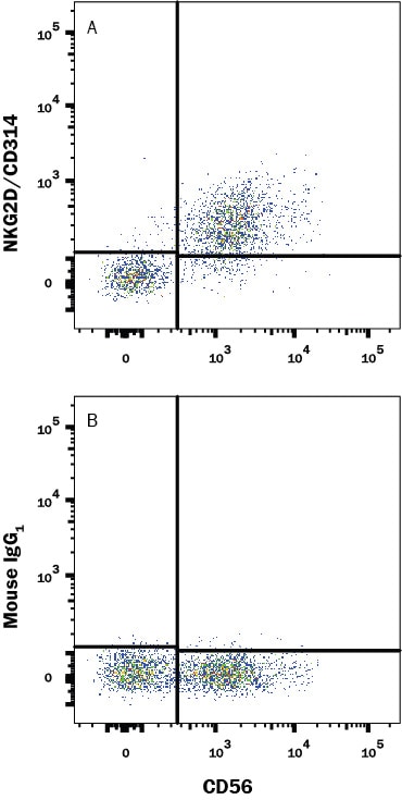
Detection of NKG2D/CD314 in Human PBMCs gated on CD3- cells by Flow Cytometry.
Human peripheral blood mononuclear cells (PBMCs) gated on CD3- cells were stained with Mouse Anti-Human NCAM-1/CD56 APC-conjugated Monoclonal Antibody (Catalog # FAB2408A) and either (A) Mouse Anti-Human NKG2D/CD314 Monoclonal Antibody (Catalog # MAB139) or (B) Mouse IgG1 Isotype Control (Catalog # MAB002) followed by anti-Mouse IgG PE-conjugated Monoclonal Antibody (Catalog # F0102B). View our protocol for Staining Membrane-associated Proteins.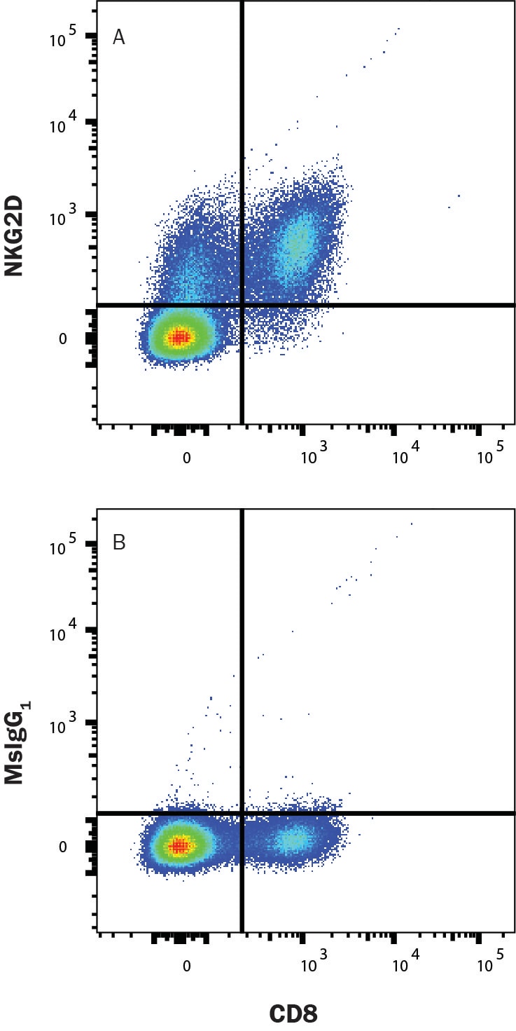
Detection of NKG2D/CD314 in Human PBMCs by Flow Cytometry.
Human peripheral blood mononuclear cells (PBMCs) were stained with Mouse Anti-Human CD8 APC-conjugated Monoclonal Antibody (FAB1509A) and either (A) Mouse Anti-Human NKG2D/CD314 Monoclonal Antibody (Catalog # MAB139) or (B) Mouse IgG1 Isotype Control (MAB002) followed by anti-Mouse IgG PE-conjugated Monoclonal Antibody (F0102B). Staining was performed using our Staining Membrane-associated Proteins protocol.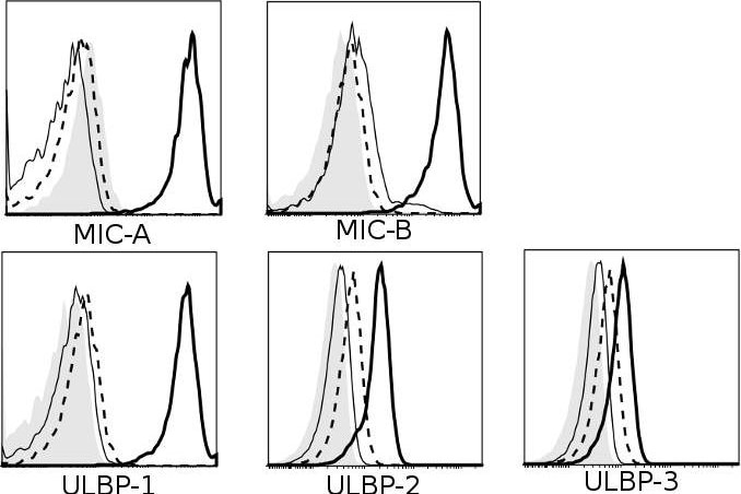
Detection of Human NKG2D/CD314 by Flow Cytometry
Multiple receptors and ligands are involved in NK cell-mediated lysis of activated CD4+ T cells.Role of (A) activating and (B) inhibitory NK receptors in NK cell degranulation. Left column: representative histograms (of n≥3) for surface expression of ligands on activated (thick black line) and resting CD4+ T cells (thin black line). Isotype-matched control Ig are represented by dashed line (activated CD4+ T) and filled histogram (resting CD4+ T). Middle- and right column: NK and CD4+ T cells were activated for 4 days in vitro as described, and co-cultured for 4 hours with 10 ug/mL mAb (or relevant isotype-matched control Ig). Degranulation is shown for CD56dim (middle column) and CD56bright (right column) NK cells. Representative histograms of surface expression of receptors on activated (thick black line) and resting NK cells (thin black line). Isotype-matched control Ig are represented by dashed line (activated NK) and filled histogram (resting NK). * P<0.05, ** P<0.005, *** P<0.001. (C) Sorted IL-2-activated CD56dim and CD56bright NK cells were co-cultured with 51Cr-labeled activated CD4+ T cells in a 51Cr-release assay with human IgG4 isotype control (•) or anti-NKG2A mAb (○). Data represents n = 3 experiments. Image collected and cropped by CiteAb from the following publication (https://pubmed.ncbi.nlm.nih.gov/22384114), licensed under a CC-BY license. Not internally tested by R&D Systems.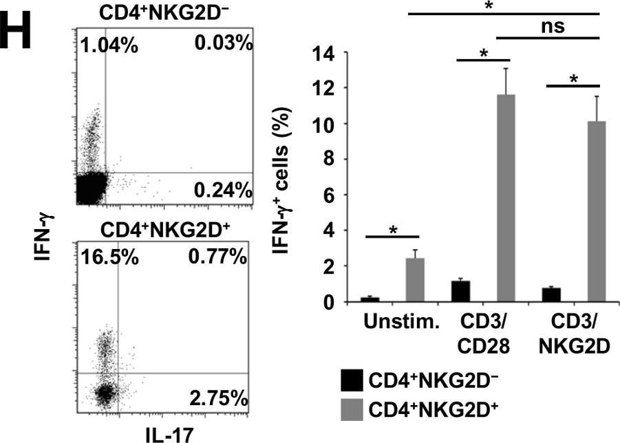
Detection of NKG2D/CD314 by Flow Cytometry
CD4+NKG2D+ T cells exert pro-migratory, cytolytic and pro-inflammatory properties.(A) Flow cytometry: The dot plot shows a representative example of NKG2D expression on CD3+CD4+CD8-CD56− T cells derived from the peripheral blood of a healthy donor (HD). The bar graph represents the mean frequency of CD4+NKG2D+ T cells in the peripheral blood of HDs (n = 20), stable RRMS (n = 15) and active RRMS patients (n = 14). (B–E) Mean fluorescence intensity (MFI) of different markers indicative for migratory capacity (B), activation (C), or cytolytic capacity (D, E) of CD4+NKG2D+ and CD4+NKG2D− T cells from the peripheral blood of HDs (n = 6) or active RRMS patients (n = 6). (F) Percentages of naive (CD45RA+CD62L+), T central memory (Tcm, CD45RA-CD62L+), T effector memory (Tem, CD45RA-CD62L−) and T effector memory RA (Tem-RA, CD45RA+CD62L-) cells in the CD4+NKG2D+ and CD4+NKG2D− T cell compartment assessed by flow cytometry (n = 6 HDs). (G) Carboxyfluorescein succinimidyl ester (CFSE) proliferation assays of CD4+NKG2D+ T cells and CD4+NKG2D− T cells under CD3/CD28, CD3/NKG2D, MOG35-55 (10 µg/ml or 100 µg/ml), MBP1-11 or PLP190-209 stimulation (n = 8 HDs). (H) Intracellular cytokine staining for IFN-gamma and IL-17 of CD4+NKG2D+ and CD4+NKG2D− T cells derived from the peripheral blood of HDs (n = 7). The dot plots depict a representative example of IFN-gamma - and IL-17-positive cells upon CD3/CD28 stimulation. The bar graphs show the frequencies of IFN-gamma or IL-17 positive cells of unstimulated, CD3/CD28- or CD3/NKG2D-stimulated cells. (I) Comparison of the proportions of IFN-gamma or IL-17 positive CD3/CD28-stimulated CD4NKG2D+ T cells derived from frozen PBMCs of HDs (n = 6) or active RRMS patients (n = 6). *P < 0.05. ns, not significant; unstim., unstimulated. Image collected and cropped by CiteAb from the following open publication (https://pubmed.ncbi.nlm.nih.gov/24282598), licensed under a CC-BY license. Not internally tested by R&D Systems.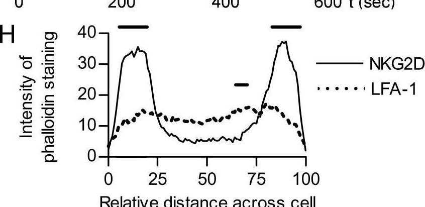
Detection of NKG2D/CD314 by Flow Cytometry
NKG2D and LFA-1 ligation trigger morphologically different responses in NK cells. (H) Quantification of f-actin (phalloidin-AlexaFluor633) across the diameter of cells stimulated on anti-NKG2D (solid line) or anti-LFA-1 (dotted line) (horizontal bars indicate p<0.05 by ANOVA, n = 20). Image collected and cropped by CiteAb from the following open publication (https://pubmed.ncbi.nlm.nih.gov/19636352), licensed under a CC-BY license. Not internally tested by R&D Systems.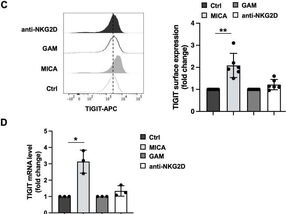
Detection of NKG2D/CD314 by Flow Cytometry
Consequences of MICA-mediated and antibody-mediated NKG2D engagement on cell activation, NKG2D and TIGIT expression. Primary cultured NK cells were incubated with plate-bound recombinant MICA Fc-chimera or anti-NKG2D mAb and goat anti-mouse antibody (GAM). GAM-stimulated NK cells were used as control of NKG2D stimulation. (A) Cells were harvested after 4 hours of stimulation, and degranulation was measured via flow cytometry by assessing CD107a endosomal marker expression. A representative experiment is shown in left panel while means ± SD of three independent experiments are shown in right panel. Paired t test was used for comparisons. *P < 0.05. (B, C) Surface expression of NKG2D and TIGIT receptors was evaluated by FACS analysis upon 18 hours of stimulation. One representative experiment is shown in left panels. Right panels: data pooled from six independent experiments (mean ± SD) are shown (the mean fluorescence intensity, MFI, value of controls NK cells in each experiment is set to 1). Paired t test was used for comparisons. *ρ < 0.05, **ρ < 0.01, ***ρ < 0.001. (D) Relative TIGIT mRNA amount, normalized with GAPDH, was expressed as arbitrary units and referred to the relative controls, considered as calibrator (the value of the calibrator in each run is set to 1). Means ± SD of five independent experiments are shown. Paired t test was used for comparisons. *ρ < 0.05. Image collected and cropped by CiteAb from the following open publication (https://pubmed.ncbi.nlm.nih.gov/40421025), licensed under a CC-BY license. Not internally tested by R&D Systems.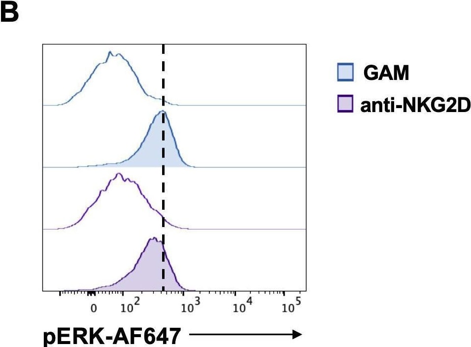
Detection of NKG2D/CD314 by Flow Cytometry
NKG2D stimulation impairs DNAM-1-triggered ERK1/2 phosphorylation. (A, B) goat anti-mouse (GAM) and anti-NKG2D-stimulated NK cells were incubated with anti-DNAM-1 mAb or isotype control Ab followed by goat anti-mouse Ab for different lengths of times. Then, cells were fixed and permeabilized, stained with anti-pAKT (panel A) and anti-pERK1/2 (panel B) and analyzed by FACS. Results from 2 minutes of stimulation with anti-DNAM-1 mAb (filled histograms) or isotype control Ab (empty histograms) from one representative of three independent experiments is shown in the left panels. Quantitative changes in protein phosphorylation at the indicated lengths of time of three independent experiments (mean ± SD) are shown in the right panels. The ratio between the mean fluorescence intensity (MFI) of anti-DNAM-1-stimulated and Ctrl-Ig samples for each time point was used to calculate fold changes. Multiple t test was used for comparisons. *ρ < 0.05. Image collected and cropped by CiteAb from the following open publication (https://pubmed.ncbi.nlm.nih.gov/40421025), licensed under a CC-BY license. Not internally tested by R&D Systems.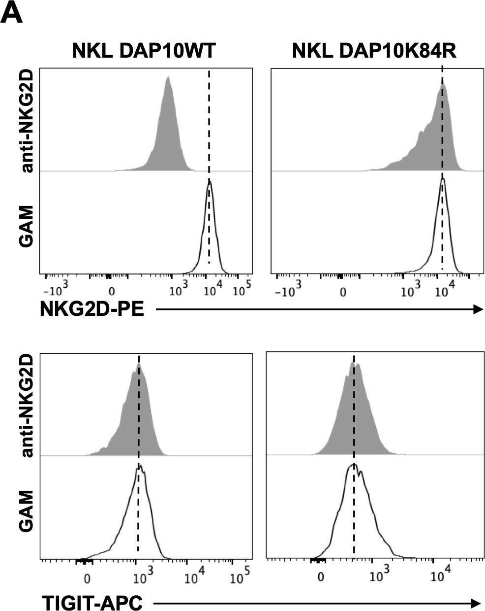
Detection of NKG2D/CD314 by Flow Cytometry
NKG2D internalization is required for impairment in DNAM-1-mediated cytotoxic granule polarization. (A) Surface expression of NKG2D (upper panels) and TIGIT (lower panels) receptors was evaluated on goat anti-mouse (GAM) or anti-NKG2D-stimulated DAP10 WT and DAP10K84R NKL. One representative of three independent experiments is shown in left panels. Data pooled from three independent experiments (mean ± SD) are shown in right panels (the mean fluorescence intensity, MFI value of GAM-stimulated cells in each experiment is set to 1). Paired t test was used for comparisons. *ρ < 0.05. (B) GAM and anti-NKG2D-stimulated DAP10 WT and DAP10K84R NKL (effector cells, E) were incubated with Ba/F3-PVR (target cells, T) for 30 minutes, fixed, permeabilized and stained with the anti-Perforin mAb followed by AlexaFluor594-conjugated GAM-IgG2b and AlexaFluor488-conjugated Phalloidin to visualize polarized actin. Cells were then counterstained with DAPI to visualize nuclei and acquired with LSM980 confocal microscope with a 63x/1.4 NA UPlanSAPO oil immersion objective (all from Zeiss). Right panel: Percentages of conjugates with polarized granules are shown as mean ± SD calculated on ≥30 conjugates for each NK cell type of three independent experiments. Paired t test was used for comparisons. *ρ < 0.05. Image collected and cropped by CiteAb from the following open publication (https://pubmed.ncbi.nlm.nih.gov/40421025), licensed under a CC-BY license. Not internally tested by R&D Systems.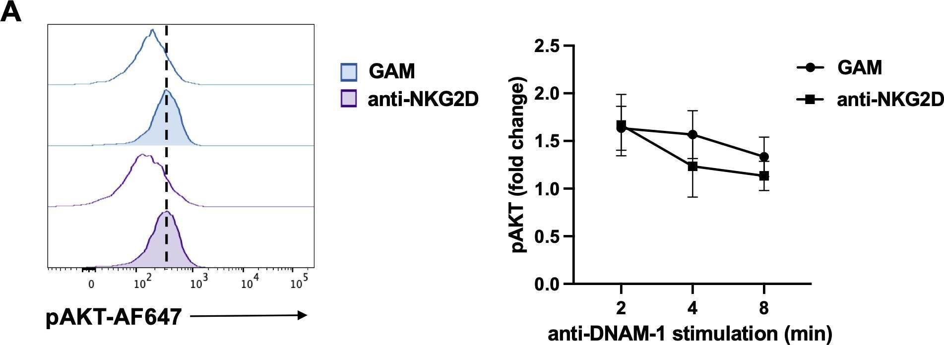
Detection of NKG2D/CD314 by Flow Cytometry
NKG2D stimulation impairs DNAM-1-triggered ERK1/2 phosphorylation. (A, B) goat anti-mouse (GAM) and anti-NKG2D-stimulated NK cells were incubated with anti-DNAM-1 mAb or isotype control Ab followed by goat anti-mouse Ab for different lengths of times. Then, cells were fixed and permeabilized, stained with anti-pAKT (panel A) and anti-pERK1/2 (panel B) and analyzed by FACS. Results from 2 minutes of stimulation with anti-DNAM-1 mAb (filled histograms) or isotype control Ab (empty histograms) from one representative of three independent experiments is shown in the left panels. Quantitative changes in protein phosphorylation at the indicated lengths of time of three independent experiments (mean ± SD) are shown in the right panels. The ratio between the mean fluorescence intensity (MFI) of anti-DNAM-1-stimulated and Ctrl-Ig samples for each time point was used to calculate fold changes. Multiple t test was used for comparisons. *ρ < 0.05. Image collected and cropped by CiteAb from the following open publication (https://pubmed.ncbi.nlm.nih.gov/40421025), licensed under a CC-BY license. Not internally tested by R&D Systems.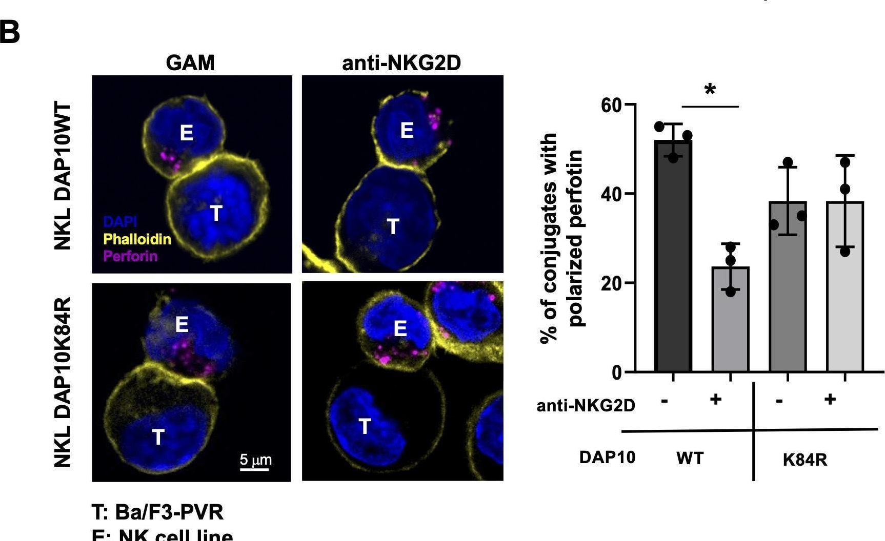
Detection of NKG2D/CD314 by Immunocytochemistry/ Immunofluorescence
NKG2D internalization is required for impairment in DNAM-1-mediated cytotoxic granule polarization. (A) Surface expression of NKG2D (upper panels) and TIGIT (lower panels) receptors was evaluated on goat anti-mouse (GAM) or anti-NKG2D-stimulated DAP10 WT and DAP10K84R NKL. One representative of three independent experiments is shown in left panels. Data pooled from three independent experiments (mean ± SD) are shown in right panels (the mean fluorescence intensity, MFI value of GAM-stimulated cells in each experiment is set to 1). Paired t test was used for comparisons. *ρ < 0.05. (B) GAM and anti-NKG2D-stimulated DAP10 WT and DAP10K84R NKL (effector cells, E) were incubated with Ba/F3-PVR (target cells, T) for 30 minutes, fixed, permeabilized and stained with the anti-Perforin mAb followed by AlexaFluor594-conjugated GAM-IgG2b and AlexaFluor488-conjugated Phalloidin to visualize polarized actin. Cells were then counterstained with DAPI to visualize nuclei and acquired with LSM980 confocal microscope with a 63x/1.4 NA UPlanSAPO oil immersion objective (all from Zeiss). Right panel: Percentages of conjugates with polarized granules are shown as mean ± SD calculated on ≥30 conjugates for each NK cell type of three independent experiments. Paired t test was used for comparisons. *ρ < 0.05. Image collected and cropped by CiteAb from the following open publication (https://pubmed.ncbi.nlm.nih.gov/40421025), licensed under a CC-BY license. Not internally tested by R&D Systems.Applications for Human NKG2D/CD314 Antibody (149810)
Costimulation of T Cells
CyTOF-ready
Flow Cytometry
Sample: Human PBMC
Western Blot
Sample: Recombinant Human NKG2D/CD314 Fc Chimera (Catalog # 1299-NK)
under non-reducing conditions only
Neutralization
Reviewed Applications
Read 3 reviews rated 5 using MAB139 in the following applications:
Flow Cytometry Panel Builder
Bio-Techne Knows Flow Cytometry
Save time and reduce costly mistakes by quickly finding compatible reagents using the Panel Builder Tool.
Advanced Features
- Spectra Viewer - Custom analysis of spectra from multiple fluorochromes
- Spillover Popups - Visualize the spectra of individual fluorochromes
- Antigen Density Selector - Match fluorochrome brightness with antigen density

Formulation, Preparation, and Storage
Purification
Reconstitution
Reconstitute at 0.5 mg/mL in sterile PBS. For liquid material, refer to CoA for concentration.
Formulation
Shipping
Stability & Storage
- 12 months from date of receipt, -20 to -70 °C as supplied.
- 1 month, 2 to 8 °C under sterile conditions after reconstitution.
- 6 months, -20 to -70 °C under sterile conditions after reconstitution.
Calculators
Background: NKG2D/CD314
References
- Li, P. et al. (2001) Nature Immunol. 2:443.
- Steinle, A. et al. (2001) Immunogenetics 53:279.
- Cosman, D. et al. (2001) Immunity 14:123.
- Cerwenka, A. and L. Lanier (2001) Immunol. Rev. 181:158.
- Wu, J. et al. (1999) Science 285:730.
- Diefenbach, A. et al. (2002) Nature Immunol. 3:1142.
- Gilfillan, S. et al. (2002) Nature Immunol. 3:1150.
- Groh, V. et al. (2001) Nature Immunol. 2:255.
- Cerwenka, A. et al. (2001) Proc. Natl. Acad. Sci. USA 98:11521.
- Diefenbach, A. et al. (2001) Nature 413:165.
- NKG2D and its Ligands (2002) www.RnDSystems.com.
Long Name
Alternate Names
Gene Symbol
Additional NKG2D/CD314 Products
Product Documents for Human NKG2D/CD314 Antibody (149810)
Certificate of Analysis
To download a Certificate of Analysis, please enter a lot or batch number in the search box below.
Note: Certificate of Analysis not available for kit components.
Product Specific Notices for Human NKG2D/CD314 Antibody (149810)
For research use only
Related Research Areas
Citations for Human NKG2D/CD314 Antibody (149810)
 Powered by Bioz
Customer Reviews for Human NKG2D/CD314 Antibody (149810) (3)
Have you used Human NKG2D/CD314 Antibody (149810)?
Submit a review and receive an Amazon gift card!
$25/€18/£15/$25CAN/¥2500 for a review with an image
$10/€7/£6/$10CAN/¥1110 for a review without an image
Submit a review
Customer Images
-
Application: Western BlotSample Tested: Peripheral blood mononuclear cells (PBMCs)Species: HumanVerified Customer | Posted 01/28/2022Peripheral blood mononuclear cells (PBMCs)

-
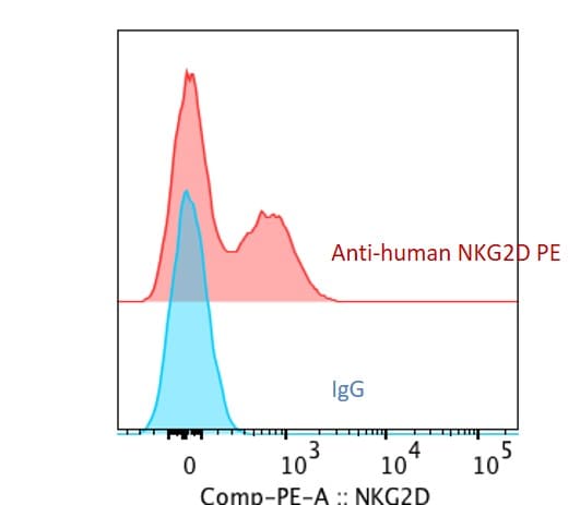
Application: Flow CytometrySample Tested: Peripheral blood mononuclear cells (PBMCs)Species: HumanVerified Customer | Posted 01/24/2021
-
Application: ImmunohistochemistrySample Tested: Paraffined tumor tissue from HCC xenograftSpecies: HumanVerified Customer | Posted 04/13/2018
There are no reviews that match your criteria.
Protocols
Find general support by application which include: protocols, troubleshooting, illustrated assays, videos and webinars.
- 7-Amino Actinomycin D (7-AAD) Cell Viability Flow Cytometry Protocol
- Cellular Response to Hypoxia Protocols
- Extracellular Membrane Flow Cytometry Protocol
- Flow Cytometry Protocol for Cell Surface Markers
- Flow Cytometry Protocol for Staining Membrane Associated Proteins
- Flow Cytometry Staining Protocols
- Flow Cytometry Troubleshooting Guide
- Intracellular Flow Cytometry Protocol Using Alcohol (Methanol)
- Intracellular Flow Cytometry Protocol Using Detergents
- Intracellular Nuclear Staining Flow Cytometry Protocol Using Detergents
- Intracellular Staining Flow Cytometry Protocol Using Alcohol Permeabilization
- Intracellular Staining Flow Cytometry Protocol Using Detergents to Permeabilize Cells
- Propidium Iodide Cell Viability Flow Cytometry Protocol
- Protocol for Liperfluo
- Protocol for the Characterization of Human Th22 Cells
- Protocol for the Characterization of Human Th9 Cells
- Protocol: Annexin V and PI Staining by Flow Cytometry
- Protocol: Annexin V and PI Staining for Apoptosis by Flow Cytometry
- R&D Systems Quality Control Western Blot Protocol
- Troubleshooting Guide: Fluorokine Flow Cytometry Kits
- Troubleshooting Guide: Western Blot Figures
- Western Blot Conditions
- Western Blot Protocol
- Western Blot Protocol for Cell Lysates
- Western Blot Troubleshooting
- Western Blot Troubleshooting Guide
- View all Protocols, Troubleshooting, Illustrated assays and Webinars
Associated Pathways

