
Heat shock proteins (HSPs) are a family of highly conserved stress response proteins. Heat shock proteins function primarily as molecular chaperones by facilitating the folding of other cellular proteins, preventing protein aggregation or targeting improperly folded proteins to specific degradative pathways. HSPs are typically expressed at low levels under normal physiological conditions but are dramatically up-regulated in response to cellular stress. Elevated levels of HSPs have been observed in association with ischemia/reperfusion, cancer, and chronic heart failure. HSP27 is a member of the small heat shock protein family, which also includes HSP25 and the alpha -crystallins. Ser78 and Ser82 of HSP27 are phosphorylated in vivo in response to growth factors or heat shock, and the extent of phosphorylation plays a role in determining specific functions. HSP27 forms a large oligomer and functions as an anti-apoptotic molecule, regulating apoptosis through direct interaction with key components of the apoptotic pathway. HSP27 binds and sequesters cytochrome c released from the mitochondria in response to an apoptotic stimulus. This prevents the proper assembly of the apoptosome and subsequently, the activation of procaspase-9 and procaspase-3.
Human phospho-HSP27 (S78/S82) Antibody (1026B)
R&D Systems | Catalog # MAB23141

Scientific images may be resized, cropped, or adjusted for brightness or contrast for presentation purposes. These updates are limited to presentation and do not affect the underlying scientific interpretation of the data. Where available, additional source data may be provided upon request.
Key Product Details
Validated by
Species Reactivity
Validated:
Cited:
Applications
Validated:
Cited:
Label
Antibody Source
Product Specifications
Immunogen
Accession # P04792
Specificity
Clonality
Host
Isotype
Scientific Data Images for Human phospho-HSP27 (S78/S82) Antibody (1026B)
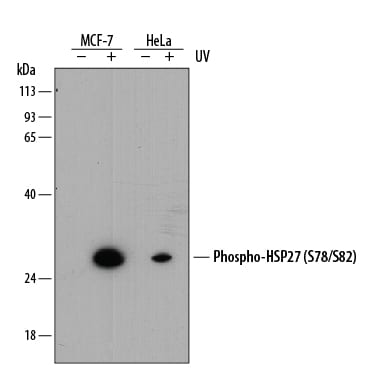
Detection of Human Phospho-HSP27 (S78/S82) by Western Blot.
Western blot shows lysates of MCF-7 human breast cancer cell line and HeLa human cervical epithelial carcinoma cell line untreated (-) or treated (+) with 20 mJ/cm2ultraviolet light (UV) with a 30 minute recovery. PVDF membrane was probed with 1 µg/mL of Rabbit Anti-Human Phospho-HSP27 (S78/S82) Monoclonal Antibody (Catalog # MAB23141) followed by HRP-conjugated Anti-Rabbit IgG Secondary Antibody (Catalog # HAF008). A specific band was detected for Phospho-HSP27 (S78/S82) at approximately 27 kDa (as indicated). This experiment was conducted under reducing conditions and using Immunoblot Buffer Group 1.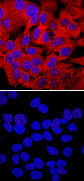
Phospho-HSP27 (S78/S82) in HeLa Human Cell Line.
HSP27 phosphorylated at S78/S82 was detected in immersion fixed HeLa human cervical epithelial carcinoma cell line unstimulated (lower panel) or stimulated with 20 mJ/cm2ultraviolet radiation (upper panel) using Rabbit Anti-Human Phospho-HSP27 (S78/S82) Monoclonal Antibody (Catalog # MAB23141) at a 10 µg/mL for 3 hours at room temperature. Cells were stained using the NorthernLights™ 557-conjugated Anti-Rabbit IgG Secondary Antibody (red; Catalog # NL004) and counterstained with DAPI (blue). Specific staining was localized to cytoplasm and nuclei. View our protocol for Fluorescent ICC Staining of Cells on Coverslips.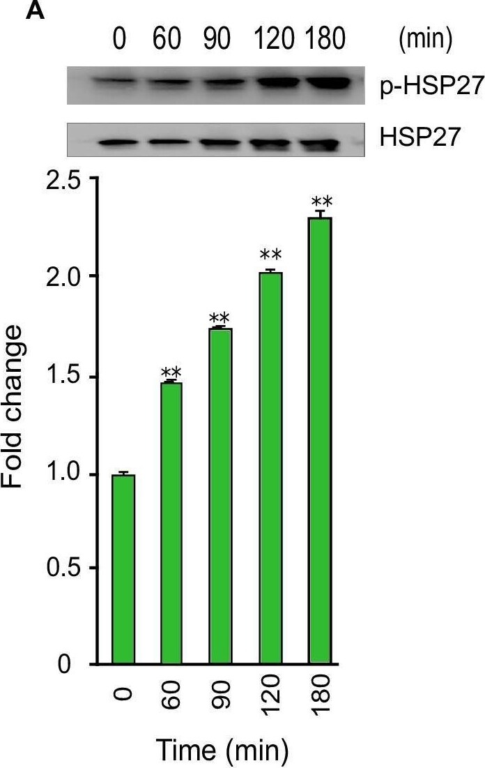
Detection of Phospho-HSP27 (S78/S82) by Western Blot
Phosphorylation of Akt, RSK, and CREB during T. cruzi infection. Lysates from primary HCoEpiC infected with T. cruzi at multiple time points were resolved by SDS-PAGE, blotted, and probed with antibodies against (A) p-HSP27 (B) p-Akt T308, (C) p-Akt S473, (D) p-RSK, and (E) p-CREB, and developed as described. The blots were stripped and reprobed with antibodies against the respective total proteins. The blots were developed by chemiluminescence and scanned. The normalized fold change in the level of each phosphorylated protein was determined and plotted in the bar graph below the respective blot. The bar graphs represent mean values ± SE from three independent biological replicates. The value of p<0.05 was considered significant; *p<0.05 **p<0.001. Image collected and cropped by CiteAb from the following open publication (https://pubmed.ncbi.nlm.nih.gov/30222739), licensed under a CC-BY license. Not internally tested by R&D Systems.Applications for Human phospho-HSP27 (S78/S82) Antibody (1026B)
Immunocytochemistry
Sample: Immersion fixed HeLa human cervical epithelial carcinoma cell line unstimulated or stimulated with 20 mJ/cm2 ultraviolet radiation
Western Blot
Sample: MCF‑7 human breast cancer cell line and HeLa human cervical epithelial carcinoma cell line treated with ultraviolet light (UV)
Reviewed Applications
Read 1 review rated 5 using MAB23141 in the following applications:
Formulation, Preparation, and Storage
Purification
Reconstitution
Reconstitute at 0.5 mg/mL in sterile PBS. For liquid material, refer to CoA for concentration.
Formulation
Shipping
Stability & Storage
Calculators
Background: HSP27
References
- Gusev, N.B. et al. (2002) Biochemistry (Moscow) 67:511.
- Garrido, C. et al. (2001) Biochem. Biophys. Res. Commun. 286:433.
- Garrido, C. (2002) Cell Death Diffr. 9:483.
- Brvey, J-M. et al. (2000) Nat. Cell Biol. 2:645.
Long Name
Alternate Names
Gene Symbol
UniProt
Additional HSP27 Products
Product Documents for Human phospho-HSP27 (S78/S82) Antibody (1026B)
Certificate of Analysis
To download a Certificate of Analysis, please enter a lot or batch number in the search box below.
Note: Certificate of Analysis not available for kit components.
Product Specific Notices for Human phospho-HSP27 (S78/S82) Antibody (1026B)
For research use only
Related Research Areas
Citations for Human phospho-HSP27 (S78/S82) Antibody (1026B)
 Powered by Bioz
Customer Reviews for Human phospho-HSP27 (S78/S82) Antibody (1026B) (1)
Have you used Human phospho-HSP27 (S78/S82) Antibody (1026B)?
Submit a review and receive an Amazon gift card!
$25/€18/£15/$25CAN/¥2500 for a review with an image
$10/€7/£6/$10CAN/¥1110 for a review without an image
Submit a review
Customer Images
-
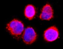
Application: Immunocytochemistry/ImmunofluorescenceSample Tested: HeLa cellsSpecies: HumanVerified Customer | Posted 05/17/2022

There are no reviews that match your criteria.
Protocols
Find general support by application which include: protocols, troubleshooting, illustrated assays, videos and webinars.
- Appropriate Fixation of IHC/ICC Samples
- Cellular Response to Hypoxia Protocols
- ClariTSA™ Fluorophore Kits
- Detection & Visualization of Antibody Binding
- ICC Cell Smear Protocol for Suspension Cells
- ICC Immunocytochemistry Protocol Videos
- ICC for Adherent Cells
- Immunocytochemistry (ICC) Protocol
- Immunocytochemistry Troubleshooting
- Immunofluorescence of Organoids Embedded in Cultrex Basement Membrane Extract
- Immunohistochemistry (IHC) and Immunocytochemistry (ICC) Protocols
- Preparing Samples for IHC/ICC Experiments
- Preventing Non-Specific Staining (Non-Specific Binding)
- Primary Antibody Selection & Optimization
- Protocol for VisUCyte™ HRP Polymer Detection Reagent
- Protocol for the Fluorescent ICC Staining of Cell Smears - Graphic
- Protocol for the Fluorescent ICC Staining of Cultured Cells on Coverslips - Graphic
- Protocol for the Preparation and Fluorescent ICC Staining of Cells on Coverslips
- Protocol for the Preparation and Fluorescent ICC Staining of Non-adherent Cells
- Protocol for the Preparation and Fluorescent ICC Staining of Stem Cells on Coverslips
- Protocol for the Preparation of a Cell Smear for Non-adherent Cell ICC - Graphic
- R&D Systems Quality Control Western Blot Protocol
- TUNEL and Active Caspase-3 Detection by IHC/ICC Protocol
- The Importance of IHC/ICC Controls
- Troubleshooting Guide: Western Blot Figures
- Western Blot Conditions
- Western Blot Protocol
- Western Blot Protocol for Cell Lysates
- Western Blot Troubleshooting
- Western Blot Troubleshooting Guide
- View all Protocols, Troubleshooting, Illustrated assays and Webinars


