
Human ROBO3 is a 200 kDa member of the ROBO family of guidance molecules (1‑3). The term ROBO derives from round-about, a description of the circuitous pathway axons take in the absence of a functional ROBO gene (3, 4). Human ROBO3 is a type I transmembrane glycoprotein that is synthesized as a 1386 amino acid (aa) precursor. It contains a 20 aa signal sequence, an 871 aa extracellular domain (ECD), a 21 aa transmembrane segment, and a 474 aa cytoplasmic region (5, 6). The ECD contains five C2-type Ig-like domains (aa 64‑531) and three fibronectin (FN) type III domains (aa 555‑863). The cytoplasmic region contains three of four possible 15‑20 aa long CC (conserved cytoplasmic) motifs that are found in ROBO1 (7, 8). Human ROBO3 has multiple isoforms. An alternate start site generates a 1365 aa A isoform and a 1341 aa B isoform. These two mature forms differ only over the first 26 and 2 amino acids of the N-terminus, respectively. There are multiple point mutations and insertions in the ROBO3 gene. Three result in truncated forms. One is 456 aa in length and ends after the Ig-like domain # 4. A second is 770 aa in length and ends in the third FN domain. A third isoform is truncated after aa 1108 in the cytoplasmic region after CC2. At least one alternate splice form is also reported. It shows a 10 aa substitution between aa 1025‑1034, followed by truncation. Human ROBO3 ECD is 84% and 91% aa identical to the ECD in mouse and canine ROBO3, respectively. Normally, axons originating on one side of the spinal cord are inhibited from crossing to the other side by a SLIT2-ROBO1 interaction at the midline. ROBO3 is permissive for this event. It is unclear how this is accomplished. One possibility is that it binds directly to ROBO1, blocking SLIT activation. A second possibility involves ROBO3 binding to SLIT2 in a nonproductive interaction. However, only ROBO3 Form B is known to bind to SLIT2 (9‑11).

Scientific images may be resized, cropped, or adjusted for brightness or contrast for presentation purposes. These updates are limited to presentation and do not affect the underlying scientific interpretation of the data. Where available, additional source data may be provided upon request.
Key Product Details
Species Reactivity
Validated:
Cited:
Applications
Validated:
Cited:
Label
Antibody Source
Product Specifications
Immunogen
Leu40-Ser545
Accession # Q96MS0
Specificity
Clonality
Host
Isotype
Scientific Data Images for Human ROBO3 Antibody
Detection of ROBO3 by Immunohistochemistry
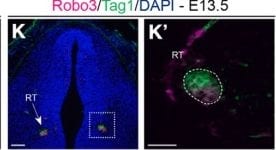
Histology and labeling of axon tracts in the brain of control and Ftm−/− fetuses. A–D, Nissl staining on coronal sections of the brain at two distinct anteroposterior levels of thalamic and hypothalamic regions in E18.5 WT (A, C) and Ftm−/− (B, D) fetuses. C, D, More posterior sections than A and B. Both levels of sections correspond to the ventral hypothalamus and the alar thalamus. Black arrowheads in B–D point to axon fascicles of the IC and RT. Double black arrows in B and D point to the dysmorphic hypothalamus in Ftm mutants. E–F′, Carbocyanine dye staining of corticothalamic (DiI, magenta) and thalamocortical (DiA, green) axons in E18.5 WT (E, E′) and Ftm−/− (F, F′) brains. E′, F′, Higher-magnification of the boxed regions in E and F, respectively. G, H, Neurofilament (NF) immunostaining of axon tracts in E18.5 control (G) and Ftm−/− (H) brains. I–L′, Immunofluorescence for Tuj1 and NF (I–J′) and for Robo3 and Tag1 (K–L′) in E13.5 control (K, K′) and Ftm−/− (L, L′) brains. M–N′, Nissl staining on coronal sections at the level of the eyes of the head of E18.5 WT (M, M′) and Ftm−/− (N, N′) fetuses. M′, N′, Higher-magnification of the boxed regions in M and N, respectively. The arrowhead in N′ points to remnants of the RPE. AN, Anteroventral nucleus; CX, cortex; HB, Habenula; NR, neural retina; V3 third ventricle. Scale bars: (in A, M) A–D, M, N, 1 mm; (in E, G) E–H, 0.5 mm; (in E′, I, K′), E′, F′, I–J′, K′, L′, 0.1 mm; (in K, M′) K, L, M′, N′, 0.2 mm. Image collected and cropped by CiteAb from the following open publication (https://pubmed.ncbi.nlm.nih.gov/30692221), licensed under a CC-BY license. Not internally tested by R&D Systems.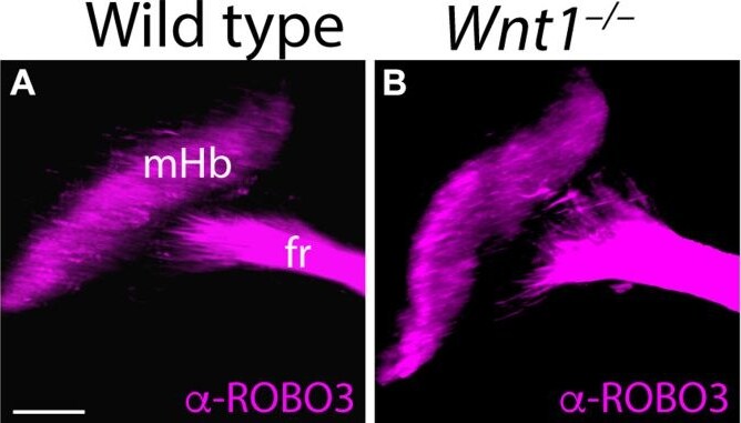
Detection of ROBO3 by Immunohistochemistry
Medial habenular volumes analysis in Wnt1–/–. Lateral views of E18.5 wt (A) and Wnt1–/–(B) brains analyzing ROBO3 distribution by iDISCO. We can observe the mHB and the initial fr portion. (C) Significance among wt (n = 6) and Wnt1–/– mHb volumes (n = 4) was analyzed by an unpaired t-test. Significant differences were found (****< 0.0001), being the mutant mHb volume larger than the wt. fr, fasciculus retroflexus; mHb, medial habenula. Scale bar: 200 μm. Image collected and cropped by CiteAb from the following open publication (https://pubmed.ncbi.nlm.nih.gov/34722541), licensed under a CC-BY license. Not internally tested by R&D Systems.Applications for Human ROBO3 Antibody
Immunohistochemistry
Sample: Immersion fixed frozen sections of embryonic mouse spinal cord (E11.5)
Western Blot
Sample: Recombinant Human ROBO3 Fc Chimera (Catalog # 3076-RB)
Reviewed Applications
Read 1 review rated 5 using AF3076 in the following applications:
Formulation, Preparation, and Storage
Purification
Reconstitution
Reconstitute at 0.2 mg/mL in sterile PBS. For liquid material, refer to CoA for concentration.
Formulation
Shipping
Stability & Storage
- 12 months from date of receipt, -20 to -70 °C as supplied.
- 1 month, 2 to 8 °C under sterile conditions after reconstitution.
- 6 months, -20 to -70 °C under sterile conditions after reconstitution.
Calculators
Background: ROBO3
References
- Rajagopalan, S. et al. (2000) Neuron 28:767.
- Guthrie, S. (2004) Curr. Biol. 14:R632.
- Guthrie, S. (2001) Curr. Biol. 11:R300.
- Seeger, M. et al. (1993) Neuron 10:409.
- Jen, J.C. et al. (2004) Science 304:1509.
- Swiss-Prot. Accession # Q96MS0.
- Bashaw, G.J. et al. (2000) Cell 101:703.
- Kidd, T. et al. (1998) Cell 92:205.
- Camurri, L. et al. (2005) Mol. Cell. Neurosci. 30:485.
- Sabatier, C. et al. (2004) Cell 117:157.
- Mambetisaeva, E.T. et al. (2005) Dev. Dyn. 233:41.
Long Name
Alternate Names
Gene Symbol
UniProt
Additional ROBO3 Products
Product Documents for Human ROBO3 Antibody
Certificate of Analysis
To download a Certificate of Analysis, please enter a lot or batch number in the search box below.
Note: Certificate of Analysis not available for kit components.
Product Specific Notices for Human ROBO3 Antibody
For research use only
Related Research Areas
Citations for Human ROBO3 Antibody
 Powered by Bioz
Customer Reviews for Human ROBO3 Antibody (1)
Have you used Human ROBO3 Antibody?
Submit a review and receive an Amazon gift card!
$25/€18/£15/$25CAN/¥2500 for a review with an image
$10/€7/£6/$10CAN/¥1110 for a review without an image
Submit a review
Customer Images
-
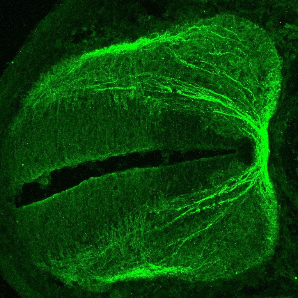
Application: Immunohistochemistry-FrozenSample Tested: Embryonic spinal cordSpecies: MouseVerified Customer | Posted 05/08/2019E11.5 Mouse spinal cord. In green, Comissural neurons are labelled using the antibody against Robo-3.

There are no reviews that match your criteria.
Protocols
Find general support by application which include: protocols, troubleshooting, illustrated assays, videos and webinars.
- Antigen Retrieval Protocol (PIER)
- Antigen Retrieval for Frozen Sections Protocol
- Appropriate Fixation of IHC/ICC Samples
- Cellular Response to Hypoxia Protocols
- Chromogenic IHC Staining of Formalin-Fixed Paraffin-Embedded (FFPE) Tissue Protocol
- Chromogenic Immunohistochemistry Staining of Frozen Tissue
- ClariTSA™ Fluorophore Kits
- Detection & Visualization of Antibody Binding
- Fluorescent IHC Staining of Frozen Tissue Protocol
- Graphic Protocol for Heat-induced Epitope Retrieval
- Graphic Protocol for the Preparation and Fluorescent IHC Staining of Frozen Tissue Sections
- Graphic Protocol for the Preparation and Fluorescent IHC Staining of Paraffin-embedded Tissue Sections
- Graphic Protocol for the Preparation of Gelatin-coated Slides for Histological Tissue Sections
- IHC Sample Preparation (Frozen sections vs Paraffin)
- Immunofluorescent IHC Staining of Formalin-Fixed Paraffin-Embedded (FFPE) Tissue Protocol
- Immunohistochemistry (IHC) and Immunocytochemistry (ICC) Protocols
- Immunohistochemistry Frozen Troubleshooting
- Immunohistochemistry Paraffin Troubleshooting
- Preparing Samples for IHC/ICC Experiments
- Preventing Non-Specific Staining (Non-Specific Binding)
- Primary Antibody Selection & Optimization
- Protocol for Heat-Induced Epitope Retrieval (HIER)
- Protocol for Making a 4% Formaldehyde Solution in PBS
- Protocol for VisUCyte™ HRP Polymer Detection Reagent
- Protocol for the Preparation & Fixation of Cells on Coverslips
- Protocol for the Preparation and Chromogenic IHC Staining of Frozen Tissue Sections
- Protocol for the Preparation and Chromogenic IHC Staining of Frozen Tissue Sections - Graphic
- Protocol for the Preparation and Chromogenic IHC Staining of Paraffin-embedded Tissue Sections
- Protocol for the Preparation and Chromogenic IHC Staining of Paraffin-embedded Tissue Sections - Graphic
- Protocol for the Preparation and Fluorescent IHC Staining of Frozen Tissue Sections
- Protocol for the Preparation and Fluorescent IHC Staining of Paraffin-embedded Tissue Sections
- Protocol for the Preparation of Gelatin-coated Slides for Histological Tissue Sections
- R&D Systems Quality Control Western Blot Protocol
- TUNEL and Active Caspase-3 Detection by IHC/ICC Protocol
- The Importance of IHC/ICC Controls
- Troubleshooting Guide: Immunohistochemistry
- Troubleshooting Guide: Western Blot Figures
- Western Blot Conditions
- Western Blot Protocol
- Western Blot Protocol for Cell Lysates
- Western Blot Troubleshooting
- Western Blot Troubleshooting Guide
- View all Protocols, Troubleshooting, Illustrated assays and Webinars