
Twist-1 (Twist-related protein 1; also H-Twist and bHLHa38) is a 25‑27 kDa class B member of the bHLH transcription factor family of proteins. It is widely expressed in embryo, and select adult cells such as white adipocytes. In fat, Twist-1 induces fatty acid oxidation via PGC-1 alpha and CPT-1, and promotes MCP-1 and TNF-alpha secretion by adipocytes. In epithelium, Twist-1 dysregulation represses E-Cadherin and induces N-Cadherin expression, resulting in an epithelial-to-mesenchymal transition that can lead to cancer. Human Twist-1 is 202 amino acids (aa) in length. It contains a bHLH domain (aa 109-164) with an embedded DNA-binding motif. Twist-1 forms homodimers, and heterodimerizes with TCF3, HAND 1 and HAND 2. Two distinct mutations exist that may impact its dimerization pattern. There is one seven aa insertion after Ile135, and a second seven aa insertion after Pro139. A third unrelated variant shows a four Gly insert after Gly92. Full-length human Twist-1 shares 98% aa identity with mouse Twist-1.

Scientific images may be resized, cropped, or adjusted for brightness or contrast for presentation purposes. These updates are limited to presentation and do not affect the underlying scientific interpretation of the data. Where available, additional source data may be provided upon request.
Key Product Details
Species Reactivity
Validated:
Cited:
Applications
Validated:
Cited:
Label
Antibody Source
Product Specifications
Immunogen
Met1-His202
Accession # Q15672
Specificity
Clonality
Host
Isotype
Scientific Data Images for Human Twist-1 Antibody (927403)
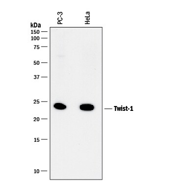
Detection of Human Twist‑1 by Western Blot.
Western blot shows lysates of PC-3 human prostate cancer cell line and HeLa human cervical epithelial carcinoma cell line. PVDF membrane was probed with 2 µg/mL of Mouse Anti-Human Twist-1 Monoclonal Antibody (Catalog # MAB6230) followed by HRP-conjugated Anti-Mouse IgG Secondary Antibody (Catalog # HAF018). A specific band was detected for Twist-1 at approximately 24 kDa (as indicated). This experiment was conducted under reducing conditions and using Immunoblot Buffer Group 1.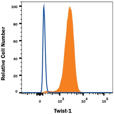
Detection of Twist‑1 in Human PBMCs by Flow Cytometry.
Human peripheral blood mononuclear cell (PBMCs) were stained with Mouse Anti-Human Twist-1 Monoclonal Antibody (Catalog # MAB6230, filled histogram) or isotype control antibody (Catalog # MAB002, open histogram), followed by Phycoerythrin-conjugated Anti-Mouse IgG Secondary Antibody (Catalog # F0102B). To facilitate intracellular staining, cells were fixed with Flow Cytometry Fixation Buffer (Catalog # FC004) and permeabilized with Flow Cytometry Permeabilization/Wash Buffer I (Catalog # FC005). View our protocol for Staining Intracellular Molecules.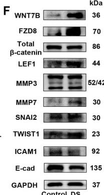
Detection of Twist-1 by Western Blot
DS regulated the adhesion of the aggregates of the H9 cells via canonical Wnt signaling. A GSEA analyses of Wnt-associated genes enriched in the aggregates of the H9 cells treated with DS. B Gene expression analysis by qRT-PCR for Wnt-associated genes, relative gene expression represents data normalized to GADPH and expressed relative to untreated aggregates. C Immunofluorescence analysis of beta -catenin (red) on the aggregates of the H9 cells treated with or without DS; the nuclei were stained with DAPI. Scale bars = 50 μm. D The protein levels of total beta -catenin in nuclei and cytoplasm were determined by western blotting in the aggregates of the H9 cells after treatment with or without DS; the purity of nuclear and cytosolic fractions was verified using laminB1 and GAPDH, respectively. E Gene expression analysis by qRT-PCR for Wnt-targeted genes; relative gene expression represents data normalized to GADPH and expressed relative to untreated aggregates. F The protein levels of WNT7B, FZD8, total beta -catenin, LEF1, MMP3, MMP7, TWIST1, SNAI2, and E-cad were determined by western blotting in the aggregates of the H9 cells after treatment with or without DS; GAPDH was used as a loading control. Data represent the mean ± SD. *P < 0.05 and **P < 0.01 and ***P < 0.001. FZD, frizzled class receptor; LEF1, lymphoid enhancer-binding factor 1; TWIST, twist family bHLH transcription factor; SNAI, snail family transcriptional repressor; MMP, matrix metallopeptidase Image collected and cropped by CiteAb from the following open publication (https://pubmed.ncbi.nlm.nih.gov/35619172), licensed under a CC-BY license. Not internally tested by R&D Systems.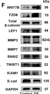
Detection of Twist-1 by Western Blot
DS regulated the adhesion of the aggregates of the H9 cells via canonical Wnt signaling. A GSEA analyses of Wnt-associated genes enriched in the aggregates of the H9 cells treated with DS. B Gene expression analysis by qRT-PCR for Wnt-associated genes, relative gene expression represents data normalized to GADPH and expressed relative to untreated aggregates. C Immunofluorescence analysis of beta -catenin (red) on the aggregates of the H9 cells treated with or without DS; the nuclei were stained with DAPI. Scale bars = 50 μm. D The protein levels of total beta -catenin in nuclei and cytoplasm were determined by western blotting in the aggregates of the H9 cells after treatment with or without DS; the purity of nuclear and cytosolic fractions was verified using laminB1 and GAPDH, respectively. E Gene expression analysis by qRT-PCR for Wnt-targeted genes; relative gene expression represents data normalized to GADPH and expressed relative to untreated aggregates. F The protein levels of WNT7B, FZD8, total beta -catenin, LEF1, MMP3, MMP7, TWIST1, SNAI2, and E-cad were determined by western blotting in the aggregates of the H9 cells after treatment with or without DS; GAPDH was used as a loading control. Data represent the mean ± SD. *P < 0.05 and **P < 0.01 and ***P < 0.001. FZD, frizzled class receptor; LEF1, lymphoid enhancer-binding factor 1; TWIST, twist family bHLH transcription factor; SNAI, snail family transcriptional repressor; MMP, matrix metallopeptidase Image collected and cropped by CiteAb from the following open publication (https://pubmed.ncbi.nlm.nih.gov/35619172), licensed under a CC-BY license. Not internally tested by R&D Systems.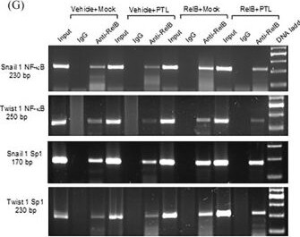
Detection of Twist-1 by Western Blot
Sp1‐RelB cooperation in regulating SNAL1 and TWIST1 genes. (A) Sp1 and NF‐ kappa B binding sites were identified in a 5′ flanking region of the human Snall 1 and Twist 1 genes. PCR primer sets were designed to amplify DNA fragments containing the binding sites, the sequence numbers are according to the transcriptional start site as +1. (B) PLT suppressed the Sp1 and NF‐ kappa B binding activity in PC‐3 cells. (C–E) The Sp1 and NF‐ kappa B interaction in PC‐3 cells was analyzed using Duolink PLA assay. The interaction of p50:RelA and p52:RelB dimers served as positive controls, and PLT was used as an inhibiter (C). RelA‐Sp1 and RelB‐Sp1 interactions were determined at 24 and 48 h (D). PLT inhibited the RelB‐Sp1 interaction (E). (F‐I) The Sp1 and NF‐ kappa B binding sites were precipitated from chromatins derived from RelB‐overexpressed LNCaP cells by ChIP using a RelB antibody. Chromatins without pulldown served as input controls, pulldown with IgG served as negative antibody controls, and PLT served as an inhibiter. Sp1 and RelB proteins in the precipitated complexes were quantified by immunoblotting using specific antibodies, and IgG served as a loading control (F). Precipitated Sp1 and NF‐ kappa B binding sites were amplified by regular PCR (G) and qPCR (H, I) with specific primers shown in (A) and Table S1. Data are shown as the mean ± SD. n = 4, **p < .01, ***p < .001; t‐text (B, D, E), one‐way ANOVA (H, I). Scale bar, 20 µm in (C). Image collected and cropped by CiteAb from the following open publication (https://pubmed.ncbi.nlm.nih.gov/39415352), licensed under a CC-BY license. Not internally tested by R&D Systems.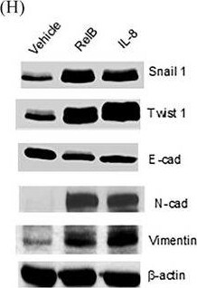
Detection of Twist-1 by Western Blot
RelB and IL‐8 reciprocity in promoting PCa expansibility. (A) RelB and IL‐8 were ectopically expressed in LNCaP cells, and no transfection served as a control. (B) The concentrations of IL‐8 in established cell lines were measured using a relative ELISA kit (n = 4). (C, D) Cell mineralization in RelB‐ or IL‐8‐overexpressed LNCaP cells was quantified with equal cell numbers (upper panel). C4‐2B cell line, an LNCaP cell‐derived bone metastatic lineage, serves as a positive control (n = 6). (E) Cell migration in the established LNCaP cell lines (n = 20). (F, G) Cell invasion in the established LNCaP cell lines (n = 6). (H) Expression of EMT‐associated proteins in the established LNCaP cell lines. (I) Scheme of orthotopic implantation of RelB‐ or IL‐8‐overexpressed LNCaP cells into nude mice prostates. (J, K) Formed tumours in mouse prostates were imaged by IVIS (n = 4). Data are shown as the mean ± SD. *p < .05, **p < .01, ***p < .001, ns, no significance; one‐way ANOVA. Image collected and cropped by CiteAb from the following open publication (https://pubmed.ncbi.nlm.nih.gov/39415352), licensed under a CC-BY license. Not internally tested by R&D Systems.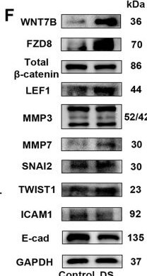
Detection of Twist-1 by Western Blot
DS regulated the adhesion of the aggregates of the H9 cells via canonical Wnt signaling. A GSEA analyses of Wnt-associated genes enriched in the aggregates of the H9 cells treated with DS. B Gene expression analysis by qRT-PCR for Wnt-associated genes, relative gene expression represents data normalized to GADPH and expressed relative to untreated aggregates. C Immunofluorescence analysis of beta -catenin (red) on the aggregates of the H9 cells treated with or without DS; the nuclei were stained with DAPI. Scale bars = 50 μm. D The protein levels of total beta -catenin in nuclei and cytoplasm were determined by western blotting in the aggregates of the H9 cells after treatment with or without DS; the purity of nuclear and cytosolic fractions was verified using laminB1 and GAPDH, respectively. E Gene expression analysis by qRT-PCR for Wnt-targeted genes; relative gene expression represents data normalized to GADPH and expressed relative to untreated aggregates. F The protein levels of WNT7B, FZD8, total beta -catenin, LEF1, MMP3, MMP7, TWIST1, SNAI2, and E-cad were determined by western blotting in the aggregates of the H9 cells after treatment with or without DS; GAPDH was used as a loading control. Data represent the mean ± SD. *P < 0.05 and **P < 0.01 and ***P < 0.001. FZD, frizzled class receptor; LEF1, lymphoid enhancer-binding factor 1; TWIST, twist family bHLH transcription factor; SNAI, snail family transcriptional repressor; MMP, matrix metallopeptidase. Image collected and cropped by CiteAb from the following open publication (https://stemcellres.biomedcentral.com/articles/10.1186/s13287-022-02890…), licensed under a CC-BY license. Not internally tested by R&D Systems.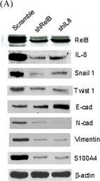
Detection of Twist-1 by Western Blot
RelB and NFATc1 cooperation in upregulating S100A4. (A) RelB and IL‐8 were silenced in PC‐3 cells, and the expression levels of S100A4 and EMT‐associated proteins were quantified. (B, C) The levels of IL‐8 and S100A4 in PC‐3 cell culture media (n = 4). (D) The levels of S100A4 in PTL‐ and FK506‐treated PC‐3 cells (n = 4). (E, F) PTL and FK506 inhibited the S100A4 enhancer‐driven reporter response in PC‐3 cells (n = 4). (G–I) The NF‐ kappa B‐ and NFAT‐binding sites in the S100A4 enhancer were precipitated from PC‐3 cell‐derived chromatins by ChIP using a RelB antibody and amplified by regular PCR (G) or qPCR (H). RelB and NFATc1 proteins in the precipitated complexes were quantified by immunoblotting. (I). (J) The RelB‐NFATc1 interaction was determined using a two‐hybrid system as indicated. (K) The integration of RelB and NFATc1 was examined by luciferase response driven by conjugating the VA16‐activation domain (AD) and the GAL4‐binding domain (BD). P53‐BD/VA40‐AD served as the positive control (n = 4). Data are shown as the mean ± SD. ***p < .001, ns, no significance; t‐test (H, K), one‐way ANOVA (B–E). Image collected and cropped by CiteAb from the following open publication (https://pubmed.ncbi.nlm.nih.gov/39415352), licensed under a CC-BY license. Not internally tested by R&D Systems.Applications for Human Twist-1 Antibody (927403)
CyTOF-ready
Intracellular Staining by Flow Cytometry
Sample: Human peripheral blood mononuclear cell (PBMCs) fixed with Flow Cytometry Fixation Buffer (Catalog # FC004) and permeabilized with Flow Cytometry Permeabilization/Wash Buffer I (Catalog # FC005).
Western Blot
Sample: PC‑3 human prostate cancer cell line and HeLa human cervical epithelial carcinoma cell line
Reviewed Applications
Read 1 review rated 2 using MAB6230 in the following applications:
Flow Cytometry Panel Builder
Bio-Techne Knows Flow Cytometry
Save time and reduce costly mistakes by quickly finding compatible reagents using the Panel Builder Tool.
Advanced Features
- Spectra Viewer - Custom analysis of spectra from multiple fluorochromes
- Spillover Popups - Visualize the spectra of individual fluorochromes
- Antigen Density Selector - Match fluorochrome brightness with antigen density

Formulation, Preparation, and Storage
Purification
Reconstitution
Reconstitute at 0.5 mg/mL in sterile PBS. For liquid material, refer to CoA for concentration.
Formulation
Shipping
Stability & Storage
- 12 months from date of receipt, -20 to -70 °C as supplied.
- 1 month, 2 to 8 °C under sterile conditions after reconstitution.
- 6 months, -20 to -70 °C under sterile conditions after reconstitution.
Calculators
Background: Twist-1
Long Name
Alternate Names
Gene Symbol
UniProt
Additional Twist-1 Products
Product Documents for Human Twist-1 Antibody (927403)
Certificate of Analysis
To download a Certificate of Analysis, please enter a lot or batch number in the search box below.
Note: Certificate of Analysis not available for kit components.
Product Specific Notices for Human Twist-1 Antibody (927403)
For research use only
Citations for Human Twist-1 Antibody (927403)
 Powered by Bioz
Customer Reviews for Human Twist-1 Antibody (927403) (1)
Have you used Human Twist-1 Antibody (927403)?
Submit a review and receive an Amazon gift card!
$25/€18/£15/$25CAN/¥2500 for a review with an image
$10/€7/£6/$10CAN/¥1110 for a review without an image
Submit a review
Customer Images

-
Application: Western BlotSample Tested: mel505Species: HumanVerified Customer | Posted 03/05/20191:500 primary 1:1000 secondary ON incubation with primary 5 min exposure during imaging several non-specific bands
 Bio-Techne ResponseThank you for reviewing our product. We are sorry to hear that this antibody did not perform as expected. We have been in touch with the customer to resolve this issue according to our Product Guarantee and to the customer’s satisfaction.
There are no reviews that match your criteria.
Protocols
Find general support by application which include: protocols, troubleshooting, illustrated assays, videos and webinars.
- 7-Amino Actinomycin D (7-AAD) Cell Viability Flow Cytometry Protocol
- Cellular Response to Hypoxia Protocols
- Extracellular Membrane Flow Cytometry Protocol
- Flow Cytometry Protocol for Cell Surface Markers
- Flow Cytometry Protocol for Staining Membrane Associated Proteins
- Flow Cytometry Staining Protocols
- Flow Cytometry Troubleshooting Guide
- Intracellular Flow Cytometry Protocol Using Alcohol (Methanol)
- Intracellular Flow Cytometry Protocol Using Detergents
- Intracellular Nuclear Staining Flow Cytometry Protocol Using Detergents
- Intracellular Staining Flow Cytometry Protocol Using Alcohol Permeabilization
- Intracellular Staining Flow Cytometry Protocol Using Detergents to Permeabilize Cells
- Propidium Iodide Cell Viability Flow Cytometry Protocol
- Protocol for Liperfluo
- Protocol for the Characterization of Human Th22 Cells
- Protocol for the Characterization of Human Th9 Cells
- Protocol: Annexin V and PI Staining by Flow Cytometry
- Protocol: Annexin V and PI Staining for Apoptosis by Flow Cytometry
- R&D Systems Quality Control Western Blot Protocol
- Troubleshooting Guide: Fluorokine Flow Cytometry Kits
- Troubleshooting Guide: Western Blot Figures
- Western Blot Conditions
- Western Blot Protocol
- Western Blot Protocol for Cell Lysates
- Western Blot Troubleshooting
- Western Blot Troubleshooting Guide
- View all Protocols, Troubleshooting, Illustrated assays and Webinars