VEGFR2 (KDR/Flk-1), VEGFR1 (Flt-1) and VEGFR3 (Flt-4) belong to the class III subfamily of receptor tyrosine kinases (RTKs). All three receptors contain seven immunoglobulin-like repeats in their extracellular domains and kinase insert domains in their intracellular regions. The expression of VEGFR1, 2, and 3 is almost exclusively restricted to the endothelial cells. These receptors are likely to play essential roles in vasculogenesis and angiogenesis. VEGFR3 cDNA encodes a 1298 amino acid (aa) precursor with a 24 aa signal peptide. Mature VEGFR3 is composed of a 751 aa extracellular domain, a 22 aa transmembrane domain and a 482 aa cytoplasmic domain. Both VEGF-C and VEGF-D have been shown to bind and activate VEGFR3 (Flt-4). VEGFR3 is widely expressed in the early embryo but becomes restricted to lymphatic endothelia at later stages of development. It is likely that VEGFR3 may be important for lymph angiogenesis.
Human VEGFR3/Flt-4 Antibody (54703)
R&D Systems | Catalog # MAB3491

Discontinued Product
MAB3491 has been discontinued.
View all VEGFR3/Flt-4 products.
Key Product Details
Species Reactivity
Validated:
Human
Cited:
Human, Mouse
Applications
Validated:
Immunohistochemistry, Western Blot, Immunocytochemistry
Cited:
Immunohistochemistry, Immunohistochemistry-Paraffin, Western Blot, Flow Cytometry, Immunocytochemistry
Label
Unconjugated
Antibody Source
Monoclonal Mouse IgG1 Clone # 54703
Loading...
Product Specifications
Immunogen
Mouse myeloma cell line NS0-derived recombinant human VEGFR3/Flt‑4
Tyr25-Ile776
Accession # P35916
Tyr25-Ile776
Accession # P35916
Specificity
Detects human VEGFR3/Flt‑4 in direct ELISAs and Western blots. In direct ELISAs and Western blots, approximately 25‑30% cross‑reactivity with recombinant mouse VEGFR3 is observed and no cross-reactivity with recombinant human (rh) VEGFR1 or rhVEGFR2 is observed.
Clonality
Monoclonal
Host
Mouse
Isotype
IgG1
Scientific Data Images for Human VEGFR3/Flt-4 Antibody (54703)
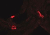
VEGFR3/Flt‑4 in HUVEC Human Cells.
VEGFR3/Flt-4 was detected in immersion fixed HUVEC human umbilical vein endothelial cells using Mouse Anti-Human VEGFR3/Flt-4 Monoclonal Antibody (Catalog # MAB3491) at 10 µg/mL for 3 hours at room temperature. Cells were stained using the NorthernLights™ 557-conjugated Anti-Mouse IgG Secondary Antibody (red; Catalog # NL007) and counterstained with DAPI (blue). Specific staining was localized to cell surfaces and cytoplasm. View our protocol for Fluorescent ICC Staining of Cells on Coverslips.Applications for Human VEGFR3/Flt-4 Antibody (54703)
Application
Recommended Usage
Immunocytochemistry
8-25 µg/mL
Sample: Immersion fixed HUVEC human umbilical vein endothelial cells
Sample: Immersion fixed HUVEC human umbilical vein endothelial cells
Immunohistochemistry
8-25 µg/mL
Sample: Immersion fixed paraffin-embedded sections of human lung
Sample: Immersion fixed paraffin-embedded sections of human lung
Western Blot
1 µg/mL
Sample: Recombinant Human VEGFR3/Flt-4 Fc Chimera (Catalog # 349-F4) under non-reducing conditions only
Sample: Recombinant Human VEGFR3/Flt-4 Fc Chimera (Catalog # 349-F4) under non-reducing conditions only
Reviewed Applications
Read 1 review rated 5 using MAB3491 in the following applications:
Formulation, Preparation, and Storage
Purification
Protein A or G purified from hybridoma culture supernatant
Reconstitution
Reconstitute at 0.5 mg/mL in sterile PBS. For liquid material, refer to CoA for concentration.
Formulation
Lyophilized from a 0.2 μm filtered solution in PBS with Trehalose. *Small pack size (SP) is supplied either lyophilized or as a 0.2 µm filtered solution in PBS.
Shipping
Lyophilized product is shipped at ambient temperature. Liquid small pack size (-SP) is shipped with polar packs. Upon receipt, store immediately at the temperature recommended below.
Stability & Storage
Use a manual defrost freezer and avoid repeated freeze-thaw cycles.
- 12 months from date of receipt, -20 to -70 °C as supplied.
- 1 month, 2 to 8 °C under sterile conditions after reconstitution.
- 6 months, -20 to -70 °C under sterile conditions after reconstitution.
Calculators
Background: VEGFR3/Flt-4
References
- Ferra, N. and R. Davis-Smyth (1997) Endocrine Reviews 18:4.
Long Name
Vascular Endothelial Growth Factor Receptor 3
Alternate Names
Flt-4, FLT4, VEGF R3
Gene Symbol
FLT4
UniProt
Additional VEGFR3/Flt-4 Products
Product Documents for Human VEGFR3/Flt-4 Antibody (54703)
Certificate of Analysis
To download a Certificate of Analysis, please enter a lot or batch number in the search box below.
Note: Certificate of Analysis not available for kit components.
Product Specific Notices for Human VEGFR3/Flt-4 Antibody (54703)
For research use only
Citations for Human VEGFR3/Flt-4 Antibody (54703)
 Powered by Bioz
Customer Reviews for Human VEGFR3/Flt-4 Antibody (54703) (1)
5 out of 5
1 Customer Rating
Have you used Human VEGFR3/Flt-4 Antibody (54703)?
Submit a review and receive an Amazon gift card!
$25/€18/£15/$25CAN/¥2500 Yen for a review with an image
$10/€7/£6/$10CAN/¥1110 Yen for a review without an image
Submit a review
Customer Images
Showing
1
-
1 的
1 review
Showing All
Filter By:
-
Application: Immunocytochemistry/ImmunofluorescenceSample Tested: Kidney tissueSpecies: HumanVerified Customer | Posted 11/02/2021

There are no reviews that match your criteria.
Protocols
Find general support by application which include: protocols, troubleshooting, illustrated assays, videos and webinars.
- Antigen Retrieval Protocol (PIER)
- Antigen Retrieval for Frozen Sections Protocol
- Appropriate Fixation of IHC/ICC Samples
- Cellular Response to Hypoxia Protocols
- Chromogenic IHC Staining of Formalin-Fixed Paraffin-Embedded (FFPE) Tissue Protocol
- Chromogenic Immunohistochemistry Staining of Frozen Tissue
- ClariTSA™ Fluorophore Kits
- Detection & Visualization of Antibody Binding
- Fluorescent IHC Staining of Frozen Tissue Protocol
- Graphic Protocol for Heat-induced Epitope Retrieval
- Graphic Protocol for the Preparation and Fluorescent IHC Staining of Frozen Tissue Sections
- Graphic Protocol for the Preparation and Fluorescent IHC Staining of Paraffin-embedded Tissue Sections
- Graphic Protocol for the Preparation of Gelatin-coated Slides for Histological Tissue Sections
- ICC Cell Smear Protocol for Suspension Cells
- ICC Immunocytochemistry Protocol Videos
- ICC for Adherent Cells
- IHC Sample Preparation (Frozen sections vs Paraffin)
- Immunocytochemistry (ICC) Protocol
- Immunocytochemistry Troubleshooting
- Immunofluorescence of Organoids Embedded in Cultrex Basement Membrane Extract
- Immunofluorescent IHC Staining of Formalin-Fixed Paraffin-Embedded (FFPE) Tissue Protocol
- Immunohistochemistry (IHC) and Immunocytochemistry (ICC) Protocols
- Immunohistochemistry Frozen Troubleshooting
- Immunohistochemistry Paraffin Troubleshooting
- Preparing Samples for IHC/ICC Experiments
- Preventing Non-Specific Staining (Non-Specific Binding)
- Primary Antibody Selection & Optimization
- Protocol for Heat-Induced Epitope Retrieval (HIER)
- Protocol for Making a 4% Formaldehyde Solution in PBS
- Protocol for VisUCyte™ HRP Polymer Detection Reagent
- Protocol for the Fluorescent ICC Staining of Cell Smears - Graphic
- Protocol for the Fluorescent ICC Staining of Cultured Cells on Coverslips - Graphic
- Protocol for the Preparation & Fixation of Cells on Coverslips
- Protocol for the Preparation and Chromogenic IHC Staining of Frozen Tissue Sections
- Protocol for the Preparation and Chromogenic IHC Staining of Frozen Tissue Sections - Graphic
- Protocol for the Preparation and Chromogenic IHC Staining of Paraffin-embedded Tissue Sections
- Protocol for the Preparation and Chromogenic IHC Staining of Paraffin-embedded Tissue Sections - Graphic
- Protocol for the Preparation and Fluorescent ICC Staining of Cells on Coverslips
- Protocol for the Preparation and Fluorescent ICC Staining of Non-adherent Cells
- Protocol for the Preparation and Fluorescent ICC Staining of Stem Cells on Coverslips
- Protocol for the Preparation and Fluorescent IHC Staining of Frozen Tissue Sections
- Protocol for the Preparation and Fluorescent IHC Staining of Paraffin-embedded Tissue Sections
- Protocol for the Preparation of Gelatin-coated Slides for Histological Tissue Sections
- Protocol for the Preparation of a Cell Smear for Non-adherent Cell ICC - Graphic
- R&D Systems Quality Control Western Blot Protocol
- TUNEL and Active Caspase-3 Detection by IHC/ICC Protocol
- The Importance of IHC/ICC Controls
- Troubleshooting Guide: Immunohistochemistry
- Troubleshooting Guide: Western Blot Figures
- Western Blot Conditions
- Western Blot Protocol
- Western Blot Protocol for Cell Lysates
- Western Blot Troubleshooting
- Western Blot Troubleshooting Guide
- View all Protocols, Troubleshooting, Illustrated assays and Webinars
Loading...
Associated Pathways


