KDELR1 Antibody (KR-10) - BSA Free
Novus Biologicals | Catalog # NBP2-12873
![Western Blot: KDELR1 Antibody (KR-10) [NBP2-12873]](https://resources.rndsystems.com/images/products/KDELR1-Antibody-KR-10-Western-Blot-NBP2-12873-img0005.jpg "Western Blot: KDELR1 Antibody (KR-10) [NBP2-12873]")
Key Product Details
Species Reactivity
Validated:
Human, Mouse, Rat, Porcine, Bovine, Canine, Chicken, Drosophila, Hamster, Mammal, Monkey, Rabbit, Sheep, Xenopus
Cited:
Mammal
Applications
Validated:
Immunohistochemistry, Immunohistochemistry-Paraffin, Western Blot, Immunocytochemistry/ Immunofluorescence, Immunoprecipitation
Cited:
Western Blot
Label
Unconjugated
Antibody Source
Monoclonal Mouse IgG1 Clone # KR-10
Format
BSA Free
Loading...
Product Specifications
Immunogen
A 21 residue synthetic peptide (amino acids 192-212) based on the bovine KDEL receptor and the peptide coupled to KLH
Reactivity Notes
Use in Mammal reported in scientific literature (PMID:31653700).
Localization
Endoplasmic Reticulum
Specificity
Detects approx 25kDa.
Clonality
Monoclonal
Host
Mouse
Isotype
IgG1
Scientific Data Images for KDELR1 Antibody (KR-10) - BSA Free
Western Blot: KDELR1 Antibody (KR-10) [NBP2-12873]
Western Blot: KDELR1 Antibody (KR-10) [NBP2-12873] - analysis of Rat tissue lysate showing detection of KDEL Receptor protein using Mouse Anti-KDEL Receptor Monoclonal Antibody, Clone KR-10. Load: 15 ug protein. Block: 1.5% BSA for 30 minutes at RT. Primary Antibody: Mouse Anti-KDEL Receptor Monoclonal Antibody at 1:1000 for 2 hours at RT. Secondary Antibody: Sheep Anti-Mouse IgG: HRP for 1 hour at RT.![Immunocytochemistry/ Immunofluorescence: KDELR1 Antibody (KR-10) [NBP2-12873]](https://resources.rndsystems.com/images/products/KDELR1-Antibody-KR-10-Immunocytochemistry-Immunofluorescence-NBP2-12873-img0006.jpg "Immunocytochemistry/ Immunofluorescence: KDELR1 Antibody (KR-10) [NBP2-12873]")
Immunocytochemistry/ Immunofluorescence: KDELR1 Antibody (KR-10) [NBP2-12873]
Immunocytochemistry/Immunofluorescence: KDELR1 Antibody (KR-10) [NBP2-12873] - Immunocytochemistry/Immunofluorescence analysis using Mouse Anti-KDELR1 Monoclonal Antibody, Clone KR-10 (NBP2-12873). Tissue: NRK cells. Species: Rat. Primary Antibody: Mouse Anti-KDELR1 Monoclonal Antibody (NBP2-12873) at 1:1000. Secondary Antibody: APC Goat Anti-Mouse (red). Counterstain: DAPI (blue) nuclear stain. KR-10 staining red; DAPI staining blue. Merged images. Courtesy of: Institute of Mol. and Cell Bio, Singapore.Applications for KDELR1 Antibody (KR-10) - BSA Free
Application
Recommended Usage
Immunocytochemistry/ Immunofluorescence
1:1000
Immunohistochemistry-Paraffin
1:10-1:500
Western Blot
1:1000
Application Notes
1 ug/ml was sufficient for detection of KDEL receptor in 20 ug monkey Vero cell lysate by colorimetric immunoblot analysis using Goat Anti-Mouse IgG:AP as the secondary.
Reviewed Applications
Read 1 review rated 4 using NBP2-12873 in the following applications:
Formulation, Preparation, and Storage
Purification
Protein G purified
Formulation
PBS (pH 7.2), 50% Glycerol
Format
BSA Free
Preservative
0.09% Sodium Azide
Concentration
1 mg/ml
Shipping
The product is shipped with polar packs. Upon receipt, store it immediately at the temperature recommended below.
Stability & Storage
Store at 4C short term. Aliquot and store at -20C long term. Avoid freeze-thaw cycles.
Background: KDELR1
Alternate Names
ER lumen protein retaining receptor 1, ERD2, ERD2.1KDEL receptor 1, HDEL, KDEL (Lys-Asp-Glu-Leu) endoplasmic reticulum protein retention receptor 1, KDEL endoplasmic reticulum protein retention receptor 1, PM23, Putative MAPK-activating protein PM23
Gene Symbol
KDELR1
UniProt
Additional KDELR1 Products
Product Documents for KDELR1 Antibody (KR-10) - BSA Free
Certificate of Analysis
To download a Certificate of Analysis, please enter a lot or batch number in the search box below.
Product Specific Notices for KDELR1 Antibody (KR-10) - BSA Free
This product is for research use only and is not approved for use in humans or in clinical diagnosis. Primary Antibodies are guaranteed for 1 year from date of receipt.
Citations for KDELR1 Antibody (KR-10) - BSA Free
 Powered by Bioz
Customer Reviews for KDELR1 Antibody (KR-10) - BSA Free (1)
4 out of 5
1 Customer Rating
Have you used KDELR1 Antibody (KR-10) - BSA Free?
Submit a review and receive an Amazon gift card!
$25/€18/£15/$25CAN/¥2500 Yen for a review with an image
$10/€7/£6/$10CAN/¥1110 Yen for a review without an image
Submit a review
Customer Images
Showing
1
-
1 的
1 review
Showing All
Filter By:
-
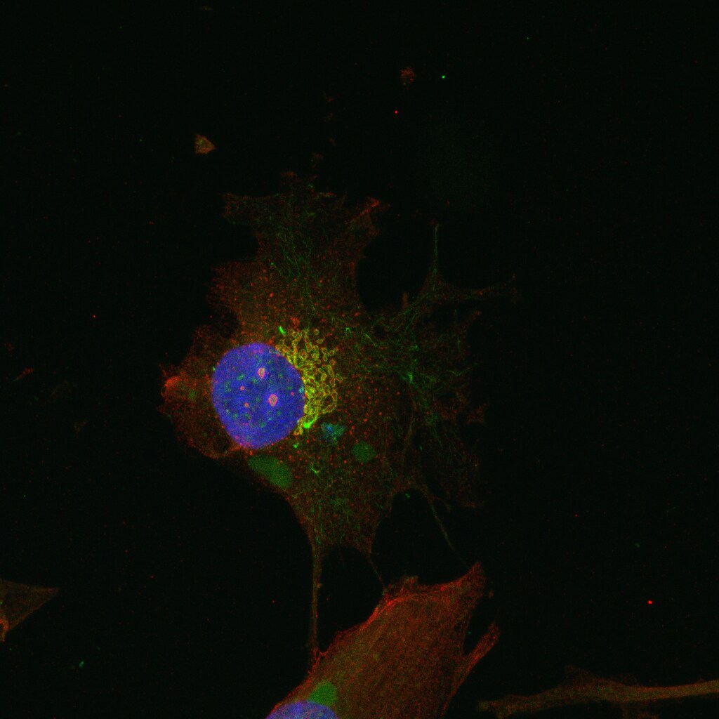
Application: ImmunocytochemistrySample Tested: RPE1 cellsSpecies: HumanVerified Customer | Posted 09/07/2021RPE1 cells were methanol fixed, co-stained with KDELR1 (red) and Giantin (green).

There are no reviews that match your criteria.
Protocols
Find general support by application which include: protocols, troubleshooting, illustrated assays, videos and webinars.
- Antigen Retrieval Protocol (PIER)
- Antigen Retrieval for Frozen Sections Protocol
- Appropriate Fixation of IHC/ICC Samples
- Cellular Response to Hypoxia Protocols
- Chromogenic IHC Staining of Formalin-Fixed Paraffin-Embedded (FFPE) Tissue Protocol
- Chromogenic Immunohistochemistry Staining of Frozen Tissue
- ClariTSA™ Fluorophore Kits
- Detection & Visualization of Antibody Binding
- Fluorescent IHC Staining of Frozen Tissue Protocol
- Graphic Protocol for Heat-induced Epitope Retrieval
- Graphic Protocol for the Preparation and Fluorescent IHC Staining of Frozen Tissue Sections
- Graphic Protocol for the Preparation and Fluorescent IHC Staining of Paraffin-embedded Tissue Sections
- Graphic Protocol for the Preparation of Gelatin-coated Slides for Histological Tissue Sections
- ICC Cell Smear Protocol for Suspension Cells
- ICC Immunocytochemistry Protocol Videos
- ICC for Adherent Cells
- IHC Sample Preparation (Frozen sections vs Paraffin)
- Immunocytochemistry (ICC) Protocol
- Immunocytochemistry Troubleshooting
- Immunofluorescence of Organoids Embedded in Cultrex Basement Membrane Extract
- Immunofluorescent IHC Staining of Formalin-Fixed Paraffin-Embedded (FFPE) Tissue Protocol
- Immunohistochemistry (IHC) and Immunocytochemistry (ICC) Protocols
- Immunohistochemistry Frozen Troubleshooting
- Immunohistochemistry Paraffin Troubleshooting
- Immunoprecipitation Protocol
- Preparing Samples for IHC/ICC Experiments
- Preventing Non-Specific Staining (Non-Specific Binding)
- Primary Antibody Selection & Optimization
- Protocol for Heat-Induced Epitope Retrieval (HIER)
- Protocol for Making a 4% Formaldehyde Solution in PBS
- Protocol for VisUCyte™ HRP Polymer Detection Reagent
- Protocol for the Fluorescent ICC Staining of Cell Smears - Graphic
- Protocol for the Fluorescent ICC Staining of Cultured Cells on Coverslips - Graphic
- Protocol for the Preparation & Fixation of Cells on Coverslips
- Protocol for the Preparation and Chromogenic IHC Staining of Frozen Tissue Sections
- Protocol for the Preparation and Chromogenic IHC Staining of Frozen Tissue Sections - Graphic
- Protocol for the Preparation and Chromogenic IHC Staining of Paraffin-embedded Tissue Sections
- Protocol for the Preparation and Chromogenic IHC Staining of Paraffin-embedded Tissue Sections - Graphic
- Protocol for the Preparation and Fluorescent ICC Staining of Cells on Coverslips
- Protocol for the Preparation and Fluorescent ICC Staining of Non-adherent Cells
- Protocol for the Preparation and Fluorescent ICC Staining of Stem Cells on Coverslips
- Protocol for the Preparation and Fluorescent IHC Staining of Frozen Tissue Sections
- Protocol for the Preparation and Fluorescent IHC Staining of Paraffin-embedded Tissue Sections
- Protocol for the Preparation of Gelatin-coated Slides for Histological Tissue Sections
- Protocol for the Preparation of a Cell Smear for Non-adherent Cell ICC - Graphic
- R&D Systems Quality Control Western Blot Protocol
- TUNEL and Active Caspase-3 Detection by IHC/ICC Protocol
- The Importance of IHC/ICC Controls
- Troubleshooting Guide: Immunohistochemistry
- Troubleshooting Guide: Western Blot Figures
- Western Blot Conditions
- Western Blot Protocol
- Western Blot Protocol for Cell Lysates
- Western Blot Troubleshooting
- Western Blot Troubleshooting Guide
- View all Protocols, Troubleshooting, Illustrated assays and Webinars
Loading...