

![Western Blot: LINE-1 ORF1p Antibody [NBP2-66934]](https://resources.rndsystems.com/images/products/LINE-1-ORF1p-Antibody-Western-Blot-NBP2-66934-img0001.jpg "Western Blot: LINE-1 ORF1p Antibody [NBP2-66934]")
Loading...
Key Product Details
Species Reactivity
Mouse
Applications
Validated:
Immunohistochemistry-Paraffin, Western Blot, Immunocytochemistry/ Immunofluorescence
Cited:
Western Blot
Label
Unconjugated
Antibody Source
Polyclonal Rabbit IgG
Loading...
Product Specifications
Immunogen
Synthetic peptide within mouse LINE-1 ORF1p aa 308-357 / 357. (SwissProt: P11260 Mouse)
Localization
Cell membrane, nucleus
Clonality
Polyclonal
Host
Rabbit
Isotype
IgG
Scientific Data Images for LINE-1 ORF1p Antibody
Western Blot: LINE-1 ORF1p Antibody [NBP2-66934]
Western Blot: LINE-1 ORF1p Antibody [NBP2-66934] - Analysis on different tissue lysates using anti- L1ORF1p rabbit polyclonal antibody.![Immunocytochemistry/ Immunofluorescence: LINE-1 ORF1p Antibody [NBP2-66934]](https://resources.rndsystems.com/images/products/LINE-1-ORF1p-Antibody-Immunocytochemistry-Immunofluorescence-NBP2-66934-img0002.jpg "Immunocytochemistry/ Immunofluorescence: LINE-1 ORF1p Antibody [NBP2-66934]")
Immunocytochemistry/ Immunofluorescence: LINE-1 ORF1p Antibody [NBP2-66934]
Immunocytochemistry/Immunofluorescence: LINE-1 ORF1p Antibody [NBP2-66934] - Staining L1ORF1p in F9 cells (green). Cells were fixed in paraformaldehyde, permeabilised with 0.25% Triton X100/PBS.Applications for LINE-1 ORF1p Antibody
Application
Recommended Usage
Immunocytochemistry/ Immunofluorescence
1:50-1:100
Immunohistochemistry-Paraffin
Validated from a verified customer review
Western Blot
1:1000
Reviewed Applications
Read 2 reviews rated 1 using NBP2-66934 in the following applications:
Formulation, Preparation, and Storage
Purification
Immunogen affinity purified
Formulation
PBS (pH7.4), 0.2% BSA, 40% Glycerol
Preservative
0.05% Sodium Azide
Concentration
1 mg/ml
Shipping
The product is shipped with polar packs. Upon receipt, store it immediately at the temperature recommended below.
Stability & Storage
Store at 4C short term. Aliquot and store at -20C long term. Avoid freeze-thaw cycles.
Background: LINE-1 ORF1p
Alternate Names
L LINE-1 retrotransposable element ORF1 protein, L1-ORF1p, LINE retrotransposable element 1, LINE1 retrotransposable element 1, Transposase element L1Md-A101/L1Md-A102/L1Md-A2
Additional LINE-1 ORF1p Products
Product Documents for LINE-1 ORF1p Antibody
Certificate of Analysis
To download a Certificate of Analysis, please enter a lot or batch number in the search box below.
Product Specific Notices for LINE-1 ORF1p Antibody
This product is for research use only and is not approved for use in humans or in clinical diagnosis. Primary Antibodies are guaranteed for 1 year from date of receipt.
Citations for LINE-1 ORF1p Antibody
 Powered by Bioz
Customer Reviews for LINE-1 ORF1p Antibody (2)
1 out of 5
2 Customer Ratings
Have you used LINE-1 ORF1p Antibody?
Submit a review and receive an Amazon gift card!
$25/€18/£15/$25CAN/¥2500 Yen for a review with an image
$10/€7/£6/$10CAN/¥1110 Yen for a review without an image
Submit a review
Customer Images
Showing
1
-
2 的
2 reviews
Showing All
Filter By:
-
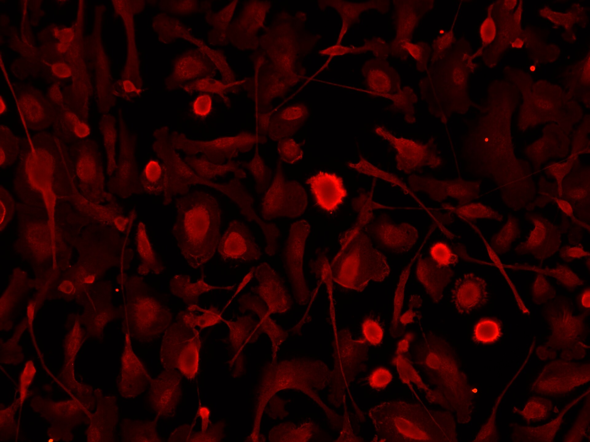
Application: ImmunocytochemistrySample Tested: human ipsc-derived microgliaSpecies: HumanVerified Customer | Posted 03/12/2026Microglia staining for ORF1P was non-specific and we confirmed that this was not due to background signal. We have previously also had issues with this antibody staining only the nuclei of SH-SY5Y cells and iPSCs.
 Bio-Techne ResponseThank you for reviewing our product. We are sorry to hear that this product did not perform as expected. We have been in touch with the customer to resolve this issue according to our Product Guarantee and to the customer’s satisfaction.
 -
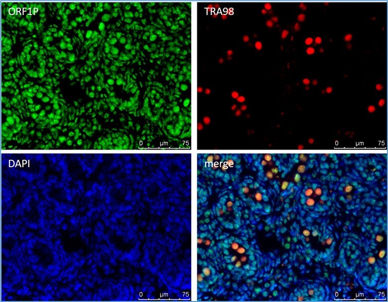
Application: Immunohistochemistry-ParaffinSample Tested: TestisSpecies: MouseVerified Customer | Posted 01/09/2021ORF1p should be mainly expressed in the cytoplasm of germ cells in testis. This antibody is expressed everywhere, and it's mainly in nuclei. It’s not appropriate for immunofluorescence.
Bio-Techne ResponseThank you for reviewing our product. We are sorry to hear that this product did not perform as expected. We have been in touch with the customer to resolve this issue according to our Product Guarantee and to the customer’s satisfaction.
There are no reviews that match your criteria.
Protocols
Find general support by application which include: protocols, troubleshooting, illustrated assays, videos and webinars.
- Antigen Retrieval Protocol (PIER)
- Antigen Retrieval for Frozen Sections Protocol
- Appropriate Fixation of IHC/ICC Samples
- Cellular Response to Hypoxia Protocols
- Chromogenic IHC Staining of Formalin-Fixed Paraffin-Embedded (FFPE) Tissue Protocol
- Chromogenic Immunohistochemistry Staining of Frozen Tissue
- ClariTSA™ Fluorophore Kits
- Detection & Visualization of Antibody Binding
- Fluorescent IHC Staining of Frozen Tissue Protocol
- Graphic Protocol for Heat-induced Epitope Retrieval
- Graphic Protocol for the Preparation and Fluorescent IHC Staining of Frozen Tissue Sections
- Graphic Protocol for the Preparation and Fluorescent IHC Staining of Paraffin-embedded Tissue Sections
- Graphic Protocol for the Preparation of Gelatin-coated Slides for Histological Tissue Sections
- ICC Cell Smear Protocol for Suspension Cells
- ICC Immunocytochemistry Protocol Videos
- ICC for Adherent Cells
- IHC Sample Preparation (Frozen sections vs Paraffin)
- Immunocytochemistry (ICC) Protocol
- Immunocytochemistry Troubleshooting
- Immunofluorescence of Organoids Embedded in Cultrex Basement Membrane Extract
- Immunofluorescent IHC Staining of Formalin-Fixed Paraffin-Embedded (FFPE) Tissue Protocol
- Immunohistochemistry (IHC) and Immunocytochemistry (ICC) Protocols
- Immunohistochemistry Frozen Troubleshooting
- Immunohistochemistry Paraffin Troubleshooting
- Preparing Samples for IHC/ICC Experiments
- Preventing Non-Specific Staining (Non-Specific Binding)
- Primary Antibody Selection & Optimization
- Protocol for Heat-Induced Epitope Retrieval (HIER)
- Protocol for Making a 4% Formaldehyde Solution in PBS
- Protocol for VisUCyte™ HRP Polymer Detection Reagent
- Protocol for the Fluorescent ICC Staining of Cell Smears - Graphic
- Protocol for the Fluorescent ICC Staining of Cultured Cells on Coverslips - Graphic
- Protocol for the Preparation & Fixation of Cells on Coverslips
- Protocol for the Preparation and Chromogenic IHC Staining of Frozen Tissue Sections
- Protocol for the Preparation and Chromogenic IHC Staining of Frozen Tissue Sections - Graphic
- Protocol for the Preparation and Chromogenic IHC Staining of Paraffin-embedded Tissue Sections
- Protocol for the Preparation and Chromogenic IHC Staining of Paraffin-embedded Tissue Sections - Graphic
- Protocol for the Preparation and Fluorescent ICC Staining of Cells on Coverslips
- Protocol for the Preparation and Fluorescent ICC Staining of Non-adherent Cells
- Protocol for the Preparation and Fluorescent ICC Staining of Stem Cells on Coverslips
- Protocol for the Preparation and Fluorescent IHC Staining of Frozen Tissue Sections
- Protocol for the Preparation and Fluorescent IHC Staining of Paraffin-embedded Tissue Sections
- Protocol for the Preparation of Gelatin-coated Slides for Histological Tissue Sections
- Protocol for the Preparation of a Cell Smear for Non-adherent Cell ICC - Graphic
- R&D Systems Quality Control Western Blot Protocol
- TUNEL and Active Caspase-3 Detection by IHC/ICC Protocol
- The Importance of IHC/ICC Controls
- Troubleshooting Guide: Immunohistochemistry
- Troubleshooting Guide: Western Blot Figures
- Western Blot Conditions
- Western Blot Protocol
- Western Blot Protocol for Cell Lysates
- Western Blot Troubleshooting
- Western Blot Troubleshooting Guide
- View all Protocols, Troubleshooting, Illustrated assays and Webinars
Loading...