Lymph Node Tissue Slides (Normal)- Paraffin
Novus Biologicals | Catalog # NBP2-30183
Key Product Details
Species
Applications
Product Summary for Lymph Node Tissue Slides (Normal)- Paraffin
Diameter: 4
Thickness:Single Tissue Slides
Pos:A01
Sl No:1
Age:42
Sex:M
Organ:Lymph node
Tissue Status:Normal
Species:Human
Product Specifications
Type
Tissue Condition
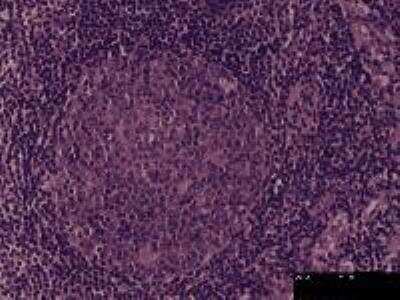
Scientific Data Images for Lymph Node Tissue Slides (Normal)- Paraffin
Formulation, Preparation, and Storage
Preparation Method
Concentration
Shipping
Storage
Product Documents for Lymph Node Tissue Slides (Normal)- Paraffin
Certificate of Analysis
To download a Certificate of Analysis, please enter a lot or batch number in the search box below.
Product Specific Notices for Lymph Node Tissue Slides (Normal)- Paraffin
This product is for research use only and is not approved for use in humans or in clinical diagnosis. Slides are guaranteed for 3 months from date of receipt.
Citations for Lymph Node Tissue Slides (Normal)- Paraffin
 Powered by Bioz
Customer Reviews for Lymph Node Tissue Slides (Normal)- Paraffin
There are currently no reviews for this product. Be the first to review Lymph Node Tissue Slides (Normal)- Paraffin and earn rewards!
Have you used Lymph Node Tissue Slides (Normal)- Paraffin?
Submit a review and receive an Amazon gift card!
$25/€18/£15/$25CAN/¥2500 Yen for a review with an image
$10/€7/£6/$10CAN/¥1110 Yen for a review without an image
Submit a review
Protocols
Find general support by application which include: protocols, troubleshooting, illustrated assays, videos and webinars.
- Antigen Retrieval Protocol (PIER)
- Antigen Retrieval for Frozen Sections Protocol
- Appropriate Fixation of IHC/ICC Samples
- Cellular Response to Hypoxia Protocols
- Chromogenic IHC Staining of Formalin-Fixed Paraffin-Embedded (FFPE) Tissue Protocol
- Chromogenic Immunohistochemistry Staining of Frozen Tissue
- ClariTSA™ Fluorophore Kits
- Detection & Visualization of Antibody Binding
- Fluorescent IHC Staining of Frozen Tissue Protocol
- Graphic Protocol for Heat-induced Epitope Retrieval
- Graphic Protocol for the Preparation and Fluorescent IHC Staining of Frozen Tissue Sections
- Graphic Protocol for the Preparation and Fluorescent IHC Staining of Paraffin-embedded Tissue Sections
- Graphic Protocol for the Preparation of Gelatin-coated Slides for Histological Tissue Sections
- IHC Sample Preparation (Frozen sections vs Paraffin)
- Immunofluorescent IHC Staining of Formalin-Fixed Paraffin-Embedded (FFPE) Tissue Protocol
- Immunohistochemistry (IHC) and Immunocytochemistry (ICC) Protocols
- Immunohistochemistry Frozen Troubleshooting
- Immunohistochemistry Paraffin Troubleshooting
- Preparing Samples for IHC/ICC Experiments
- Preventing Non-Specific Staining (Non-Specific Binding)
- Primary Antibody Selection & Optimization
- Protocol for Heat-Induced Epitope Retrieval (HIER)
- Protocol for Making a 4% Formaldehyde Solution in PBS
- Protocol for VisUCyte™ HRP Polymer Detection Reagent
- Protocol for the Preparation & Fixation of Cells on Coverslips
- Protocol for the Preparation and Chromogenic IHC Staining of Frozen Tissue Sections
- Protocol for the Preparation and Chromogenic IHC Staining of Frozen Tissue Sections - Graphic
- Protocol for the Preparation and Chromogenic IHC Staining of Paraffin-embedded Tissue Sections
- Protocol for the Preparation and Chromogenic IHC Staining of Paraffin-embedded Tissue Sections - Graphic
- Protocol for the Preparation and Fluorescent IHC Staining of Frozen Tissue Sections
- Protocol for the Preparation and Fluorescent IHC Staining of Paraffin-embedded Tissue Sections
- Protocol for the Preparation of Gelatin-coated Slides for Histological Tissue Sections
- TUNEL and Active Caspase-3 Detection by IHC/ICC Protocol
- The Importance of IHC/ICC Controls
- Troubleshooting Guide: Immunohistochemistry
- View all Protocols, Troubleshooting, Illustrated assays and Webinars
FAQs for Lymph Node Tissue Slides (Normal)- Paraffin
-
Q: How many sections are provided per slide? What is the thickness of each section? Is it embedded in paraffin?
A: There is one section/slide. The sections are infiltrated with 60°C paraffin with three changes for 1 hour each. They are sectioned by microtome in 4 um thickness.