![Western Blot: MEIS1 Antibody [NBP2-24597]](https://resources.rndsystems.com/images/products/MEIS1-Antibody-Western-Blot-NBP2-24597-img0002.jpg "Western Blot: MEIS1 Antibody [NBP2-24597]")
Loading...
Key Product Details
Species Reactivity
Validated:
Human, Mouse, Rat, Chicken, Primate
Predicted:
Bovine (100%), Orangutan (100%). Backed by our 100% Guarantee.
Applications
Immunohistochemistry, Immunohistochemistry-Paraffin, Western Blot
Label
Unconjugated
Antibody Source
Polyclonal Rabbit IgG
Loading...
Product Specifications
Immunogen
A portion of amino acids 170-220 of MEIS1 was used as the immunogen.
Reactivity Notes
The amino acid sequence used as immunogen is 88% homologous in Xenopus.
Clonality
Polyclonal
Host
Rabbit
Isotype
IgG
Scientific Data Images for MEIS1 Antibody
Western Blot: MEIS1 Antibody [NBP2-24597]
Western Blot: MEIS1 Antibody [NBP2-24597] - Analysis of MEIS1 in mouse embryo brain lysate in the 1) absence and 2) presence of immunizing peptide using NBP2-24597 at 3 ug/ml. Goat anti-rabbit Ig HRP secondary antibody and PicoTect ECL substrate solution were used for this test.![Immunohistochemistry-Paraffin: MEIS1 Antibody [NBP2-24597]](https://resources.rndsystems.com/images/products/MEIS1-Antibody-Immunohistochemistry-Paraffin-NBP2-24597-img0001.jpg "Immunohistochemistry-Paraffin: MEIS1 Antibody [NBP2-24597]")
Immunohistochemistry-Paraffin: MEIS1 Antibody [NBP2-24597]
Immunohistochemistry-Paraffin: MEIS1 Antibody [NBP2-24597] - Analysis of MEIS1 in formalin-fixed, paraffin-embedded human brain tissue using an isotype control (top left) and this antibody (bottom left, right) at 5 ug/ml.Applications for MEIS1 Antibody
Application
Recommended Usage
Immunohistochemistry-Paraffin
5 ug/ml
Western Blot
2-4 ug/ml
Reviewed Applications
Read 1 review rated 1 using NBP2-24597 in the following applications:
Formulation, Preparation, and Storage
Purification
Protein A purified
Formulation
PBS and 0.05% BSA
Preservative
0.05% Sodium Azide
Concentration
0.5 mg/ml
Shipping
The product is shipped with polar packs. Upon receipt, store it immediately at the temperature recommended below.
Stability & Storage
Store at 4C short term. Aliquot and store at -20C long term. Avoid freeze-thaw cycles.
Background: MEIS1
Alternate Names
homeobox protein Meis1, leukemogenic homolog protein, Meis homeobox 1, MGC43380, myeloid ecotropic viral integration site 1 homolog, myeloid ecotropic viral integration site 1 homolog (mouse)
Gene Symbol
MEIS1
UniProt
Additional MEIS1 Products
Product Documents for MEIS1 Antibody
Certificate of Analysis
To download a Certificate of Analysis, please enter a lot or batch number in the search box below.
Product Specific Notices for MEIS1 Antibody
This product is for research use only and is not approved for use in humans or in clinical diagnosis. Primary Antibodies are guaranteed for 1 year from date of receipt.
Customer Reviews for MEIS1 Antibody (1)
1 out of 5
1 Customer Rating
Have you used MEIS1 Antibody?
Submit a review and receive an Amazon gift card!
$25/€18/£15/$25CAN/¥2500 Yen for a review with an image
$10/€7/£6/$10CAN/¥1110 Yen for a review without an image
Submit a review
Customer Images
Showing
1
-
1 的
1 review
Showing All
Filter By:
-
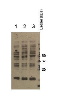
Application: Western BlotSample Tested: LNCaP whole cell lysateSpecies: HumanVerified Customer | Posted 11/04/2016Western blot analysis of MEIS1 expression in LNCaP cells transfected with CMV driven GFP (1), MEIS1 (2) and MEIS2 (3). Overnight incubation was performed in 1:200 dilution

There are no reviews that match your criteria.
Protocols
Find general support by application which include: protocols, troubleshooting, illustrated assays, videos and webinars.
- Antigen Retrieval Protocol (PIER)
- Antigen Retrieval for Frozen Sections Protocol
- Appropriate Fixation of IHC/ICC Samples
- Cellular Response to Hypoxia Protocols
- Chromogenic IHC Staining of Formalin-Fixed Paraffin-Embedded (FFPE) Tissue Protocol
- Chromogenic Immunohistochemistry Staining of Frozen Tissue
- ClariTSA™ Fluorophore Kits
- Detection & Visualization of Antibody Binding
- Fluorescent IHC Staining of Frozen Tissue Protocol
- Graphic Protocol for Heat-induced Epitope Retrieval
- Graphic Protocol for the Preparation and Fluorescent IHC Staining of Frozen Tissue Sections
- Graphic Protocol for the Preparation and Fluorescent IHC Staining of Paraffin-embedded Tissue Sections
- Graphic Protocol for the Preparation of Gelatin-coated Slides for Histological Tissue Sections
- IHC Sample Preparation (Frozen sections vs Paraffin)
- Immunofluorescent IHC Staining of Formalin-Fixed Paraffin-Embedded (FFPE) Tissue Protocol
- Immunohistochemistry (IHC) and Immunocytochemistry (ICC) Protocols
- Immunohistochemistry Frozen Troubleshooting
- Immunohistochemistry Paraffin Troubleshooting
- Preparing Samples for IHC/ICC Experiments
- Preventing Non-Specific Staining (Non-Specific Binding)
- Primary Antibody Selection & Optimization
- Protocol for Heat-Induced Epitope Retrieval (HIER)
- Protocol for Making a 4% Formaldehyde Solution in PBS
- Protocol for VisUCyte™ HRP Polymer Detection Reagent
- Protocol for the Preparation & Fixation of Cells on Coverslips
- Protocol for the Preparation and Chromogenic IHC Staining of Frozen Tissue Sections
- Protocol for the Preparation and Chromogenic IHC Staining of Frozen Tissue Sections - Graphic
- Protocol for the Preparation and Chromogenic IHC Staining of Paraffin-embedded Tissue Sections
- Protocol for the Preparation and Chromogenic IHC Staining of Paraffin-embedded Tissue Sections - Graphic
- Protocol for the Preparation and Fluorescent IHC Staining of Frozen Tissue Sections
- Protocol for the Preparation and Fluorescent IHC Staining of Paraffin-embedded Tissue Sections
- Protocol for the Preparation of Gelatin-coated Slides for Histological Tissue Sections
- R&D Systems Quality Control Western Blot Protocol
- TUNEL and Active Caspase-3 Detection by IHC/ICC Protocol
- The Importance of IHC/ICC Controls
- Troubleshooting Guide: Immunohistochemistry
- Troubleshooting Guide: Western Blot Figures
- Western Blot Conditions
- Western Blot Protocol
- Western Blot Protocol for Cell Lysates
- Western Blot Troubleshooting
- Western Blot Troubleshooting Guide
- View all Protocols, Troubleshooting, Illustrated assays and Webinars
Loading...