Dickkopf related protein 2 (Dkk-2) is a member of the Dickkopf family of secreted Wnt modulators (1-3). Dkk proteins contain a signal peptide and two conserved cysteine-rich domains that are separated by a linker region. The second cysteine-rich domain, which shows a configuration of cysteines conserved in prokineticin and colipase families, mediates Dkk-2 binding activities (2-4). The 226 amino acid (aa), ~35 kDa mature mouse Dkk-2 shares 41% and 34% aa identity with mouse Dkk-1 and Dkk-4, respectively. It also shares 99%, 96%, 96%, 96% and 94% aa identity with rat, human, canine, equine and bovine Dkk-2, respectively, and can activate the canonical Wnt signaling pathway in Xenopus embryos (5). Dkk proteins modify Wnt engagement of a receptor complex composed of a Frizzled protein and a low-density lipoprotein receptor-related protein, either LRP5 or LRP6 (3). When LRP6 is overexpressed, direct high-affinity binding of Dkk-2 to LRP can enhance canonical Wnt signaling (6-8). However, when Dkk-2 and LRP6 form a ternary complex with Kremen2, Wnt signaling is inhibited due to internalization of Dkk-2/LRP6/Krm2 complexes (9, 10). Thus, depending on the cellular context, Dkk-2 can either activate or inhibit canonical Wnt signaling (3). In contrast, binding of Dkk-1 or Dkk-4 to LRP is consistently antagonistic (3). Dkk proteins are expressed in mesenchymal tissues and control epithelial transformations. Dkk-2 expression has been studied most in bone and eye. Mouse Dkk-1 or Dkk-2 deficiencies have opposite effects on bone homeostasis, despite downregulating Wnt antagonism in both cases (11, 12). Dkk-2 expression is induced by Wnts in bone, and is thought to enhance bone density by promoting terminal differentiation of osteoblasts and mineral deposition (11). In contrast, Dkk-1 negatively regulates late osteoblast proliferation, which limits bone density (12). Dkk-2-deficient mice are blind due to faulty differentiation of corneal epithelium (13).

Key Product Details
Species Reactivity
Validated:
Mouse
Cited:
Human, Mouse
Applications
Validated:
Immunohistochemistry, Western Blot
Cited:
Immunohistochemistry, Western Blot
Label
Unconjugated
Antibody Source
Polyclonal Goat IgG
Loading...
Product Specifications
Immunogen
Mouse myeloma cell line NS0-derived recombinant mouse Dkk-2
Ser26-Ile259
Accession # Q9QYZ8
Ser26-Ile259
Accession # Q9QYZ8
Specificity
Detects mouse Dkk‑2 in Western blots.
Clonality
Polyclonal
Host
Goat
Isotype
IgG
Scientific Data Images for Mouse Dkk-2 Antibody
Dkk‑2 in Mouse Embryo.
Dkk‑2 was detected in immersion fixed frozen sections of mouse embryo (15 d.p.c.) using 15 µg/mL Mouse Dkk‑2 Antigen Affinity-purified Polyclonal Antibody (Catalog # AF2435) overnight at 4 °C. Tissue was stained with the Anti-Goat HRP-DAB Cell & Tissue Staining Kit (brown; Catalog # CTS008) and counterstained with hematoxylin (blue). View our protocol for Chromogenic IHC Staining of Frozen Tissue Sections.Applications for Mouse Dkk-2 Antibody
Application
Recommended Usage
Immunohistochemistry
5-15 µg/mL
Sample: Immersion fixed frozen sections of mouse embryo (15 d.p.c.)
Sample: Immersion fixed frozen sections of mouse embryo (15 d.p.c.)
Western Blot
0.1 µg/mL
Sample: Recombinant Mouse Dkk‑2
Sample: Recombinant Mouse Dkk‑2
Reviewed Applications
Read 1 review rated 5 using AF2435 in the following applications:
Formulation, Preparation, and Storage
Purification
Antigen Affinity-purified
Reconstitution
Reconstitute at 0.2 mg/mL in sterile PBS. For liquid material, refer to CoA for concentration.
Loading...
Formulation
Lyophilized from a 0.2 μm filtered solution in PBS with Trehalose. *Small pack size (SP) is supplied either lyophilized or as a 0.2 µm filtered solution in PBS.
Shipping
Lyophilized product is shipped at ambient temperature. Liquid small pack size (-SP) is shipped with polar packs. Upon receipt, store immediately at the temperature recommended below.
Stability & Storage
Use a manual defrost freezer and avoid repeated freeze-thaw cycles.
- 12 months from date of receipt, -20 to -70 °C as supplied.
- 1 month, 2 to 8 °C under sterile conditions after reconstitution.
- 6 months, -20 to -70 °C under sterile conditions after reconstitution.
Calculators
Background: Dkk-2
References
- Monaghan, A.P. et al. (1999) Mech. Dev. 87:45.
- Krupnik, V.E. et al. (1999) Gene 238:301.
- Niehrs, C. (2006) Oncogene 25:7469.
- Bullock, C.M. et al. (2004) Mol. Pharmacol. 65:582.
- Wu, W. et al. (2000) Current Biol. 10:1611.
- Mao, B. et al. (2001) Nature 411:321.
- Li, L. et al. (2002) J. Biol. Chem. 277:5977.
- Brott, B. and S.Y. Sokol (2002) Mol. Cell. Biol. 22:6100.
- Mao, B. et al. (2002) Nature 417:664.
- Mao, B. and C. Niehrs (2003) Gene 302:179.
- Li, X. et al. (2005) Nat. Genet. 37:945.
- Morvan, F. et al. (2006) J. Bone Miner. Res. 21:934.
- Mukhopadhyay, M. et al. (2006) Development 133:2149.
Long Name
Dickkopf-2
Alternate Names
Dkk2
Gene Symbol
DKK2
UniProt
Additional Dkk-2 Products
Product Documents for Mouse Dkk-2 Antibody
Certificate of Analysis
To download a Certificate of Analysis, please enter a lot or batch number in the search box below.
Note: Certificate of Analysis not available for kit components.
Product Specific Notices for Mouse Dkk-2 Antibody
For research use only
Related Research Areas
Citations for Mouse Dkk-2 Antibody
 Powered by Bioz
Customer Reviews for Mouse Dkk-2 Antibody (1)
5 out of 5
1 Customer Rating
Have you used Mouse Dkk-2 Antibody?
Submit a review and receive an Amazon gift card!
$25/€18/£15/$25CAN/¥2500 Yen for a review with an image
$10/€7/£6/$10CAN/¥1110 Yen for a review without an image
Submit a review
Customer Images
Showing
1
-
1 的
1 review
Showing All
Filter By:
-
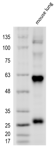
Application: Western BlotSample Tested: Lung tissueSpecies: MouseVerified Customer | Posted 03/14/2017Mouse lung lysate probed with 1:1,000 DKK-2 Antibody showing two bands corresponding to fragments 60 kDa and 28 kDa. Buffer: 5% BSA in PBS. Secondary Ab: anti-Goat IgG 1:5,000.

There are no reviews that match your criteria.
Protocols
Find general support by application which include: protocols, troubleshooting, illustrated assays, videos and webinars.
- Antigen Retrieval Protocol (PIER)
- Antigen Retrieval for Frozen Sections Protocol
- Appropriate Fixation of IHC/ICC Samples
- Cellular Response to Hypoxia Protocols
- Chromogenic IHC Staining of Formalin-Fixed Paraffin-Embedded (FFPE) Tissue Protocol
- Chromogenic Immunohistochemistry Staining of Frozen Tissue
- ClariTSA™ Fluorophore Kits
- Detection & Visualization of Antibody Binding
- Fluorescent IHC Staining of Frozen Tissue Protocol
- Graphic Protocol for Heat-induced Epitope Retrieval
- Graphic Protocol for the Preparation and Fluorescent IHC Staining of Frozen Tissue Sections
- Graphic Protocol for the Preparation and Fluorescent IHC Staining of Paraffin-embedded Tissue Sections
- Graphic Protocol for the Preparation of Gelatin-coated Slides for Histological Tissue Sections
- IHC Sample Preparation (Frozen sections vs Paraffin)
- Immunofluorescent IHC Staining of Formalin-Fixed Paraffin-Embedded (FFPE) Tissue Protocol
- Immunohistochemistry (IHC) and Immunocytochemistry (ICC) Protocols
- Immunohistochemistry Frozen Troubleshooting
- Immunohistochemistry Paraffin Troubleshooting
- Preparing Samples for IHC/ICC Experiments
- Preventing Non-Specific Staining (Non-Specific Binding)
- Primary Antibody Selection & Optimization
- Protocol for Heat-Induced Epitope Retrieval (HIER)
- Protocol for Making a 4% Formaldehyde Solution in PBS
- Protocol for VisUCyte™ HRP Polymer Detection Reagent
- Protocol for the Preparation & Fixation of Cells on Coverslips
- Protocol for the Preparation and Chromogenic IHC Staining of Frozen Tissue Sections
- Protocol for the Preparation and Chromogenic IHC Staining of Frozen Tissue Sections - Graphic
- Protocol for the Preparation and Chromogenic IHC Staining of Paraffin-embedded Tissue Sections
- Protocol for the Preparation and Chromogenic IHC Staining of Paraffin-embedded Tissue Sections - Graphic
- Protocol for the Preparation and Fluorescent IHC Staining of Frozen Tissue Sections
- Protocol for the Preparation and Fluorescent IHC Staining of Paraffin-embedded Tissue Sections
- Protocol for the Preparation of Gelatin-coated Slides for Histological Tissue Sections
- R&D Systems Quality Control Western Blot Protocol
- TUNEL and Active Caspase-3 Detection by IHC/ICC Protocol
- The Importance of IHC/ICC Controls
- Troubleshooting Guide: Immunohistochemistry
- Troubleshooting Guide: Western Blot Figures
- Western Blot Conditions
- Western Blot Protocol
- Western Blot Protocol for Cell Lysates
- Western Blot Troubleshooting
- Western Blot Troubleshooting Guide
- View all Protocols, Troubleshooting, Illustrated assays and Webinars
Loading...