EOMES (Eomesodermin), also TBR2, is a 72 kDa member of the TBR1 subfamily, T-box family of transcription factors. It is expressed in NK and CD8+ T cells, where CTLA4 activation suppresses EOMES activation of IFN-gamma and granzyme B genes. It is also found in the embryo, where it occurs in forebrain floorplate and migrating neuroblasts at 12.5 weeks gestation. Notably, it is reported to undergo intercellular transfer in fetal Xenopus tissue destined to become mesoderm. Here, it synchronizes a multicellular commitment to a cell lineage. Human EOMES is 686 amino acids (aa) in length. It contains short poly-Ala, -Gly and -Asn motifs, and a DNA-binding T box (aa 276-456). There is one isoform that shows a 19 aa insertion after Ser460. Over aa 471‑686, human EOMES shares 91% aa identity with mouse EOMES.
Mouse EOMES Antibody (1219A)
R&D Systems | Catalog # MAB8889
Recombinant Monoclonal Antibody.

Key Product Details
Species Reactivity
Validated:
Mouse
Cited:
Human, Mouse, Transgenic Mouse
Applications
Validated:
Flow Cytometry
Cited:
Immunohistochemistry, Flow Cytometry
Label
Unconjugated
Antibody Source
Recombinant Monoclonal Rabbit IgG Clone # 1219A
Loading...
Product Specifications
Immunogen
E. coli-derived recombinant mouse EOMES
Met1-Ser126
Accession # O54839
Met1-Ser126
Accession # O54839
Specificity
Detects mouse EOMES in direct ELISAs.
Clonality
Monoclonal
Host
Rabbit
Isotype
IgG
Scientific Data Images for Mouse EOMES Antibody (1219A)
Detection of EOMES in Mouse Splenocytes by Flow Cytometry.
Mouse splenocytes were stained with Rat Anti-Mouse NKp46/NCR1 APC-conjugated Monoclonal Antibody (Catalog # FAB22252A) and either (A) Rabbit Anti-Mouse EOMES Monoclonal Antibody (Catalog # MAB8889) or (B) Normal Rabbit IgG Control (Catalog # AB-105-C) followed by Phycoerythrin-conjugated Anti-Rabbit IgG Secondary Antibody (Catalog # F0110).Applications for Mouse EOMES Antibody (1219A)
Application
Recommended Usage
Flow Cytometry
0.25 µg/106 cells
Sample: Mouse splenocytes
Sample: Mouse splenocytes
Reviewed Applications
Read 1 review rated 5 using MAB8889 in the following applications:
Flow Cytometry Panel Builder
Bio-Techne Knows Flow Cytometry
Save time and reduce costly mistakes by quickly finding compatible reagents using the Panel Builder Tool.
Advanced Features
- Spectra Viewer - Custom analysis of spectra from multiple fluorochromes
- Spillover Popups - Visualize the spectra of individual fluorochromes
- Antigen Density Selector - Match fluorochrome brightness with antigen density

Formulation, Preparation, and Storage
Purification
Protein A or G purified from cell culture supernatant
Reconstitution
Reconstitute at 0.5 mg/mL in sterile PBS. For liquid material, refer to CoA for concentration.
Loading...
Formulation
Lyophilized from a 0.2 μm filtered solution in PBS with Trehalose. *Small pack size (SP) is supplied either lyophilized or as a 0.2 µm filtered solution in PBS.
Shipping
Lyophilized product is shipped at ambient temperature. Liquid small pack size (-SP) is shipped with polar packs. Upon receipt, store immediately at the temperature recommended below.
Stability & Storage
Use a manual defrost freezer and avoid repeated freeze-thaw cycles.
- 12 months from date of receipt, -20 to -70 °C as supplied.
- 1 month, 2 to 8 °C under sterile conditions after reconstitution.
- 6 months, -20 to -70 °C under sterile conditions after reconstitution.
Calculators
Background: EOMES
Long Name
Eomesodermin Homolog
Alternate Names
Eomesodermin, TBR2
Gene Symbol
EOMES
UniProt
Additional EOMES Products
Product Documents for Mouse EOMES Antibody (1219A)
Certificate of Analysis
To download a Certificate of Analysis, please enter a lot or batch number in the search box below.
Note: Certificate of Analysis not available for kit components.
Product Specific Notices for Mouse EOMES Antibody (1219A)
For research use only
Citations for Mouse EOMES Antibody (1219A)
 Powered by Bioz
Customer Reviews for Mouse EOMES Antibody (1219A) (1)
5 out of 5
1 Customer Rating
Have you used Mouse EOMES Antibody (1219A)?
Submit a review and receive an Amazon gift card!
$25/€18/£15/$25CAN/¥2500 Yen for a review with an image
$10/€7/£6/$10CAN/¥1110 Yen for a review without an image
Submit a review
Customer Images
Showing
1
-
1 的
1 review
Showing All
Filter By:
-
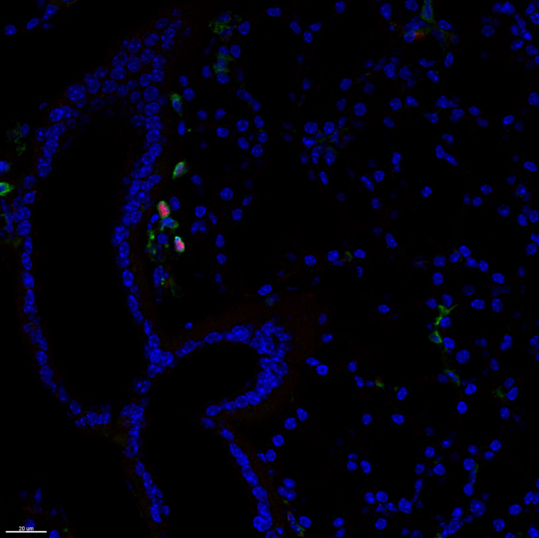
Application: Immunocytochemistry/ImmunofluorescenceSample Tested: Salivary gland tissueSpecies: MouseVerified Customer | Posted 07/09/2019Cryosections from PLP immersion fixed tissues were stained for EOMES (1:500) for 4h @ RT. After washing, tissue was stained with Goat anti-Rabbit alexa555 2ndary antibody (1:500) for 1h at RT. CD45 in green, EOMES in red, nuclear staining (Hoechst) in blue.

There are no reviews that match your criteria.
Protocols
Find general support by application which include: protocols, troubleshooting, illustrated assays, videos and webinars.
- 7-Amino Actinomycin D (7-AAD) Cell Viability Flow Cytometry Protocol
- Extracellular Membrane Flow Cytometry Protocol
- Flow Cytometry Protocol for Cell Surface Markers
- Flow Cytometry Protocol for Staining Membrane Associated Proteins
- Flow Cytometry Staining Protocols
- Flow Cytometry Troubleshooting Guide
- Intracellular Flow Cytometry Protocol Using Alcohol (Methanol)
- Intracellular Flow Cytometry Protocol Using Detergents
- Intracellular Nuclear Staining Flow Cytometry Protocol Using Detergents
- Intracellular Staining Flow Cytometry Protocol Using Alcohol Permeabilization
- Intracellular Staining Flow Cytometry Protocol Using Detergents to Permeabilize Cells
- Propidium Iodide Cell Viability Flow Cytometry Protocol
- Protocol for Liperfluo
- Protocol for the Characterization of Human Th22 Cells
- Protocol for the Characterization of Human Th9 Cells
- Protocol: Annexin V and PI Staining by Flow Cytometry
- Protocol: Annexin V and PI Staining for Apoptosis by Flow Cytometry
- Troubleshooting Guide: Fluorokine Flow Cytometry Kits
- View all Protocols, Troubleshooting, Illustrated assays and Webinars


