
Growth hormone (GH), also known as somatotropin, is a member of a family of growth factors that includes prolactin, placental lactogens, proliferins and somatolactin (1, 2). It is synthesized primarily by somatotropes in the anterior pituitary and is released as an endocrine hormone. Other cells and tissues, including lymphoid tissues, can also produce GH (3). GH is a pleiotropic molecule which can act directly or indirectly via IGF-I to regulate growth and metabolism as well as enhance T cell survival and thymic functions (1, 2, 4). GH exerts its biological actions by binding to the GH receptor (GHR) that is present in many cell types (1, 2). Mouse GHR cDNA encodes a 650 amino acid (aa) residue type I transmembrane protein with a 24 aa signal peptide, a 249 aa extracellular domain, a 24 aa transmembrane domain, and a 353 aa cytoplasmic domain (5). An alternatively spliced secreted isoform of mouse GHR also exists (6). This variant corresponds to the serum GH‑binding protein. Ligation of GHR by GH has been shown to result in receptor dimerization and activation of the JAK/STAT signaling cascade (7). The soluble GHBP has been shown to interfere with GH signaling by competing with the transmembrane receptor of GH. Alternatively, the GHBP has also been shown to enhance GH action by slowing GH clearance (8).
Mouse Growth Hormone R/GHR Antibody
R&D Systems | Catalog # AF1360

Scientific images may be resized, cropped, or adjusted for brightness or contrast for presentation purposes. These updates are limited to presentation and do not affect the underlying scientific interpretation of the data. Where available, additional source data may be provided upon request.
Key Product Details
Species Reactivity
Validated:
Cited:
Applications
Validated:
Cited:
Label
Antibody Source
Product Specifications
Immunogen
Thr25-Gln273
Accession # Q3UP14
Specificity
Clonality
Host
Isotype
Scientific Data Images for Mouse Growth Hormone R/GHR Antibody
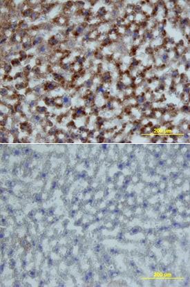
Growth Hormone R/GHR in Mouse Liver.
Growth Hormone R/GHR was detected in perfusion fixed frozen sections of mouse liver using Mouse Growth Hormone R/GHR Antigen Affinity-purified Polyclonal Antibody (Catalog # AF1360) at 15 µg/mL overnight at 4 °C. Tissue was stained using the Anti-Goat HRP-DAB Cell & Tissue Staining Kit (brown; Catalog # CTS008) and counterstained with hematoxylin (blue). Lower panel shows a lack of labeling if primary antibodies are omitted and tissue is stained only with secondary antibody followed by incubation with detection reagents. View our protocol for Chromogenic IHC Staining of Frozen Tissue Sections.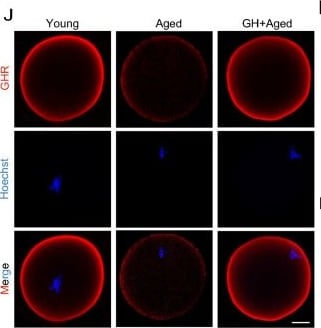
Detection of Growth Hormone R (GHR) by Immunocytochemistry/ Immunofluorescence
Effect of GH supplementation on ovary aging and the quality of aged oocytes. A Schedule of GH treatment and hormone administration to induce superovulation. B and C Morphological features of ovaries. Ovarian index = (ovary weight/body weight) × 100. D H&E staining of ovarian sections from young, aged, and GH + aged mice. PA, preantral follicle; AF, antral follicle; AT, atretic follicle; CL, corpus luteum. The bottom panel is the enlarged view of the box area in the top panel. Scale bar, 200 μm. E Numbers of follicles at the indicated stages in young (n = 3), aged (n = 3), and GH + aged (n = 3) mice. F Representative images of in vivo matured oocytes from young, aged, and GH + aged mice. Black arrows, oocytes with fragmentation. Scale bar, 80 μm. G Numbers of oocytes after superovulation in young (n = 10), aged (n = 10), and GH + aged (n = 10) mice. H and I Fragmentation and first polar body (PB1) extrusion rates in young (n = 39), aged (n = 35), and GH + aged (n = 42) oocytes. J Representative images of GHR in young, aged, and GH + aged oocytes. Scale bar, 20 μm. K and L Protein and mRNA levels of GHR revealed by immunofluorescence (n = 15) and real-time PCR (n = 25), respectively. M Cumulative number of pups over 12 weeks as a measure of the fertility of young (n = 6), aged (n = 6), and GH + aged (n = 6) mice. N Body weights of newborns (n = 24, 10, and 12 in the young, aged, and GH + aged groups, respectively) over 4 weeks. Data are means ± SEM of at least three independent experiments. *p < 0.05, **p < 0.01, ***p < 0.001, ****p < 0.0001 Image collected and cropped by CiteAb from the following open publication (https://pubmed.ncbi.nlm.nih.gov/37386516), licensed under a CC-BY license. Not internally tested by R&D Systems.Applications for Mouse Growth Hormone R/GHR Antibody
Immunohistochemistry
Sample: Perfusion fixed frozen sections of mouse kidney, liver, lung, thymus, and muscle
Western Blot
Sample: Recombinant Mouse Growth Hormone R/GHR Fc Chimera (Catalog # 1360-GR)
Reviewed Applications
Read 1 review rated 4 using AF1360 in the following applications:
Formulation, Preparation, and Storage
Purification
Reconstitution
Reconstitute at 0.2 mg/mL in sterile PBS. For liquid material, refer to CoA for concentration.
Formulation
Shipping
Stability & Storage
- 12 months from date of receipt, -20 to -70 °C as supplied.
- 1 month, 2 to 8 °C under sterile conditions after reconstitution.
- 6 months, -20 to -70 °C under sterile conditions after reconstitution.
Calculators
Background: Growth Hormone R/GHR
References
- Goffin, V. et al. (1996) Endocrine Rev. 17:385.
- Le Roith, D. et al. (2001) Endocrine Rev. 22:53.
- Clark, R. (1997) Endocr. Rev. 18:157.
- Welniak, L.A. et al. (2002) J. Leukoc. Biol. 71:381.
- Smith, W.C. et al. (1989) Mol. Endocrinol. 3:984.
- Edens, A. et al. (1994) Endocrinol. 135:2802.
- Carter-Su, C. et al. (1996) Annu. Rev. Physiol. 58:187.
- Postel-Vinay, M.C. and J. Finidori (1995) Eur. J. Endocrinol. 133:654.
Long Name
Alternate Names
Gene Symbol
UniProt
Additional Growth Hormone R/GHR Products
Product Documents for Mouse Growth Hormone R/GHR Antibody
Certificate of Analysis
To download a Certificate of Analysis, please enter a lot or batch number in the search box below.
Note: Certificate of Analysis not available for kit components.
Product Specific Notices for Mouse Growth Hormone R/GHR Antibody
For research use only
Citations for Mouse Growth Hormone R/GHR Antibody
 Powered by Bioz
Customer Reviews for Mouse Growth Hormone R/GHR Antibody (1)
Have you used Mouse Growth Hormone R/GHR Antibody?
Submit a review and receive an Amazon gift card!
$25/€18/£15/$25CAN/¥2500 for a review with an image
$10/€7/£6/$10CAN/¥1110 for a review without an image
Submit a review
Customer Images
-
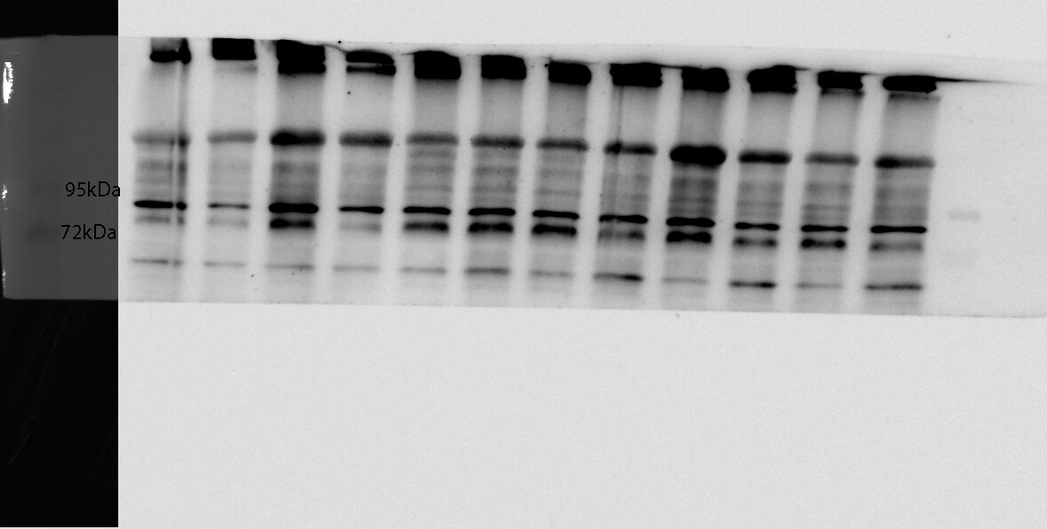
Application: Western BlotSample Tested: Liver tissueSpecies: MouseVerified Customer | Posted 04/18/2019

There are no reviews that match your criteria.
Protocols
Find general support by application which include: protocols, troubleshooting, illustrated assays, videos and webinars.
- Antigen Retrieval Protocol (PIER)
- Antigen Retrieval for Frozen Sections Protocol
- Appropriate Fixation of IHC/ICC Samples
- Cellular Response to Hypoxia Protocols
- Chromogenic IHC Staining of Formalin-Fixed Paraffin-Embedded (FFPE) Tissue Protocol
- Chromogenic Immunohistochemistry Staining of Frozen Tissue
- ClariTSA™ Fluorophore Kits
- Detection & Visualization of Antibody Binding
- Fluorescent IHC Staining of Frozen Tissue Protocol
- Graphic Protocol for Heat-induced Epitope Retrieval
- Graphic Protocol for the Preparation and Fluorescent IHC Staining of Frozen Tissue Sections
- Graphic Protocol for the Preparation and Fluorescent IHC Staining of Paraffin-embedded Tissue Sections
- Graphic Protocol for the Preparation of Gelatin-coated Slides for Histological Tissue Sections
- IHC Sample Preparation (Frozen sections vs Paraffin)
- Immunofluorescent IHC Staining of Formalin-Fixed Paraffin-Embedded (FFPE) Tissue Protocol
- Immunohistochemistry (IHC) and Immunocytochemistry (ICC) Protocols
- Immunohistochemistry Frozen Troubleshooting
- Immunohistochemistry Paraffin Troubleshooting
- Preparing Samples for IHC/ICC Experiments
- Preventing Non-Specific Staining (Non-Specific Binding)
- Primary Antibody Selection & Optimization
- Protocol for Heat-Induced Epitope Retrieval (HIER)
- Protocol for Making a 4% Formaldehyde Solution in PBS
- Protocol for VisUCyte™ HRP Polymer Detection Reagent
- Protocol for the Preparation & Fixation of Cells on Coverslips
- Protocol for the Preparation and Chromogenic IHC Staining of Frozen Tissue Sections
- Protocol for the Preparation and Chromogenic IHC Staining of Frozen Tissue Sections - Graphic
- Protocol for the Preparation and Chromogenic IHC Staining of Paraffin-embedded Tissue Sections
- Protocol for the Preparation and Chromogenic IHC Staining of Paraffin-embedded Tissue Sections - Graphic
- Protocol for the Preparation and Fluorescent IHC Staining of Frozen Tissue Sections
- Protocol for the Preparation and Fluorescent IHC Staining of Paraffin-embedded Tissue Sections
- Protocol for the Preparation of Gelatin-coated Slides for Histological Tissue Sections
- R&D Systems Quality Control Western Blot Protocol
- TUNEL and Active Caspase-3 Detection by IHC/ICC Protocol
- The Importance of IHC/ICC Controls
- Troubleshooting Guide: Immunohistochemistry
- Troubleshooting Guide: Western Blot Figures
- Western Blot Conditions
- Western Blot Protocol
- Western Blot Protocol for Cell Lysates
- Western Blot Troubleshooting
- Western Blot Troubleshooting Guide
- View all Protocols, Troubleshooting, Illustrated assays and Webinars