


Scientific images may be resized, cropped, or adjusted for brightness or contrast for presentation purposes. These updates are limited to presentation and do not affect the underlying scientific interpretation of the data. Where available, additional source data may be provided upon request.
Key Product Details
Validated by
Species Reactivity
Validated:
Cited:
Applications
Validated:
Cited:
Label
Antibody Source
Product Specifications
Immunogen
Accession # P18893
Specificity
Clonality
Host
Isotype
Endotoxin Level
Scientific Data Images for Mouse IL-10 Antibody (JES052A5)
Detection of Recombinant Mouse IL‑10 by Western Blot.
Western blot shows 25 ng of Recombinant Mouse IL-10 (Catalog # 417-ML), Recombinant Human IL-10 (Catalog # 217-IL), Recombinant Rat IL-10 (Catalog # 522-RLB), and Recombinant Human IL-26/AK155 Monomer (Catalog # 1375-IL). PVDF Membrane was probed with 1 µg/mL of Rat Anti-Mouse IL-10 Monoclonal Antibody (Catalog # MAB417) followed by HRP-conjugated Anti-Rat IgG Secondary Antibody (Catalog # HAF005). A specific band was detected for IL-10 at approximately 15 kDa (as indicated). This experiment was conducted under reducing conditions and using Immunoblot Buffer Group 3.
Cell Proliferation Induced by IL‑10 and Neutralization by Mouse IL‑10 Antibody.
Recombinant Mouse IL-10 (Catalog # 417-ML) stimulates proliferation in the MC/9-2 mouse mast cell line in a dose-dependent manner (orange line). Proliferation elicited by Recombinant Mouse IL-10 (2.5 ng/mL) is neutralized (green line) by increasing concen-trations of Rat Anti-Mouse IL-10 Monoclonal Antibody (Catalog # MAB417). The ND50 is typically 0.005-0.015 µg/mL.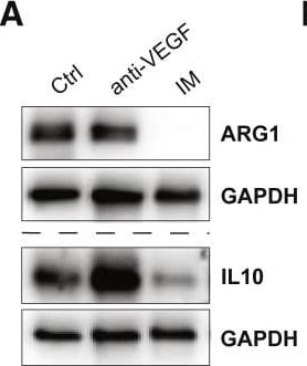
Detection of Mouse IL-10 by Western Blot
Imipramine downregulates an M2-like program in TAMs(A) Western blot analysis of ARG1 and IL-10 in single tumors treated or not with IM or anti-VEGF.(B) Flow cytometry analysis of ARG1 and IL-10 expression in GBM tumors treated for 1 week. Ctrl (n = 9 tumors), IM (n = 6), anti-VEGF (n = 6), anti-VEGFR2 (n = 6), Axitinib (n = 6). Macrophages were gated as CD45+CD11b+Ly6C-Ly6G−.(C) Expression of ARG1 and IL-10 in microglia (CD49d−) and MDMs (CD49d+) assessed by FACS in untreated tumors (n = 5).(D) Expression of MHC-II within microglia as assessed by flow cytometry. Ctrl (n = 4 tumors) and IM + anti-VEGF (n = 4).(E) Ex vivo co-cultures of tumoral CD11b cells and activated splenic CFSE-labeled CD8 or CD4 T cells. Each dot represents the average of two or three technical replicates. T cells alone (n = 4), Ctrl co-culture (n = 5), anti-VEGF (n = 3), IM (n = 4), IM + anti-VEGF (n = 4).(F) Analysis of the M2-like program in cytokine-polarized macrophages as assessed by qRT-PCR analysis of Ctrl and IM-treated M2-like BMDMs. Expression is normalized to 18S statistics by Welch’s t test. Each dot represents an individual sample. Data are representative of three independent experiments.(G) Analysis of the M1-like program in BMDMs assessed by FACS. Each dot represents an individual replicate.(H) Expression of Hrh1 mRNA normalized to 18S in ex vivo M1-and M2-polarized BMDMs, either untreated or IM treated for 24 h. Each dot represents an individual sample. Data are representative of three independent experiments.(I) mRNA expression of Hrh1 in FACS-sorted microglia or MDMs from Ctrl and IM-treated tumors.(J) mRNA expression of Arg1, Chil3, and Il10 in M2-polarized macrophages that were transfected with siCtrl or two different siHrh1 constructs. Cells were treated with 40 μm IM for 24 h. Data are representative of two independent experiments.(K) Western blot analysis of MRC1 and ARG1 expression of siRNA-transfected M2 BMDMs. Data are representative of two independent experiments.(L)
Detection of Mouse IL-10 by Immunocytochemistry/Immunofluorescence
Lymphocyte infiltrate in tumors from IL-10 deficient mice (A, C and D) or from anti-IL10/IL-10R neutralizing antibody treated mice (B, E and F). A. Single cell suspensions were stained with the indicated antibodies plus anti-CD45 and anti-F4/80. Lymphocytes were analyzed in the CD45+F4/80- population. B. After 6 hours incubation in tissue culture flasks, non adherent cells from total tumor suspension were harvested and stained with the indicated antibodies, tetramer and anti-CD45. C and E. Immunofluorescence of cryo-sections from tumors harvested from IL-10 KO and wild type mice or neutralizing anti-IL10/IL-1R and irrelevant IgG antibodies, as indicated. Sections were stained with anti-CD8 (in FITC, green) and PE (red) conjugated HPV16 E749-57 tetramer - E749-57 Tet, and irrelevant tetramer - Irrelevant Tet. Cells were counterstained with DAPI (blue). Arrowheads indicate double positive cells in yellow, arrows indicate cells positive only for CD8. D and F. Granzyme B expression in tumor cryo-sections from IL-10 KO and wild type mice or mice treated with anti-IL10/IL-10R neutralizing antibodies and irrelevant IgG, as indicated. Sections were stained with anti-CD8 (green), followed by fixation, permeabilization and staining with anti-Granzyme B (red). Tissue was counterstained with DAPI. Solid arrows indicate double positive cells, arrowhead indicate CD8 only positive cell. Insets show details of cells positive only for CD8 (green) or double positive CD8/tetramer (C and E), CD8/Granzyme B (D and F) in yellow or orange due to the sobreposition of emission from FITC and PE (tetramer) or alexa 594 (Granzyme B). Image collected and cropped by CiteAb from the following publication (https://bmcimmunol.biomedcentral.com/articles/10.1186/1471-2172-11-27), licensed under a CC-BY license. Not internally tested by R&D Systems.
Mouse IL-10 ELISA Standard Curve
Recombinant Mouse IL‑10 (Catalog # 417-ML) was serially diluted and captured by Rat Anti-Mouse IL‑10 Monoclonal Antibody (Catalog # MAB417) coated on a Clear Polystyrene Microplate (Catalog # DY990). Goat Anti-Mouse IL‑10 Antigen Affinity-purified Polyclonal Antibody (Catalog # AF-417-NA) was biotinylated and incubated with the protein captured on the plate. Detection of the standard curve was achieved by incubating Streptavidin-HRP (Catalog # DY998)Applications for Mouse IL-10 Antibody (JES052A5)
Western Blot
Sample: Recombinant Mouse IL‑10 (Catalog # 417-ML)
Neutralization
Mouse IL-10 Sandwich Immunoassay
Reviewed Applications
Read 2 reviews rated 5 using MAB417 in the following applications:
Formulation, Preparation, and Storage
Purification
Reconstitution
Reconstitute at 0.5 mg/mL in sterile PBS. For liquid material, refer to CoA for concentration.
Formulation
Shipping
Stability & Storage
- 12 months from date of receipt, -20 to -70 °C as supplied.
- 1 month, 2 to 8 °C under sterile conditions after reconstitution.
- 6 months, -20 to -70 °C under sterile conditions after reconstitution.
Calculators
Background: IL-10
Long Name
Alternate Names
Entrez Gene IDs
Gene Symbol
UniProt
Additional IL-10 Products
Product Documents for Mouse IL-10 Antibody (JES052A5)
Certificate of Analysis
To download a Certificate of Analysis, please enter a lot or batch number in the search box below.
Note: Certificate of Analysis not available for kit components.
Product Specific Notices for Mouse IL-10 Antibody (JES052A5)
For research use only
Related Research Areas
Citations for Mouse IL-10 Antibody (JES052A5)
 Powered by Bioz
Customer Reviews for Mouse IL-10 Antibody (JES052A5) (2)
Have you used Mouse IL-10 Antibody (JES052A5)?
Submit a review and receive an Amazon gift card!
$25/€18/£15/$25CAN/¥2500 for a review with an image
$10/€7/£6/$10CAN/¥1110 for a review without an image
Submit a review
Customer Images
-
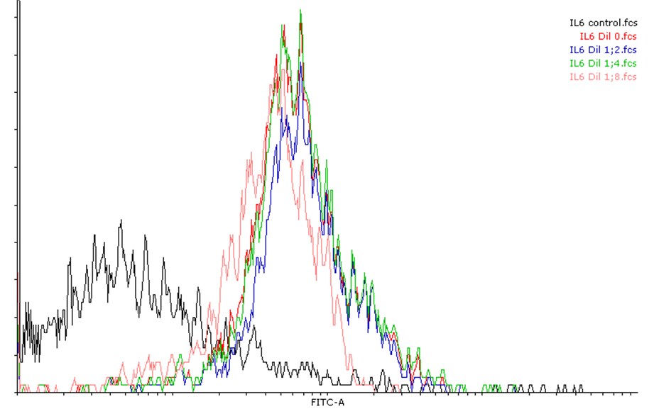
Application: Flow CytometrySample Tested: T cellsSpecies: MouseVerified Customer | Posted 03/16/2022

-
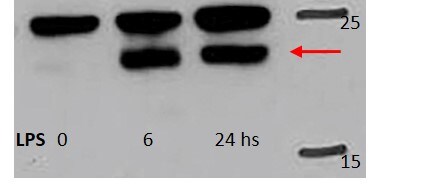
Application: Western BlotSample Tested: Peritoneal macrophagesSpecies: MouseVerified Customer | Posted 11/13/2020
There are no reviews that match your criteria.
Protocols
Find general support by application which include: protocols, troubleshooting, illustrated assays, videos and webinars.
- Cellular Response to Hypoxia Protocols
- R&D Systems Quality Control Western Blot Protocol
- Troubleshooting Guide: Western Blot Figures
- Western Blot Conditions
- Western Blot Protocol
- Western Blot Protocol for Cell Lysates
- Western Blot Troubleshooting
- Western Blot Troubleshooting Guide
- View all Protocols, Troubleshooting, Illustrated assays and Webinars
Associated Pathways



