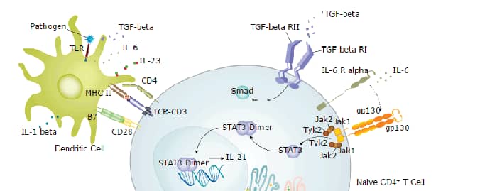
IL-12 R beta 1 is a 100 kDa type I transmembrane protein that belongs to the gp130/G‑CSF R family of cytokine receptors. IL-12 R beta 1 is a common subunit of both the IL‑12 and IL-23 receptor complexes which play distinct but related roles in T cell mediated inflammatory reactions (1, 2). Mature mouse IL-12 R beta 1 contains a 546 amino acid (aa) extracellular domain (ECD) with five fibronectin type III repeats, and a 147 aa cytoplasmic domain (3). Within the ECD, mouse IL-12 R beta 1 shares 85% and 52% aa sequence identity with rat and human IL-12 R beta 1, respectively. It shares 16%‑21% aa sequence identity with the ECDs of mouse gp130, LIF R, G‑CSF R, and IL‑23 R. IL-12 and IL-23 are disulfide linked heterodimeric cytokines that share a common p40 subunit (1, 2). IL-12 R beta 1 interacts with p40 at low affinity but does not transmit signals (3). Increased ligand binding affinity and signaling capacity are gained by association of IL-12 R beta 1 with either IL-12 R beta 2 or IL‑23 R (4‑6). IL‑12 R beta 2 and IL‑23 R are the signal transducing components of these receptor complexes (4, 7). IL-12 R beta 1 is expressed on activated T cells, NK cells, B cells, macrophages, and microglia (8‑10). IL-12 induced signaling promotes the development of naïve T cells into IFN-beta producing Th1 cells (11). IL-23 contributes to chronic inflammation by inducing the production of IL-17 by memory T cells (12). Naturally occurring homodimers of p40 can function as antagonists of IL-12 and IL‑23 and can also induce macrophage chemotaxis in the absence of IL-12 R beta 2 (13, 14).
Mouse IL-12 R beta 1 Antibody [Biotin]
R&D Systems | Catalog # BAF1998

Scientific images may be resized, cropped, or adjusted for brightness or contrast for presentation purposes. These updates are limited to presentation and do not affect the underlying scientific interpretation of the data. Where available, additional source data may be provided upon request.
Key Product Details
Species Reactivity
Validated:
Cited:
Applications
Validated:
Cited:
Label
Antibody Source
Product Specifications
Immunogen
Val32-Glu561
Accession # Q60837
Specificity
Clonality
Host
Isotype
Scientific Data Images for Mouse IL-12 R beta 1 Antibody [Biotin]
Detection of IL-12 R beta 1 by Western Blot
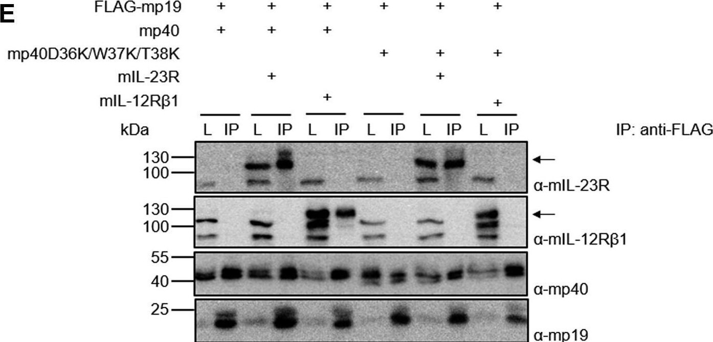
Murine p40 with D36K/W37K/T38K substitutions interacts with murine p19 but not with murine IL-12R beta 1.A, structure of hIL-23 extracted from the structure of the IL-23:IL-23R complex structure (PDB 5mzv). A flexible linker sequence connecting p40 and p19 is indicated. B, Western blot analysis of secreted murine HIL-23, mp19 and mp40 variants from transfected CHO-K1 cells. The position of mp19 and mp40 variants is indicated by arrows. C, cellular proliferation of Ba/F3-gp130-mIL-12R beta 1-mIL-23R cells. The cells were cultured for 3 days in the presence of 10 ng/ml HIL-6 or with the indicated cytokines (10% conditioned cell culture supernatant of transfected CHO-K1 cells). Parental Ba/F3-gp130 cells were used as controls. The results of one representative experiment of three are shown. Error bars represent S.D. for technical replicates. Statistical analysis used a one-way ANOVA, followed by Bonferroni correction (n = 3), ∗∗∗p ≤ 0.001. D, analysis of STAT3 and ERK1/2 activation. Ba/F3-gp130-mIL-12R beta 1-mIL-23R cells were washed, starved, and stimulated with the indicated cytokines (10% conditioned cell culture supernatant of transfected CHO-K1 cells) for 30 min. Cellular lysates were prepared, and equal amounts of total protein (50 μg/lane) were loaded on SDS-PAA gels, followed by immunoblotting using specific antibodies for phospho-STAT3, STAT3, phospho-ERK1/2, and ERK1/2. Western blotting data show results of one representative experiment of two. E, co-IP of FLAG-tagged murine mp19 coexpressed with either mp40 or mp40D36K/W37K/T38K and full-length mIL-12R beta 1 or mIL-23R. The position of mIL-23R and mIL-12R beta 1 is indicated by arrows. One of two independent experiments is shown. Image collected and cropped by CiteAb from the following open publication (https://pubmed.ncbi.nlm.nih.gov/34637790), licensed under a CC-BY license. Not internally tested by R&D Systems.Applications for Mouse IL-12 R beta 1 Antibody [Biotin]
Western Blot
Sample: Recombinant Mouse IL‑12 R beta 1 (Catalog # 1998-B1)
Formulation, Preparation, and Storage
Purification
Reconstitution
Reconstitute at 0.2 mg/mL in sterile PBS.
Formulation
Shipping
Stability & Storage
- 12 months from date of receipt, -20 to -70 °C as supplied.
- 1 month, 2 to 8 °C under sterile conditions after reconstitution.
- 6 months, -20 to -70 °C under sterile conditions after reconstitution.
Calculators
Background: IL-12 R beta 1
References
- Becker, C. et al. (2005) Inflamm. Bowel Dis. 11:755.
- Hunter, C.A. (2005) Nat. Rev. Immunol. 5:521.
- Chua, A.O. et al. (1995) J. Immunol. 155:4286.
- Parham, C. et al. (2002) J. Immunol. 168:5699.
- Wu, C. et al. (1997) J. Immunol. 159:1658.
- Zou, J. et al. (1997) J. Biol. Chem. 272:6073.
- Presky, D.H. et al. (1996) Proc. Natl. Acad. Sci. 93:14002.
- Wu, C. et al. (1997) Eur. J. Immunol. 27:147.
- Airoldi, I. et al. (2000) J. Immunol. 165:6880.
- Li, J. et al. (2003) J. Neurol. Sci. 215:95.
- Schmitt, E. et al. (1994) Eur. J. Immunol. 24:793.
- Yen, D. et al. (2006) J. Clin. Invest. 116:1310.
- Shimozato, O. et al. (2006) Immunology 117:22.
- Russell, T.D. et al. (2003) J. Immunol. 171:6866.
Long Name
Alternate Names
Gene Symbol
UniProt
Additional IL-12 R beta 1 Products
Product Documents for Mouse IL-12 R beta 1 Antibody [Biotin]
Certificate of Analysis
To download a Certificate of Analysis, please enter a lot or batch number in the search box below.
Note: Certificate of Analysis not available for kit components.
Product Specific Notices for Mouse IL-12 R beta 1 Antibody [Biotin]
For research use only
Citations for Mouse IL-12 R beta 1 Antibody [Biotin]
 Powered by Bioz
Customer Reviews for Mouse IL-12 R beta 1 Antibody [Biotin]
There are currently no reviews for this product. Be the first to review Mouse IL-12 R beta 1 Antibody [Biotin] and earn rewards!
Have you used Mouse IL-12 R beta 1 Antibody [Biotin]?
Submit a review and receive an Amazon gift card!
$25/€18/£15/$25CAN/¥2500 for a review with an image
$10/€7/£6/$10CAN/¥1110 for a review without an image
Submit a review
Protocols
Find general support by application which include: protocols, troubleshooting, illustrated assays, videos and webinars.
- Cellular Response to Hypoxia Protocols
- R&D Systems Quality Control Western Blot Protocol
- Troubleshooting Guide: Western Blot Figures
- Western Blot Conditions
- Western Blot Protocol
- Western Blot Protocol for Cell Lysates
- Western Blot Troubleshooting
- Western Blot Troubleshooting Guide
- View all Protocols, Troubleshooting, Illustrated assays and Webinars
Associated Pathways