Interleukin-21 (IL-21) and its receptor appear to play important roles in the regulation of the immune system. IL-21 is most related to IL-2, IL-4, and IL-15. IL-21 R, also called NILR (novel interleukin receptor), is a type I cytokine receptor with 4 conserved cysteine residues and an extracellular WSXWS motif. It is most closely related to IL-2 R beta and IL-4 R alpha. Mouse IL-21 is a 146 amino acid (aa) residue protein with a 24 aa signal peptide. Mouse and human IL-21 share 57% amino acid sequence identity. IL‑21 is expressed by activated T cells. Although not fully elucidated, the IL-2 R gamma ( gamma c) chain appears to play a role in IL-21 R signaling. The IL‑21/IL‑21 R interaction appear to play important roles in B and T cell proliferation after antigen stimulation and NK cell maturation.

Key Product Details
Species Reactivity
Validated:
Mouse
Cited:
Human, Mouse, Bovine, Transgenic Mouse
Applications
Validated:
Western Blot, ELISA Capture (Matched Antibody Pair), Neutralization, Immunocytochemistry
Cited:
Immunohistochemistry, Immunohistochemistry-Frozen, Western Blot, Neutralization, Flow Cytometry, Immunocytochemistry, ELISA Capture, ELISA Development, ELISpot Development
Label
Unconjugated
Antibody Source
Polyclonal Goat IgG
Loading...
Product Specifications
Immunogen
E. coli-derived recombinant mouse IL‑21 (R&D Systems, Catalog # 594-ML)
Pro25-Ser146
Accession # Q9ES17
Pro25-Ser146
Accession # Q9ES17
Specificity
Detects mouse IL-21 in ELISAs and Western blots. In sandwich ELISAs, less than 0.5% cross-reactivity with recombinant mouse (rm) IL‑2, rmIL‑4, rmIL-15, and recombinant human IL-21 is observed.
Clonality
Polyclonal
Host
Goat
Isotype
IgG
Endotoxin Level
<0.10 EU per 1 μg of the antibody by the LAL method.
Scientific Data Images for Mouse IL-21 Antibody
IL‑21 in Mouse Splenocytes.
IL‑21 was detected in immersion fixed concanavalin A-stimulated mouse splenocytes using 15 µg/mL Mouse IL‑21 Antigen Affinity-purified Polyclonal Antibody (Catalog # AF594) for 3 hours at room temperature. Cells were stained with the NorthernLights™ 557-conjugated Anti-Goat IgG Secondary Antibody (red; Catalog # NL001) and counterstained (green). View our protocol for Fluorescent ICC Staining of Non-adherent Cells.
Cell Proliferation Induced by IL-21 and Neutralization by Mouse IL-21 Antibody.
Recombinant Mouse IL-21 (Catalog # 594-ML) stimulates proliferation in the NK-92 human natural killer lymphoma cell line in a dose-dependent manner (orange line). Proliferation elicited by Recombinant Mouse IL-21 (10 ng/mL) is neutralized (green line) by increasing concentrations of Mouse IL-21 Antigen Affinity-purified Polyclonal Antibody (Catalog # AF594). The ND50 is typically 0.2-2 µg/mL.
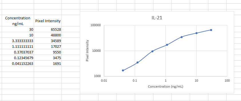
Mouse IL-21 ELISA Standard Curve
Recombinant Mouse IL‑21 (Catalog # 594-ML) was serially diluted and captured by Goat Anti-Mouse IL‑21 Antigen Affinity-purified Polyclonal Antibody (Catalog # AF594) coated on a Clear Polystyrene Microplate (Catalog # DY990). Goat Anti-Mouse IL‑21 Antigen Affinity-purified Polyclonal Antibody (Catalog # AF594) was biotinylated and incubated with the protein captured on the plate. Detection of the standard curve was achieved by incubating Streptavidin-HRP (Catalog # DY998)
Mouse IL-21 ELISA Standard Curve
Recombinant Mouse IL‑21 (Catalog # 594-ML) was serially diluted and captured by Goat Anti-Mouse IL‑21 Antigen Affinity-purified Polyclonal Antibody (Catalog # AF594) coated on a Clear Polystyrene Microplate (Catalog # DY990). Goat Anti-Mouse IL‑21 Antigen Affinity-purified Polyclonal Antibody (Catalog # AF594) was biotinylated and incubated with the protein captured on the plate. Detection of the standard curve was achieved by incubating Streptavidin-HRP (Catalog # DY998)Applications for Mouse IL-21 Antibody
Application
Recommended Usage
Immunocytochemistry
5-15 µg/mL
Sample: Immersion fixed concanavalin A-stimulated mouse splenocytes
Sample: Immersion fixed concanavalin A-stimulated mouse splenocytes
Western Blot
0.1 µg/mL
Sample: Recombinant Mouse IL‑21 (Catalog # 594-ML)
Sample: Recombinant Mouse IL‑21 (Catalog # 594-ML)
Neutralization
Measured by its ability to neutralize IL‑21-induced proliferation in the NK-92 human natural killer lymphoma cell line. The Neutralization Dose (ND50) is typically 0.2-2 µg/mL in the presence of 10 ng/mL Recombinant Mouse IL‑21.
Mouse IL-21 Sandwich Immunoassay
Please Note: Optimal dilutions of this antibody should be experimentally determined.
Reviewed Applications
Read 1 review rated 5 using AF594 in the following applications:
Formulation, Preparation, and Storage
Purification
Antigen Affinity-purified
Reconstitution
Reconstitute at 0.2 mg/mL in sterile PBS. For liquid material, refer to CoA for concentration.
Loading...
Formulation
Lyophilized from a 0.2 μm filtered solution in PBS with Trehalose. *Small pack size (SP) is supplied either lyophilized or as a 0.2 µm filtered solution in PBS.
Shipping
Lyophilized product is shipped at ambient temperature. Liquid small pack size (-SP) is shipped with polar packs. Upon receipt, store immediately at the temperature recommended below.
Stability & Storage
Use a manual defrost freezer and avoid repeated freeze-thaw cycles.
- 12 months from date of receipt, -20 to -70 °C as supplied.
- 1 month, 2 to 8 °C under sterile conditions after reconstitution.
- 6 months, -20 to -70 °C under sterile conditions after reconstitution.
Calculators
Background: IL-21
References
- Parrish-Novak, et al. (2000) Nature 408:57.
- Ozaki, K. et al. (2000) PNAS 97:11439.
- Dumoutier, L. et al. (2000) Proc. Natl. Acad. Sci. USA 97:10144.
- Asao, H. et al. (2001) J. Immunol. 167:1.
Long Name
Interleukin 21
Alternate Names
CVID11, IL21, Za11
Gene Symbol
IL21
UniProt
Additional IL-21 Products
Product Documents for Mouse IL-21 Antibody
Certificate of Analysis
To download a Certificate of Analysis, please enter a lot or batch number in the search box below.
Note: Certificate of Analysis not available for kit components.
Product Specific Notices for Mouse IL-21 Antibody
For research use only
Citations for Mouse IL-21 Antibody
 Powered by Bioz
Customer Reviews for Mouse IL-21 Antibody (1)
5 out of 5
1 Customer Rating
Have you used Mouse IL-21 Antibody?
Submit a review and receive an Amazon gift card!
$25/€18/£15/$25CAN/¥2500 Yen for a review with an image
$10/€7/£6/$10CAN/¥1110 Yen for a review without an image
Submit a review
Customer Images
Showing
1
-
1 的
1 review
Showing All
Filter By:
-
Application: ELISASample Tested: SerumSpecies: MouseVerified Customer | Posted 11/14/2022Worked well as a capture spot for our Mouse Elisa.

There are no reviews that match your criteria.
Protocols
Find general support by application which include: protocols, troubleshooting, illustrated assays, videos and webinars.
- Appropriate Fixation of IHC/ICC Samples
- Cellular Response to Hypoxia Protocols
- ClariTSA™ Fluorophore Kits
- Detection & Visualization of Antibody Binding
- ICC Cell Smear Protocol for Suspension Cells
- ICC Immunocytochemistry Protocol Videos
- ICC for Adherent Cells
- Immunocytochemistry (ICC) Protocol
- Immunocytochemistry Troubleshooting
- Immunofluorescence of Organoids Embedded in Cultrex Basement Membrane Extract
- Immunohistochemistry (IHC) and Immunocytochemistry (ICC) Protocols
- Preparing Samples for IHC/ICC Experiments
- Preventing Non-Specific Staining (Non-Specific Binding)
- Primary Antibody Selection & Optimization
- Protocol for VisUCyte™ HRP Polymer Detection Reagent
- Protocol for the Fluorescent ICC Staining of Cell Smears - Graphic
- Protocol for the Fluorescent ICC Staining of Cultured Cells on Coverslips - Graphic
- Protocol for the Preparation and Fluorescent ICC Staining of Cells on Coverslips
- Protocol for the Preparation and Fluorescent ICC Staining of Non-adherent Cells
- Protocol for the Preparation and Fluorescent ICC Staining of Stem Cells on Coverslips
- Protocol for the Preparation of a Cell Smear for Non-adherent Cell ICC - Graphic
- R&D Systems Quality Control Western Blot Protocol
- TUNEL and Active Caspase-3 Detection by IHC/ICC Protocol
- The Importance of IHC/ICC Controls
- Troubleshooting Guide: Western Blot Figures
- Western Blot Conditions
- Western Blot Protocol
- Western Blot Protocol for Cell Lysates
- Western Blot Troubleshooting
- Western Blot Troubleshooting Guide
- View all Protocols, Troubleshooting, Illustrated assays and Webinars


