
MTF1 Antibody - BSA Free
Novus Biologicals | Catalog # NBP1-86379

Scientific images may be resized, cropped, or adjusted for brightness or contrast for presentation purposes. These updates are limited to presentation and do not affect the underlying scientific interpretation of the data. Where available, additional source data may be provided upon request.
Key Product Details
Species Reactivity
Validated:
Predicted:
Applications
Label
Antibody Source
Format
Product Specifications
Immunogen
Clonality
Host
Isotype
Scientific Data Images for MTF1 Antibody - BSA Free
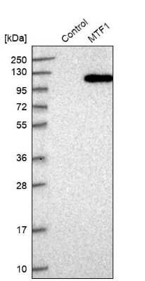
Western Blot: MTF1 Antibody [NBP1-86379]
Western Blot: MTF1 Antibody [NBP1-86379] - Analysis in control (vector only transfected HEK293T lysate) and MTF1 over-expression lysate (Co-expressed with a C-terminal myc-DDK tag (3.1 kDa) in mammalian HEK293T cells).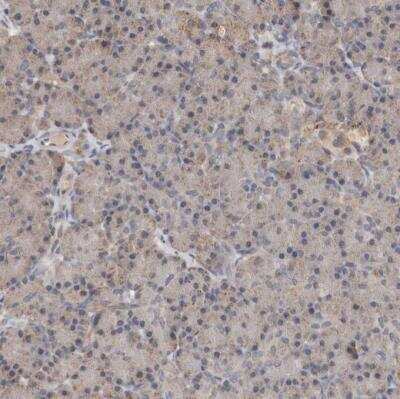
Immunohistochemistry-Paraffin: MTF1 Antibody [NBP1-86379]
Immunohistochemistry-Paraffin: MTF1 Antibody [NBP1-86379] - Staining of human pancreas shows weak cytoplasmic positivity in exocrine glandular cells.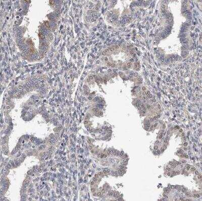
Immunohistochemistry-Paraffin: MTF1 Antibody [NBP1-86379]
Immunohistochemistry-Paraffin: MTF1 Antibody [NBP1-86379] - Staining of human endometrium shows weak positivity in nuclear membrane in glandular cells.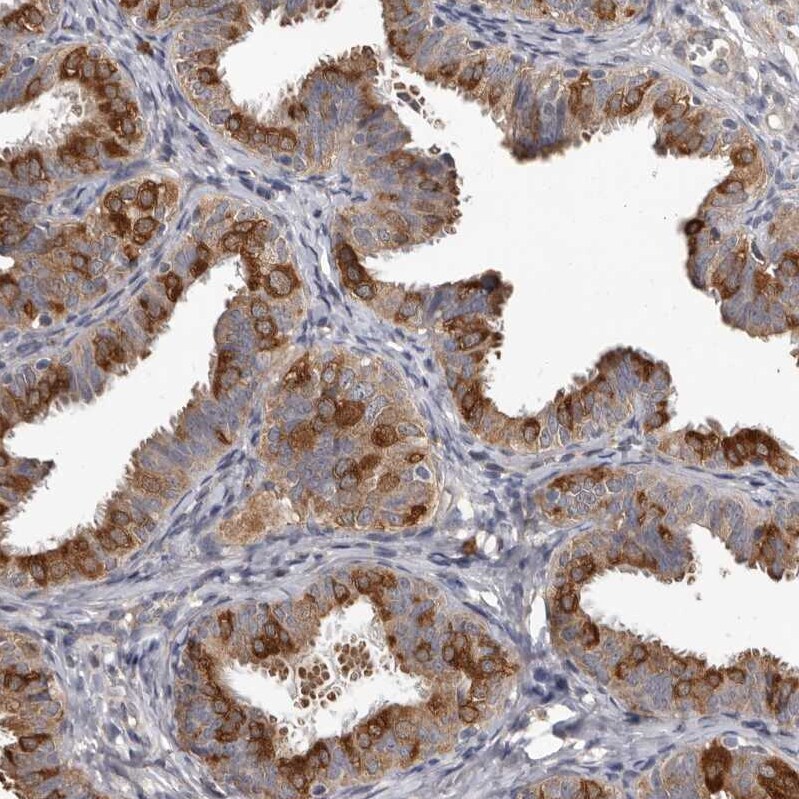
Immunohistochemistry-Paraffin: MTF1 Antibody - BSA Free [NBP1-86379]
Staining of human fallopian tube shows strong cytoplasmic positivity in glandular cells.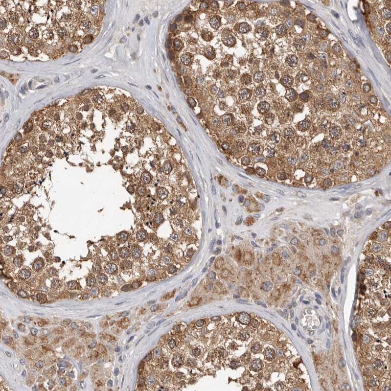
Immunohistochemistry-Paraffin: MTF1 Antibody - BSA Free [NBP1-86379]
Staining of human testis shows strong cytoplasmic positivity in cells in seminiferous ducts.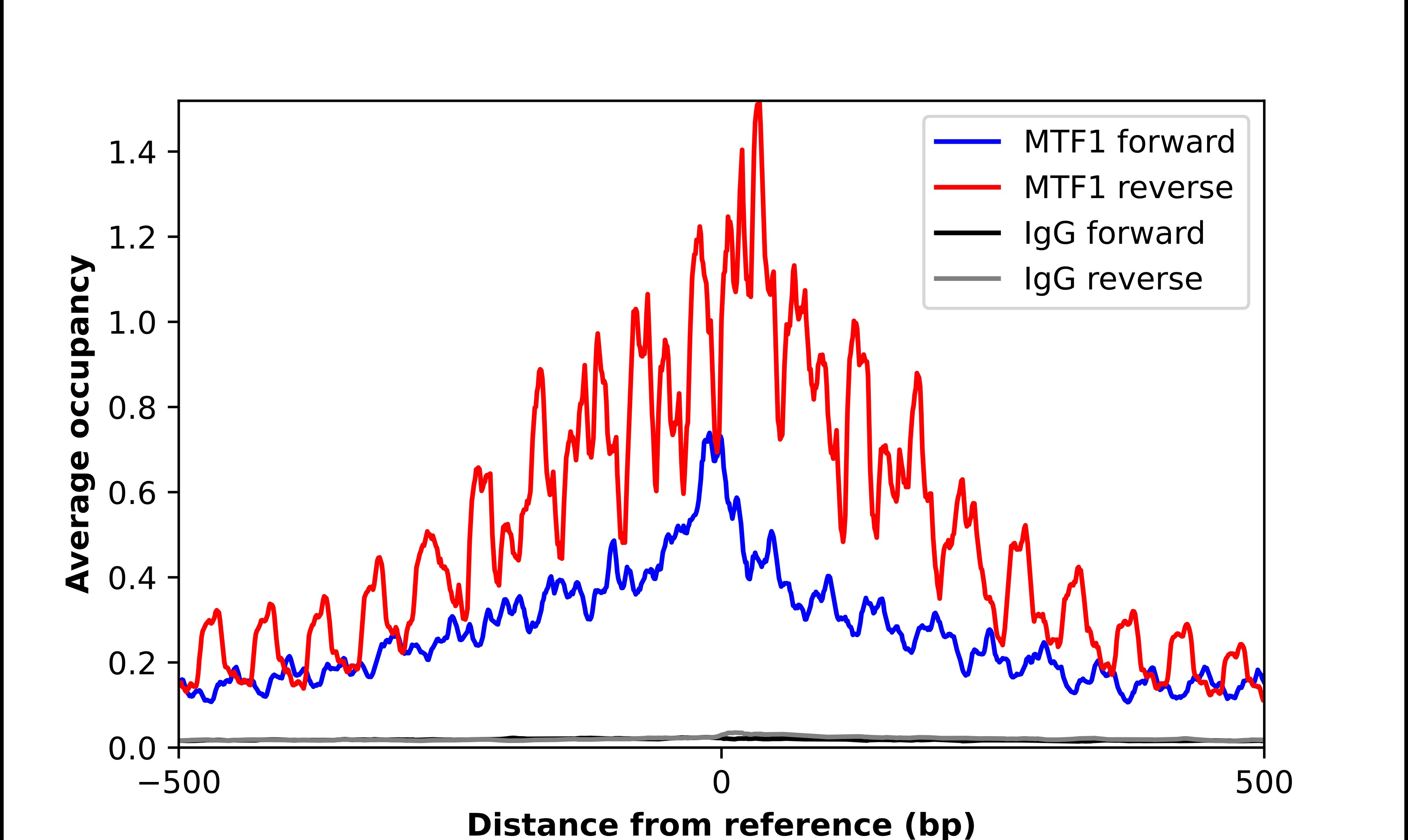
Chromatin Immunoprecipitation-exo-Seq: MTF1 Antibody - BSA Free [NBP1-86379]
ChIP-Exo-Seq composite graph for Anti-MTF1 (NBP1-86379) tested in K562 cells. Strand-specific reads (blue: forward, red: reverse) and IgG controls (black: forward, grey: reverse) are plotted against the distance from a composite set of reference binding sites. The antibody exhibits robust target enrichment compared to a non-specific IgG control and precisely reveals its structural organization around the binding site. Data generated by Prof. B. F. Pugh´s Lab at Cornell University.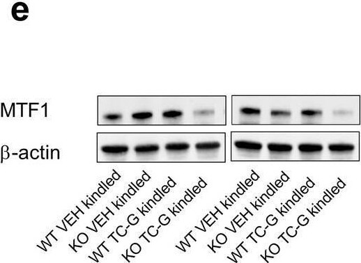
Western Blot: MTF1 Antibody - BSA Free [NBP1-86379] -
The effects of PTZ-kindling in GPR39 KO or WT mice and chronic treatment with TC-G 1008 (10 mg/kg) on zinc levels in serum and hippocampus. a The PTZ kindling in GPR39 KO or WT (C57BL/6/Tar × CBA/Tar) mice consisted of 14 injections of PTZ (25 mg/kg). The biochemical analyses were performed 24 h after the last PTZ injection. b Total zinc (protein bound and [Zn2+]I) was measured in serum by ICP-OES. Data are expressed as means +/- SEM. n = 8 WT VEH, n = 6 WT TC-G 1008, n = 7 KO VEH, n = 7 KO TC-G 1008. c Total zinc was analyzed semi-quantitatively in coronal hippocampal sections by LA-ICP-MS. Data are expressed as means +/- SEM of counts per second (CPS). n = 4 in each group. d The protein expression level of the putative [Zn2+]I sensor, metal regulatory transcription factor 1 (MTF1), was analyzed in the hippocampus. The results (means +/- SEM) are presented as the MTF1/ beta -actin ratio. n = 7 in each group. e Representative blots of MTF1. The observed band (~ 130 kDa). f–h [Zn2+]I was examined in coronal hippocampal sections by a cell-membrane permeable probe, Zinpyr-1 (ZP-1). The mean ZP-1 grey values ratio between mouse sections in the CA3, CA1, or dentate gyrus (DG) regions of the hippocampus is shown. n = 6 WT VEH, n = 6 WT TC-G 1008, n = 5 KO VEH, n = 6 KO TC-G 1008. i A greyscale image of the hippocampal section after staining with ZP-1. p values were determined by two-way ANOVA and Bonferroni’s multiple comparison test. *p < 0.05 Image collected and cropped by CiteAb from the following open publication (https://pubmed.ncbi.nlm.nih.gov/37185787), licensed under a CC-BY license. Not internally tested by Novus Biologicals.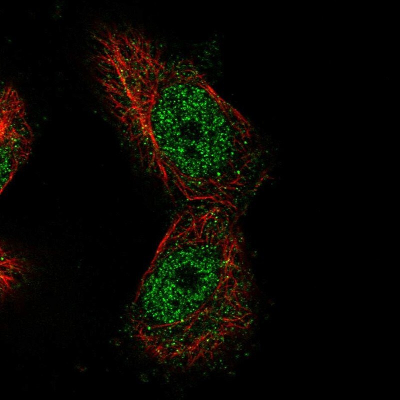
Immunocytochemistry/ Immunofluorescence: MTF1 Antibody [NBP1-86379]
Staining of human cell line U-251 MG shows localization to nucleoplasm.Applications for MTF1 Antibody - BSA Free
Chromatin Immunoprecipitation
Immunohistochemistry
Immunohistochemistry-Paraffin
Western Blot
Formulation, Preparation, and Storage
Purification
Formulation
Format
Preservative
Concentration
Shipping
Stability & Storage
Background: MTF1
Alternate Names
Gene Symbol
Additional MTF1 Products
Product Documents for MTF1 Antibody - BSA Free
Certificate of Analysis
To download a Certificate of Analysis, please enter a lot or batch number in the search box below.
Product Specific Notices for MTF1 Antibody - BSA Free
This product is for research use only and is not approved for use in humans or in clinical diagnosis. Primary Antibodies are guaranteed for 1 year from date of receipt.
Citations for MTF1 Antibody - BSA Free
 Powered by Bioz
Customer Reviews for MTF1 Antibody - BSA Free
There are currently no reviews for this product. Be the first to review MTF1 Antibody - BSA Free and earn rewards!
Have you used MTF1 Antibody - BSA Free?
Submit a review and receive an Amazon gift card!
$25/€18/£15/$25CAN/¥2500 for a review with an image
$10/€7/£6/$10CAN/¥1110 for a review without an image
Submit a review
Protocols
Find general support by application which include: protocols, troubleshooting, illustrated assays, videos and webinars.
- Antigen Retrieval Protocol (PIER)
- Antigen Retrieval for Frozen Sections Protocol
- Appropriate Fixation of IHC/ICC Samples
- Cellular Response to Hypoxia Protocols
- Chromogenic IHC Staining of Formalin-Fixed Paraffin-Embedded (FFPE) Tissue Protocol
- Chromogenic Immunohistochemistry Staining of Frozen Tissue
- ClariTSA™ Fluorophore Kits
- Detection & Visualization of Antibody Binding
- Fluorescent IHC Staining of Frozen Tissue Protocol
- Graphic Protocol for Heat-induced Epitope Retrieval
- Graphic Protocol for the Preparation and Fluorescent IHC Staining of Frozen Tissue Sections
- Graphic Protocol for the Preparation and Fluorescent IHC Staining of Paraffin-embedded Tissue Sections
- Graphic Protocol for the Preparation of Gelatin-coated Slides for Histological Tissue Sections
- IHC Sample Preparation (Frozen sections vs Paraffin)
- Immunofluorescent IHC Staining of Formalin-Fixed Paraffin-Embedded (FFPE) Tissue Protocol
- Immunohistochemistry (IHC) and Immunocytochemistry (ICC) Protocols
- Immunohistochemistry Frozen Troubleshooting
- Immunohistochemistry Paraffin Troubleshooting
- Preparing Samples for IHC/ICC Experiments
- Preventing Non-Specific Staining (Non-Specific Binding)
- Primary Antibody Selection & Optimization
- Protocol for Heat-Induced Epitope Retrieval (HIER)
- Protocol for Making a 4% Formaldehyde Solution in PBS
- Protocol for VisUCyte™ HRP Polymer Detection Reagent
- Protocol for the Preparation & Fixation of Cells on Coverslips
- Protocol for the Preparation and Chromogenic IHC Staining of Frozen Tissue Sections
- Protocol for the Preparation and Chromogenic IHC Staining of Frozen Tissue Sections - Graphic
- Protocol for the Preparation and Chromogenic IHC Staining of Paraffin-embedded Tissue Sections
- Protocol for the Preparation and Chromogenic IHC Staining of Paraffin-embedded Tissue Sections - Graphic
- Protocol for the Preparation and Fluorescent IHC Staining of Frozen Tissue Sections
- Protocol for the Preparation and Fluorescent IHC Staining of Paraffin-embedded Tissue Sections
- Protocol for the Preparation of Gelatin-coated Slides for Histological Tissue Sections
- R&D Systems Quality Control Western Blot Protocol
- TUNEL and Active Caspase-3 Detection by IHC/ICC Protocol
- The Importance of IHC/ICC Controls
- Troubleshooting Guide: Immunohistochemistry
- Troubleshooting Guide: Western Blot Figures
- Western Blot Conditions
- Western Blot Protocol
- Western Blot Protocol for Cell Lysates
- Western Blot Troubleshooting
- Western Blot Troubleshooting Guide
- View all Protocols, Troubleshooting, Illustrated assays and Webinars