NMDA R, NR1 Subunit Antibody - Splice Variant - Azide and BSA Free
Novus Biologicals | Catalog # NB300-114
![Western Blot: NMDA R, NR1 Subunit AntibodyAzide and BSA Free [NB300-114]](https://resources.rndsystems.com/images/products/NMDA-receptor-N1-Antibody-Western-Blot-NB300-114-img0004.jpg "Western Blot: NMDA R, NR1 Subunit AntibodyAzide and BSA Free [NB300-114]")
Loading...
Key Product Details
Species Reactivity
Validated:
Mouse, Rat
Predicted:
Canine (100%), Chicken (100%), Human (100%), Primate (100%), Xenopus (100%). Backed by our 100% Guarantee.
Applications
Immunohistochemistry, Immunohistochemistry-Frozen, Western Blot
Label
Unconjugated
Antibody Source
Polyclonal Rabbit IgG
Format
Azide and BSA Free
Loading...
Product Specifications
Immunogen
Synthetic peptide corresponding to amino acid residues specific to the NR1 subunit, N1 splice variant insert conjugated to KLH. Accession # P35439
Reactivity Notes
It is anticipated that the antibody will also work with canine, chicken, human, non-human primate and Xenopus tissues based on the fact that these species have 100% homology with the amino acid sequence used as antigen.
Specificity
Specific for endogenous levels of the ~120 kDa NMDA R, NR1 Subunit splice variant insert protein of the NMDA receptor. Does not recognize NR1 subunits of the NMDA receptor that do not contain the N1 insert.
Clonality
Polyclonal
Host
Rabbit
Isotype
IgG
Theoretical MW
120 kDa.
Disclaimer note: The observed molecular weight of the protein may vary from the listed predicted molecular weight due to post translational modifications, post translation cleavages, relative charges, and other experimental factors.
Disclaimer note: The observed molecular weight of the protein may vary from the listed predicted molecular weight due to post translational modifications, post translation cleavages, relative charges, and other experimental factors.
Description
The antigen is a peptide from the NR1 subunit, N1 splice variant insert of rat NMDA receptor. Recommended that the undiluted antibody be aliquoted into smaller working volumes (10-30 uL/vial depending on usage).
Scientific Data Images for NMDA R, NR1 Subunit Antibody - Splice Variant - Azide and BSA Free
Western Blot: NMDA R, NR1 Subunit AntibodyAzide and BSA Free [NB300-114]
Western Blot: NMDA receptor N1 Antibody [NB300-114] - 10 ug of HEK 293 cells expressing: Lane 1 - NR1 subunit containing the N1 and C2' Insert showing specific immunolabeling of the ~120k NR1 subunit of the NMDA receptor containing the N1 splice variant insert.; Lane 2 - NR1 subunit containing only the C2 Insert; Lane 3 - NR1 subunit containing the C1 and C2' Insert; Lane 4 - without NR1 expression (Mock).![Flow Cytometry: NMDA R, NR1 Subunit Antibody - Azide and BSA Free [NB300-114]](https://resources.rndsystems.com/images/products/NMDA-receptor-N1-Antibody-Flow-Cytometry-NB300-114-img0002.jpg "Flow Cytometry: NMDA R, NR1 Subunit Antibody - Azide and BSA Free [NB300-114]")
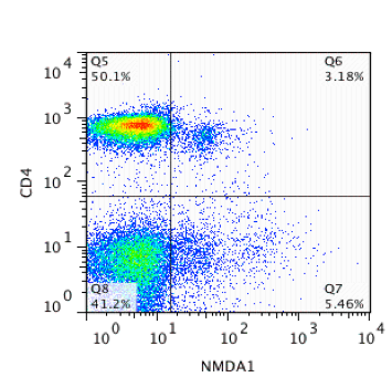
Flow Cytometry: NMDA R, NR1 Subunit Antibody - Azide and BSA Free [NB300-114]
Flow Cytometry: NMDA receptor N1 Antibody [NB300-114] - CD4+ Lymphocytes expressing NMDAR1. Image provided by verified customer.Applications for NMDA R, NR1 Subunit Antibody - Splice Variant - Azide and BSA Free
Application
Recommended Usage
Immunohistochemistry
1:10-1:500
Immunohistochemistry-Frozen
1:1000-1:2000
Western Blot
1:1000
Application Notes
NB 300-114 can be used in Western blot where a band is seen at ~ 100 kDa representing the NMDAR1 N1 splice variant.
Reviewed Applications
Read 1 review rated 4 using NB300-114 in the following applications:
Formulation, Preparation, and Storage
Purification
Antigen Affinity-purified
Reconstitution
Reconstitute with 50 uL PBS to desired concentration.
Formulation
Lyophilized
Format
Azide and BSA Free
Preservative
No Preservative
Concentration
LYOPH mg/ml
Shipping
The product is shipped with polar packs. Upon receipt, store it immediately at the temperature recommended below.
Stability & Storage
Store at -20C. Avoid freeze-thaw cycles.
Calculators
Background: NMDA R, NR1 Subunit
Long Name
Glutamate Receptor, Ionotropic, N-Methyl-D-Aspartate, Subunit 1
Alternate Names
GluN1, MRD8, NDHMSD, NDHMSR;NMD-R1, NMDA R, NR1 Subunit, NMDA1, NMDAR1, NMD-R1, NR1
Gene Symbol
GRIN1
UniProt
Additional NMDA R, NR1 Subunit Products
Product Documents for NMDA R, NR1 Subunit Antibody - Splice Variant - Azide and BSA Free
Certificate of Analysis
To download a Certificate of Analysis, please enter a lot or batch number in the search box below.
Product Specific Notices for NMDA R, NR1 Subunit Antibody - Splice Variant - Azide and BSA Free
This product is for research use only and is not approved for use in humans or in clinical diagnosis. Primary Antibodies are guaranteed for 1 year from date of receipt.
Citations for NMDA R, NR1 Subunit Antibody - Splice Variant - Azide and BSA Free
 Powered by Bioz
Customer Reviews for NMDA R, NR1 Subunit Antibody - Splice Variant - Azide and BSA Free (1)
4 out of 5
1 Customer Rating
Have you used NMDA R, NR1 Subunit Antibody - Splice Variant - Azide and BSA Free?
Submit a review and receive an Amazon gift card!
$25/€18/£15/$25CAN/¥2500 Yen for a review with an image
$10/€7/£6/$10CAN/¥1110 Yen for a review without an image
Submit a review
Customer Images
Showing
1
-
1 的
1 review
Showing All
Filter By:
-
Application: Flow CytometrySample Tested: Balb/c Mouse LymphocytesSpecies: MouseVerified Customer | Posted 01/31/2014CD4+ Lymphocytes expressing NMDAR1

There are no reviews that match your criteria.
Protocols
Find general support by application which include: protocols, troubleshooting, illustrated assays, videos and webinars.
- Antigen Retrieval Protocol (PIER)
- Antigen Retrieval for Frozen Sections Protocol
- Appropriate Fixation of IHC/ICC Samples
- Cellular Response to Hypoxia Protocols
- Chromogenic IHC Staining of Formalin-Fixed Paraffin-Embedded (FFPE) Tissue Protocol
- Chromogenic Immunohistochemistry Staining of Frozen Tissue
- ClariTSA™ Fluorophore Kits
- Detection & Visualization of Antibody Binding
- Fluorescent IHC Staining of Frozen Tissue Protocol
- Graphic Protocol for Heat-induced Epitope Retrieval
- Graphic Protocol for the Preparation and Fluorescent IHC Staining of Frozen Tissue Sections
- Graphic Protocol for the Preparation and Fluorescent IHC Staining of Paraffin-embedded Tissue Sections
- Graphic Protocol for the Preparation of Gelatin-coated Slides for Histological Tissue Sections
- IHC Sample Preparation (Frozen sections vs Paraffin)
- Immunofluorescent IHC Staining of Formalin-Fixed Paraffin-Embedded (FFPE) Tissue Protocol
- Immunohistochemistry (IHC) and Immunocytochemistry (ICC) Protocols
- Immunohistochemistry Frozen Troubleshooting
- Immunohistochemistry Paraffin Troubleshooting
- Preparing Samples for IHC/ICC Experiments
- Preventing Non-Specific Staining (Non-Specific Binding)
- Primary Antibody Selection & Optimization
- Protocol for Heat-Induced Epitope Retrieval (HIER)
- Protocol for Making a 4% Formaldehyde Solution in PBS
- Protocol for VisUCyte™ HRP Polymer Detection Reagent
- Protocol for the Preparation & Fixation of Cells on Coverslips
- Protocol for the Preparation and Chromogenic IHC Staining of Frozen Tissue Sections
- Protocol for the Preparation and Chromogenic IHC Staining of Frozen Tissue Sections - Graphic
- Protocol for the Preparation and Chromogenic IHC Staining of Paraffin-embedded Tissue Sections
- Protocol for the Preparation and Chromogenic IHC Staining of Paraffin-embedded Tissue Sections - Graphic
- Protocol for the Preparation and Fluorescent IHC Staining of Frozen Tissue Sections
- Protocol for the Preparation and Fluorescent IHC Staining of Paraffin-embedded Tissue Sections
- Protocol for the Preparation of Gelatin-coated Slides for Histological Tissue Sections
- R&D Systems Quality Control Western Blot Protocol
- TUNEL and Active Caspase-3 Detection by IHC/ICC Protocol
- The Importance of IHC/ICC Controls
- Troubleshooting Guide: Immunohistochemistry
- Troubleshooting Guide: Western Blot Figures
- Western Blot Conditions
- Western Blot Protocol
- Western Blot Protocol for Cell Lysates
- Western Blot Troubleshooting
- Western Blot Troubleshooting Guide
- View all Protocols, Troubleshooting, Illustrated assays and Webinars
Loading...